Skoči na glavni sadržaj

Kratko priopćenje

https://doi.org/10.54062/jb.3.1.4

Inheritance of the Epidermolysis Bullosa Subtypes

Anjeza Temaj ; Department of General Medicine, Faculty of Medicine, North Macedonia
Yllka Gashi ; Department of General Medicine, Faculty of Medicine, North Macedonia
Nexhibe Nuhii ; Department of Pharmacy, Faculty of medicine, North Macedonia
Nevzat Elezi ; Department of General Medicine, Faculty of Medicine, North Macedonia
Sheqibe Beadini ; Department of General Medicine, Faculty of Medicine, North Macedonia
Ron Elezi ; Department of General Medicine, Faculty of Medicine, North Macedonia
Vegim Zhaku ; Department of General Medicine, Faculty of Medicine, North Macedonia
Nexbedin Beadini ; Department of General Medicine, Faculty of Medicine, North Macedonia
Crita Uzeiri-Havziu ; Department of Pharmacy, Faculty of medicine, North Macedonia
Sefedin Bilalli ; Institute of Biochemistry, Shkup & Department of Pharmacy, Faculty of medicine, North Macedonia
Albulena Beadini ; Department of General Medicine, Faculty of Medicine, North Macedonia
Arjeta Shabani ; Department of Pharmacy, Faculty of medicine, North Macedonia
Gjylai Alija ; Department of Pharmacy, Faculty of medicine, North Macedonia
Arlinda Haxhiu- Zajmi ; Department of Pharmacy, Faculty of medicine, North Macedonia


Puni tekst: engleski pdf 833 Kb

verzije

str. 53-57

preuzimanja: 22

citiraj

Preuzmi JATS datoteku


Sažetak

Epidermolysis bullosa (EB) is a group of inherited disorders that cause skin to blister and tear easily. The disease is caused by mutations in structural proteins that are key for maintaining the integrity of the skin’s basement membrane zone or dermoepidermal junction.
EB can be inherited in two ways: autosomal dominant and autosomal recessive. The most common form of EB, epidermolysis bullosa simplex (EBS), as well as some forms of dystrophic epidermolysis bullosa (DEB) are inherited in an autosomal dominant pattern. This means they are passed down from an affected parent to half of his or her children. Other forms of EB, such as junctional epidermolysis bullosa (JEB) and some forms of DEB, are inherited in an autosomal recessive pattern. This means that two copies of the mutated gene, one from each parent, are required to develop the condition.

Ključne riječi

Epidermolysis bullosa; Inheritance; Cure; Kindler syndrome

Hrčak ID:

314612

URI

https://hrcak.srce.hr/314612

Datum izdavanja:

23.2.2024.

Posjeta: 66 *


Introduction

Hereditary diseases such as Epidermolysis bullosa (EB) is caused by mutation of approximately 16 different genes who are involved in the maintenance of the structure and function of dermo-epidermal adhesion in epithelia, and in many cases, it is fatal for patients affected (Wally et al., 2020) (see Table 1 and figure 1).

Skin blister is the main symptoms which characterized Epidermolysis bullosa. Blistering can cause minor trauma and are painful. The prevalence of EB in the United States is 8.2 per million live births (Fine et al., 2004).

The term epidermolysis bullosa (EB) was introduced in 1886 and refers to a group of rare genetic disease which are characterized by varying degrees of skin fragility caused by mutations in the protein level at the various skin structure. There are four main types: Epidermolysis bullosa simplex (EBS), Dystrophic epidermolysis bullosa (DEB), Junctional epidermolysis bullosa (JEB), and Kindler syndrome (KS) (Fine and Hintner 2021)

Intong and Murrell report on recent changes and agree for new definitions during the meeting which is take placed in Vienna, Austria, in Vienna, Austria, in May 2007 and was published in 2012 (Intong and Murrell, 2012).

Table 1. Several types and subtypes of Epidermolysis bullosa.

Major EB typeMajor EB subtypeMinor EB subtypeTarget proteins
Epidermolysis bullosa simplex

Suprabasal EBS

Basal EBS

Lethal acantholytic EB

Plakophilin deficiency

EB superficialis

EBS localized

EBS- Dowling Meara

EBS-other generalized

EBS with mottled pigmentation

EBS with muscular dystrophy

EBS pyloric atresia

EBS autosomal recessive

EBS ogna

EBS migratory circinate

Desmoplakin

Plakophilin-1

?

K5, K14

K5, K14

K5, K14

K5

Plectin

Plactin, α6β4 integrin

K14

Plectin

K5

Junctional epidermolysis bullosa

JEB-Herlitz

JEB, other

-

JEB-non-herlitz, generalized

JEB-Non-herlitz, localized

JEB-Pyloric atresia

JEB inversa

JEB late onset

LOC syndrome

Laminin -332

Laminin -332

Type XVII collagen α6β4 integrine

Laminin -332

?

Laminin -332

Dystrophic epidermolysis bullosaDominant DEB

DDEB generalized

DDEB acral

DDEB pretibial

DDEB pruriginosa

DDEB nails only

DDEB bullous dermolysis of newborn

Type VII collagen

Type VII collagen

Dystrophic epidermolysis bullosaRecessive DEB

RDEB severe generalized

RDEB inversa

RDEB pretibial

RDEB pruriginosa

RDEB centripetalis

RDEB bullous dermolysis of newborn

COL7A1
Kindler syndromeKSAutosomal recessiveFERMT1

Based on symptoms the Epidermolysis bullosa (EB) genetically and clinically is characterized by blister formation and erosions of the skin and mucous membranes after minor trauma (Laimer et al., 2009). Mayr et al., (2013) suggest the inheritance of the affected genes can occur in a dominant or recessive way depending on the subform of the disease. The EBs is caused by gene mutations which encode proteins placed in basal membrane zone of the skin. Loss function (absence) of proteins placed in this zone is shown to participate in lacking of akin stability and microarchitecture of the connection between dermis and epidermis leading to a loss of coherence. The connection between dermis and epidermis (called basal membrane) is addicted by keratinocytes and dermal fibroblasts that acts as mechanical support for the connection of both skin layers. The basal membrane also regulates the metabolic exchange between the two skin compartments.

Mayr et al., (2013) report for mutations in the genes, encoding for the keratins 5 and 14 and plectin, lead to epidermolysis bullosa simplex (EBS) characterized by the cytolysis within basal keratinocytes. Loss function of laminin – 332, collagen type XVII or integrin- β4 is shown to cause Junctional epidermolysis bullosa (JEB) which is subtype of EBs. This subtype is the most serve of EBs characterized by separation of skin within lamina lucida. Mutations in type VII collagen (encoded by COL7A1) lead to the dystrophic form of epidermolysis bullosa. The clinical manifestation depended on the mutation type (missense mutation, nonsense mutation, splice site mutation, deletion and insertion).

The aim of this paper is to present the different subtype of EBs and using of different methods for cure of them used and suggested by different research groups.

image1.png

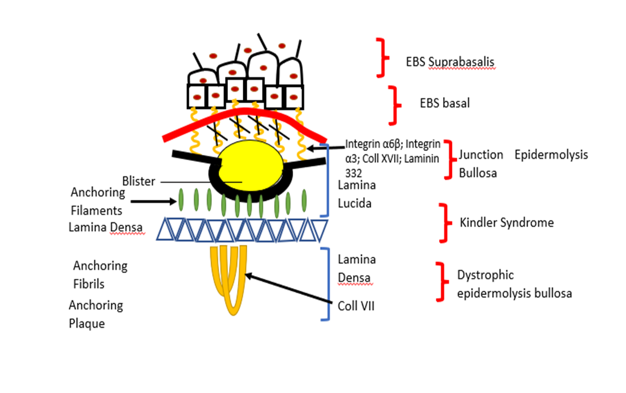
Figure 1. Here are presented different subtypes of EBs and gene mutation which are found.

The therapy suggested for cure of EBs

There is suggestion different type of therapies for EBs cure. For example, gene therapy: correction of JEB by transplantation of epidermis stem cells by Murauer et al., (2015). Using of small molecules such Topical Diacerin in cure of EBs Dowling Meara by Wally et al., (2013). Gene expression studies to identified candidate repair proteins in wound healing suggested by Breitenbach et al., (2015). Bauer et al., (2013) suggest using of specialized ribosome for cure of EBs.

Ribosomal protein has extra function

The ribosome is ribonucleoprotein complex organelle which is responsible for protein synthesis, and its synthesis is highly coordinated; this is shown in involvement of many macromolecules’ components (Temaj et al., 2022; Temaj et al., 2022; Temaj et al., 2022).

Narla and Ebert suggested that several ribosomal proteins have extraribosomal functions, including replication and repair of DNA repair, so mutations in ribosomal proteins may have effects that are independent of the protein translation machinery (Narla and Ebert, 2010).

Which criteria are needed for ribosomal protein to consider that have extra-ribosomal capacity? Warner and McIntosh suggest three criteria: 1) the ribosomal protein in question interacts specifically with some nonribosomal components of the cell, presumably RNA or protein; 2) demonstrating that such interaction in living cell have physiological effects; and 3) evidence that the latter is occurring away from the ribosome (Warner and McIntosh, 2009).

Danilova and Gazda (2015), report that DBA, often is diagnosed during the first year of life; the clinical feature is anemia, low reticulocyte count, macrocytic erythrocyte, increased expression of fetal hemoglobin and elevated activity of adenosine deaminase. Mutation of RPL5/uL18 in DBA caused cleft lips and palats, but this malformation is not observed in mutations of RPS19/eS19 (Gazda et al., 2008).

Mutations of RPL5/uL18 and RPL10/uL16 in DBA have been reported in T-cell acute leukemia (De Keersmaecker et al., 2013). Mutations of 40S subunit ribosomal proteins cause congenital aplesnia (Bolze et al., 2013). In Drosophila melanogaster haploinsuficiency 40S ribosomal proteins is characterized by delay of development, small size, small bristles, and small rough eyes (Marygold et al., 2005). In zebra fishes’ mutations in RPs are associated with delay of development, small size, small head and eyes, brain, apoptosis, reduced pigmentations, pericardial edema and hematopoietic defects (Danilova and Gazda 2015; Amsterdam et al., 2004; Zhang et al., 2014). Mutations of RPS19/eS19 and RPS20/uS10 in mice are associated with dark skin and reduced body (McGowan et al., 2008). RPL24/eL24 mutations in mice lead to the Belly Spot and Tail (Bst) phenotype which is characterized by small size, eye defects, a white ventral spot, white hind feet and various skeletal abnormalities (Oliver et al., 2004). RPL7/uL30 mutation also is manifested with skeletal abnormalities, and with ventral white spot and eye defects (Watkins-Chow et al., 2013).

JEBs (Junction Epidermolysis Bullosa)

The PTC (premature termination codon) in LAMB3 is shown to play pivotal role in JEBs as subtype of EBs. The ribosome as a complex organelle is responsible for translation of LAMB3PTC mRNA aborts protein synthesis at the PTC signal, with production of a truncated, non-functional protein. New drug development in the future must play pivotal in binding with ribosomal protein L35 (rpL35/uL29), and to modified them which will customize increase in production of full-length Lamb3 protein from a LAMB3PTC mRNA. The same authors suggest that Atazanavir and artesunate are the main drug candidate which can bind to ribosomal protein rpL35 and now may tested for their potential to trigger a rpL35 ribosomal switch to increase production of full-length Lamb3 protein from a LAMB3PTC mRNA for targeted systemic therapy in treating JEB (Rathner et al., 2021).

Conclusions

In conclusion, we can say, that in EBs suggested from clinical dermatologist, are used different strategies for cure of epidermolysis bullosa. For example, using of stem cells are shown to be very benefits in some subtypes. Employment of small molecules such as drug diacerin for treatment of EBs Dowling Meara. Gene technology for correction of mutated gene COL7A1 by trans-splicing in dystrophic epidermolysis bullosa (DEBs). Last time is suggested modification of ribosomal protein for translation repair of PTC (premature termination codon) in mutated JEBs.

References

Ribosomopathies and cancer: pharmacological implications. Expert Rev Clin Pharmacol, 15(6):729-746.

References


Amsterdam, A., Nissen, R.M., Sun, Z., Swindell, E.C., Farrington, S., Hopkins. N., 2004 Identification of 315 genes essential for early zebrafish development. Proc Natl Acad Sci USA. 101127927:


Bauer, J.W., Brandl, C., Haubenreisser, O., Wimmer, B., Weber, M., Karl, T., Klausegger, A., Breitenbach, M., Hintner, H., von der Haar, T., Tuite, M.F., Breitenbach-Koller, L., 2013 Specialized yeast ribosomes: a customized tool for selective mRNA translation. PLoS One. 8:67609


Bolze, A., Mahlaoui, N., Byun, M., Turner, B., Trede, N., Ellis, S.R., et al., 2013 Ribosomal protein SA haploinsufficiency in humans with isolated congenital asplenia. Science. 340:976–98


Breitenbach, J.S., Rinnerthaler, M., Trost, A., Weber, M., Klausegger, A., Gruber, C., Bruckner, D., Reitsamer, H.A., Bauer, J.W., Breitenbach, M., 2015 Transcriptome and ultrastructural changes in dystrophic Epidermolysis bullosa resemble skin aging. Aging (Albany NY). 7389411(6) https://doi.org/ 10.18632/aging.100755.


Danilova, N., Gazda, H.T., 2015 Ribosomopathies: how a common root can cause a tree of pathologies. Dis Model Mech. 8101326:


De Keersmaecker, K., Atak, Z.K., Li, N., Vicente, C., Patchett, S., Girardi, T., Gianfelici, V., et al. 2013 Exome sequencing identifies mutation in CNOT3 and ribosomal genes RPL5 and RPL10 in T-cell acute lymphoblastic leukemia. Nat Genet. 4518690:


Fine, J.D., Hintner, H,. 2021 Life with Epidermolysis Bullosa (EB): Etiology, Diagnosis, Multidisciplinary Care and Therapy. Springer Science & Business Media.; p. 242Archived from the original on 2021-11-03. Retrieved 2020-11-21.


Fine, J.D., Johnson, L.B., Weiner, M., Stein, A., Cash, S., DeLeoz, J., Devries, D.T., Suchindran, C., 2014 "Genitourinary Complications of Inherited Epidermolysis Bullosa:. Experience of the National Epidermylosis Bullosa Registry and Review of the Literature". J Urol. 1725120404:


Gazda, H.T., Sheen, M.R., Vlachos, A., Choesmel, V., O'Donohue, M.F., Schneider, H., et al. 2008 Ribosomal protein L5 and L11 mutations are associated with cleft palate and abnormal thumbs in Diamond-Blackfan anemia patients. Am J Hum Genet. 8376980:


Intong, L.R., Murrell, D.F., 2012 Inherited epidermolysis bullosa: new diagnostic criteria and classification. Clin Dermatol. 30707:


Laimer, M., Lanschutzer, C.M., Nischler, E., Klausegger, A., Diem, A., Pohla-Gubo, G., Bauer, J.W., Hintner, H., 2013 [Hereditary blistering diseases. Symptoms, diagnosis and treatment of epidermolysis bullosa]. Hautarzt. 6037888:


Marygold, S.J., Coelho, C.M., Leevers, S.J., 2005 Genetic analysis of RpL38 and RpL5, two-minute genes located in the centric heterochromatin of chromosome 2 of Drosophila melanogaster. Genetics. 16968395:


Mayr, E.K.U., Bauer, J.W., 2013 Gene Therapy for the COL7A1 Gene in Molina, F. M. (Ed), , editor. Gene Therapy - Tools and Potential Applications, InTech:. Rijeka, Croatia.:


McGowan, K.A., Li, J.Z., Park, C.Y., Beaudry, V., Tabor, H.K., Sabnis, A.J., et al., 2008 Ribosomal mutations cause p53-mediated dark skin and pleiotropic effects. Nat Genet. 4096370:


Murauer, E.M., Koller, U., Pellegrini, G., De Luca, M., Bauer, J.W., 2015 Advances in Gene/Cell Therapy in Epidermolysis Bullosa. Keio J Med. 64215:


Narla, A., Ebert, B.L., 2010 Ribosomopathies: human disorders of ribosome dysfunction. Blood. 1153196205:


Oliver, E.R., Saunders, T.L., Tarle, S.A., Glaser, T., 2004 Ribosomal protein L24 defect in belly spot and tail (Bst), a mouse Minute. Development. 131390720:


Rathner, A., Rathner, P., Friedrich, A., Wießner, M., Kitzler, C.M., Schernthaner, J., et al., 2021 Drug Development for Target Ribosomal Protein rpL35/uL29 for Repair of LAMB3R635X in Rare Skin Disease Epidermolysis Bullosa. Skin Pharmacol Physiol. 34167182(4)


Temaj, G., Chichiarelli, S., Eufemi, M., Altieri, F., Hadziselimovic, R., Farooqi, A.A., Yaylim, I., Saso, L., 2022 Ribosome-Directed Therapies in Cancer. Biomedicines. 102088(9)


Temaj, G., Hadziselimovic, R., Nefic, H., Nuhii, N., 2022 Ribosome biogenesis and ribosome therapy in cancer cells. Research Result in Pharmacology. 281524103897881706(4)


Temaj, G., Saha, S., Dragusha, S., Ejupi, V., Buttari, B., Profumo, E., Beqa, L., Saso, L., 2022 Ribosomopathies and cancer: pharmacological implications. Expert Rev Clin Pharmacol. 15729746(6)


Wally, V., Kitzmueller, S., Lagler, F., Moder, A., Hitzl, W., Wolkersdorfer, M., Hofbauer, P., Felder, T.K., Dornauer, M., Diem, A., Eiler, N., Bauer, B.J.W., 2013 Topical diacerein for epidermolysis bullosa: a randomized controlled pilot study. Orphanet J Rare Dis. 869:


Wally, V., Reisenberger, M., Kitzmüller, S., Laimer M., 2020 Small molecule drug development for rare genodermatoses - evaluation of the current status in epidermolysis bullosa. Orphanet J Rare Dis. 15292(1)


Warner, J.R., McIntosh, K.B., 2009 How common are extraribosomal functions of ribosomal proteins? Mol Cell. 34311:


Watkins-Chow, D.E., Cooke, J., Pidsley, R., Edwards, A., Slotkin, R., Leeds, K.E., Mullen, R., et al., 2013 Mutation of the diamond-blackfan anemia gene Rps7 in mouse results in morphological and neuroanatomical phenotypes. PLoS Genet. 9:1003094


Zhang, Y., Ear, J., Yang, Z., Morimoto, K., Zhang, B., Lin, S., 2014 Defects of protein production in erythroid cells revealed in a zebrafish Diamond-Blackfan anemia model for mutation in RPS19. Cell Death Dis. 5:1352


This display is generated from NISO JATS XML with jats-html.xsl. The XSLT engine is libxslt.