Skoči na glavni sadržaj

Pregledni rad

https://doi.org/10.5599/admet.1918

Anti-inflammatory activities of flavonoid derivates

Ysrafil Ysrafil
Zulfiayu Sapiun
Nangsih Sulastri Slamet
Fihrina Mohamad
Hartati Hartati
Sukmawati A Damiti
Francisca Diana Alexandra
Sudarman Rahman
Sri Masyeni
Harapan Harapan
Sukamto S. Mamada
Talha Bin Emran
Firzan Nainu


Puni tekst: engleski pdf 843 Kb

str. 331-359

preuzimanja: 54

citiraj

Preuzmi JATS datoteku


Sažetak

Background and purpose
Flavonoids are a group of phytochemicals found abundantly in various plants. Scientific evidence has revealed that flavonoids display potential biological activities, including their ability to alleviate inflammation. This activity is closely related to their action in blocking the inflammatory cascade and inhibiting the production of pro-inflammatory factors. However, as flavonoids typically have poor bioavailability and pharmacokinetic profile, it is quite challenging to establish these compounds as a drug. Nevertheless, progressive advancements in drug delivery systems, particularly in nanotechnology, have shown promising approaches to overcome such challenges.
Review approach
This narrative review provides an overview of scientific knowledge about the mechanism of action of flavonoids in the mitigation of inflammatory reaction prior to delivering a comprehensive discussion about the opportunity of the nanotechnology-based delivery system in the preparation of the flavonoid-based drug.
Key results
Various studies conducted in silico, in vitro, in vivo, and clinical trials have deciphered that the anti-inflammatory activities of flavonoids are closely linked to their ability to modulate various biochemical mediators, enzymes, and signalling pathways involved in the inflammatory processes. This compound could be encapsulated in nanotechnology platforms to increase the solubility, bioavailability, and pharmacological activity of flavonoids as well as reduce the toxic effects of these compounds.
Conclusion
In Summary, we conclude that flavonoids and their derivates have given promising results in their development as new anti-inflammatory drug candidates, especially if they formulate in nanoparticles.

Ključne riječi

inflammation treatment; bioactive compounds; plants-base drug; nanotechnology

Hrčak ID:

308783

URI

https://hrcak.srce.hr/308783

Datum izdavanja:

21.9.2023.

Posjeta: 196 *


Introduction

Inflammation is a complex and important protective response of the body to a stimulus in the form of stimulation by microorganisms, physical injuries, chemicals, allergic reactions or the presence of endogenous signals due to cell damage. In a physiological state, inflammation aims to eradicate inflammatory agents as well as repair the injured tissue and wounds [1-3]. In the aspect of pathomechanism, inflammation involves the body's immune system response, both natural and adaptive, although in most cases, the response is mainly initiated by the natural immune system. Inflammation is initially stimulated by the introduction of highly conserved pathogen- or damaged- associated molecular patterns (PAMPs and DAMPs) by pattern recognition receptors (PRRs) of the immune cells [4-6]. This will trigger the subsequent activation of the inflammatory cells such as neutrophils, eosinophils, mononuclear phagocytes, and macrophages, leading to the excessive production of nitric oxide (NO), prostaglandin E2 (PGE2), C-reactive protein (CRP), chemokines and proinflammatory cytokines such as interleukins (IL)-1β, IL-6, and tumour necrosis factor (TNF)-α.

The production of proinflammatory cytokines generally occurs due to the induction of nuclear factor-kappa B (NF-κB), a transcription factor that plays a key role in the regulation of inflammation. In addition to the release of several molecules above, the inflammatory process is also accompanied by the release of a number of bioactive lipid mediators such as thromboxane, prostaglandins and prostacyclins on the initiative of the phospholipid conversion cascade by phospholipase and cyclooxygenase enzymes. The release of cytokines, chemokines, and mediators and these inflammatory molecules then triggers the appearance of inflammatory signs such as calor, dolor, rubor, tumour and functio laesa, and if it continues, it can cause a worse impact [3,7].

However, the protective function of inflammation can shift to the detrimental effects caused by the uncontrolled and excessive inflammatory response. In this condition, inflammation may disturb the homeostasis of the body's physiological processes and eventually develop into an inflammatory disorder [1-3]. Several studies have demonstrated that inflammation is associated with developing and worsening various non-communicable disease-associated inflammation disorders, such as autoimmune diseases, neurodegenerative diseases, cancer, diabetes mellitus, and cardiovascular diseases [1,8]. These findings support the development of anti-inflammatory drugs to prevent the progression of the diseases.

Advances in the last few decades have revealed many facts about the benefits of various plant metabolites against inflammation. Curcumin, resveratrol, and capsaicin have been previously reported to have the ability to decrease the production of the proinflammatory cytokines IL-1β, IL-6, and TNF-α, inhibit cyclooxygenase-2 (COX-2) and the inflammatory pathways (e.g., NF-κB, mitogen-activated protein kinase (MAPK), and activator protein 1 (AP-1) [9,10].

Of several plant metabolites with potential anti-inflammatory action, flavonoids have attracted much interest. Several studies have reported the activity of flavonoids in interfering with inflammatory signalling pathways and key enzymes, leading to the inhibition of the release of pro-inflammatory proteins and mediators [8,11]. However, the current problem is that these compounds have poor bioavailability and pharmacokinetic profiles. Advances in nanotechnology have brought promising approaches in improving the bioavailability and increasing the effectiveness of these compounds as anti-inflammatory drug candidates [12,13].

In our review, we summarize the latest scientific findings regarding the benefits of flavonoids, pharmacokinetics, and their development based on nanomedicine, as this technology has become a solution in the delivery and improvement of drug effectiveness, including drugs used to impede inflammatory conditions.

Mechanisms of inflammation

As a fundamental biological process, inflammation is the protective response to tackle invading pathogens, antigens or damaged cells or tissues. It provides a defence mechanism to eliminate harmful substances and perform tissue repair [14,15]. At the molecular and cellular level, inflammation is characterized by five major signs: redness, heat, pain, swelling and functional loss. These changes can be triggered by two main causes, including infectious and non-infectious-related causes. The presence of these causes in the body will be recognized by receptors in the body that are subsequently followed by the activation of the inflammatory cascade, the release of the markers, and the recruitment of the inflammatory cells [16].

The recognition of PAMPs and DAMPs by PRRs

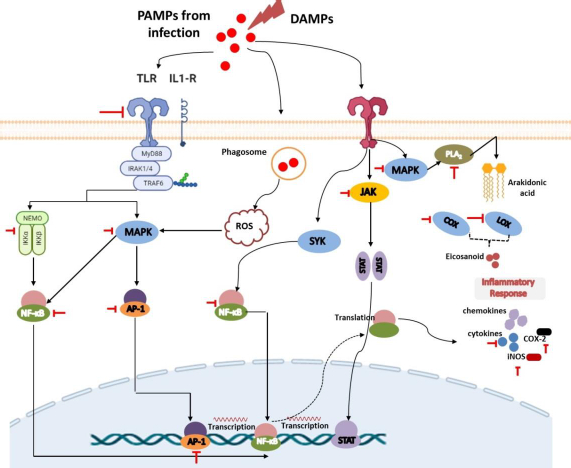
Tissue injury, cell-infected pathogens, microbial invasion, or death cells associated with necroptosis or pyroptosis will release PAMPs or DAMPs. PAMPs are unique structures in microbes or pathogens (e.g., LPS, β-glucan, flagellin, spike, DNA, RNA) that can be recognized by immune cells and trigger their activation [17,18]. Meanwhile, DAMPs are endogenous molecules that are hidden from recognition by the immune system and will only be released when cell damage occurs as a signal to activate innate immune cells Several DAMPs released by damaged cells include high mobility group box 1 (HMGB1), histones, ATP, heat shock proteins, fibrinogen, versican, uric acid, and mitochondrial components such as N-formylated peptides, C-phosphate-G (CpG), DNA repeats, and mitochondrial DNA (mtDNA) [19].

Both PAMPs and DAMPs will be recognized by PRRs presented on innate immune cells such as neutrophils, monocytes, dendritic cells, macrophages, and other inflammatory cells [18,20]. Neutrophils are the first leukocytes migrating to the injured or infected tissue to kill pathogens through the phagocytic mechanism and granule secretion. This process involves the release of reactive oxygen species (ROS), hydrolytic enzymes, antimicrobial peptides, chemokines/cytokines, lipid mediators, as well as neutrophil extracellular traps (NETs) to initiate inflammation and monocyte activation. Upon this process, monocytes and macrophages will also migrate to the site of inflammation within a few hours to further promote inflammation as well as be involved in tissue repair [17,20].

Innate immune cells have various PRRs that recognize DAMPs and PAMPs, trigger phagocytic processes, and mediate inflammation. There are 4 classes of PRRs, including Toll-like receptors (TLRs), NOD-like receptors (NLRs), C-type lectin receptors (CLRs), and RIG-I-like receptors (RLRs) [21,22]. TLRs are a highly conserved subclass of PRRs that are localized in transmembrane (TLR1, 2, 4, 5, 6, and 10) and in intracellular (TLR3, 7, 8, and 9). They can recognize various DAMPs and PAMPs to trigger further activation of the MyD88 signalling pathway or TRIF-dependent signalling pathway to induce the production of proinflammatory cytokines and interferon-gamma. NLRs and RLRs have sensors that are located in the cytoplasm. However, they play different functions. RLRs are generally more specific for recognizing genome viruses (ssRNA and dsRNA) and trigger the production of IFNs and inflammatory cytokines. Meanwhile, NLRs tend to recognize PAMPs associated with bacteria (e.g., peptidoglycans, gamma-d-glutamyl-meso-diaminopimelic acid, and muramyl dipeptide) and some DAMPs (e.g., uric acid, histones, biglycan, hyaluronan). Recognition by these NLRs triggers the activation of the NF-kB signalling pathway [19,23].

Furthermore, CLRs are unique PRRs that are located on the transmembrane and are characterized by the presence of a carbohydrate-binding domain in their receptors. They can recognize several pathogenic molecules (e.g., carbohydrates presented on bacteria, fungi, and viruses) as well as damaged cells (e.g. SAP130, F-actin). This recognition will trigger the activation of the NF-kB signalling pathway either through TLR modulation or directly through spleen tyrosine kinase (SYK) and RAF1 pathways. The interaction between CLRs and SYK also triggers the activation of MAP kinase [19,23].

Lipid-based signaling: arachidonic acid-derived eicosanoids

Recognition of DAMPs and PAMPs by TLRs leads to the rapid phosphorylation of MAPK. The presence of MAPK will lead to the activation of cytosolic phospholipase A2 (cPLA), an enzyme that can cleave membrane phospholipids to produce arachidonic acid (AA) as the primary eicosanoid precursor [24,25]. Cytoplasmic AA can further be metabolized by COX, lipoxygenases (LOX), or cytochrome enzymes (CYP) to produce oxygenated derivatives of eicosanoids including prostaglandins (PGs), thromboxanes (TXs), leukotrienes (LTs), lipoxins (LXs), hydroxyeicosatetraenoic acid (HETEs), epoxyeicosatrienoic acids (EETs), and eicosatetraenoic acids (ETEs). In addition to using arachidonic acid, eicosanoids can also be synthesized from other polyunsaturated fatty acids (PUFAs) such as di-homo-γ-linolenic acid (DGLA), α-linolenic acid (ALA), docosahexaenoic acid (DHA), or eicosapentaenoic acid (EPA). These eicosanoids can increase nerve pain signals and vasodilation in tissues that cause pain and swelling in the injured or invaded area [25-29]. COX is a highly conserved enzyme that plays an important role in the inflammatory process, making it a popular target for therapy like non-steroidal inflammatory drugs (NSAIDs). They exist in two isoforms, namely COX-1 and 2, and are responsible for the conversion of AA to various prostaglandins and thromboxanes [25,27]. Furthermore, LOX (such as 5-LOX, 8-LOX, 12-LOX, and 15-LOX) is an AA-metabolizing enzyme that works by inserting molecular oxygen on its substrate to produce hydroperoxy eicosatetraenoic acids (HPETE). HPETE is an intermediate product that can be further converted to various eicosanoids, including HETEs, LXs, and hepoxilins. Meanwhile, CYP is associated with the metabolism of AA into HETEs and EET on the regulation of inflammation in certain organs such as the heart, blood vessels, and several other organs. However, despite this, drug development through this route is in lack of interest [29]. It is noteworthy that in addition to these afore mentioned enzymes, drug development is also carried out by targeting another enzyme called cytosolic phospholipase A2.

Release of proinflammatory chemokines and cytokines

Recognition and binding of pathogen and damaged cell molecules by PRRs induce innate immune cells to release small signalling molecules, namely chemokines and cytokines, as a cascade of protective responses against invaded pathogens and repair of tissue injury. Cytokines are small proteins secreted by immune cells to facilitate interaction and communication between cells. These proteins exist in several terms based on their origin, including lymphokine (secreted by lymphocytes) and monokine (secreted by monocytes), interleukins (secreted by certain lymphocytes but acting on other lymphocytes) as well as their activity, e.g. chemokines (cytokines with chemotactic activities) [30,31]. Cytokines play an important role in inflammation both as inflammatory stimulants (proinflammatory cytokines) such as IL-1β, IL-6, and TNF-α and those inhibitions of inflammatory (anti-inflammatory cytokines), e.g., IL-10, IL-1RA, IL-4, IL-11, and IL-13. Proinflammatory cytokines are highly secreted by activated and polarized macrophages for the upregulation of inflammatory reactions and mediate remote adaptive immunity during tissue or cell injury, invasion of the pathogen, and inflammation. Some evidence reported that IL-1β can also be secreted by monocytes, fibroblasts, and endothelial cells during the stimulation of PRRs. Expressed IL-1β will be further activated by IL-1-converting enzyme (ICE) or caspase-1 and followed by forming a complex with their receptor. This situation causes signal transduction leading to the activation of MAPK and NF-κβ to produce proinflammatory cytokines. Furthermore, similar to IL-1β, TNF-α is also secreted by other cells, including monocytes, T cells, mast cells, natural killer (NK) cells, keratinocytes, fibroblasts, and neurons. Their proinflammatory effect predominantly appears after their binding to tumour necrosis factor receptor 1 (TNFR1) and further facilitates activation of the c-Jun N-terminal Kinase (JNK), NF-κB, and MAPK signalling pathways. Meanwhile, IL-6, which is also expressed by neutrophils, fibroblasts, both T and B cells, endothelial cells, keratinocytes, hepatocytes, and bone marrow cells, first binds to IL-6R to mediate inflammation through activation of signal transduction via the gp130 proteins and lead activation of the JAK/STAT signalling pathway [32].

The other highlighted cytokine in inflammation is pro-inflammatory chemokine. Chemokines, also known as chemotactic cytokines, are proteins of the cytokine family that play an important role in the chemoattraction and migration of leukocytes to various sites of tissue injury. Inflammatory chemokines will be secreted when there is an inflammatory stimulus (both pathogens or damaged cells) to mediate further the innate and adaptive immune response [30,33]. Chemokines induce the expression of integrins such as 2-integrin into leukocytes to facilitate diapedesis of these cells to the site of injury. Cytokine signalling can be transduced by binding to the seven-transmembrane G protein-coupled receptor (GPCR) found throughout the body [32,33].

Involvement of MAPK and NF-κβ signalling pathways

In response to the introduction of PRRs to PAMPs or DAMPs activating the innate immune cells like macrophages, a series of intracellular signals are also activated to produce inflammatory mediators such as IL-1β and TNFα. They will trigger a signalling cascade involving the adapter protein MyD88 to lead to activation of the host MAPK pathway, which is further followed by activation of transcription factor NF-κB. This, in turn, induces the production of proinflammatory cytokines [34-36]. Under normal conditions, the presence of IκB proteins in the cytoplasm could inhibit NF-κB. The presence of a stimulus in PRRs triggers signal transduction leading to the formation and activation of IκB kinase (IKK) that is composed of IKKα and IKKβ, as well as IKKγ as the regulatory subunit. IκB will be further phosphorylated to lead activation of NF-κβ. Taken together, both signalling pathways play an important role in the inflammatory processes because they are the suppliers of proinflammatory cytokines and thus serve as targets for drug action [15,21,36].

Flavonoid and its derivates

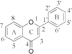
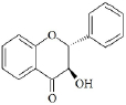
Flavonoids are a class of compounds with a low molecular weight based on the 2-phenyl-chromone nucleus (Figure 1).

The basic structure of flavonoids consists of aglycones, but their presence in nature is usually bound to glycosides and methylated derivatives. They are produced via the shikimic acid pathway using acetic acid/phenylalanine derivatives as the precursor. Flavonoids are traditionally classed by oxidation degree, ring C annularity, and ring B connectivity [1,37]. They comprise 6 subclasses, as presented inTable 2 [38,39]. Among higher plant families and genera, the flavones and flavonols are the most commonly highly conserved encountered and marked by the presence of the 2-phenylchromen-4-one (2-phenyl-1-benzopyran-4-one) structures in their backbone [40].

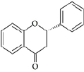
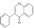
Flavones are distinct from other flavonoids because their skeleton contains a double bond between C2 and C3. Also, there is no substitution at their C3 position, while flavonols possess hydroxyl substitution at that position. Further, flavones are oxidized at the C4 position [41,42]. Flavanones and flavanols have saturated bonds between C2 and C3 and frequently coexist in plants with flavones and flavonols [37]. Flavanones have a flavan nucleus formed by two aromatic rings (A and B) linked by a dihydropyrone ring (C). The presence of a saturated C2–C3 bond, a chiral carbon atom at the C2 position, and no substitution at the C3 position of the C ring distinguish flavanones from the other two classes of flavonoids, i.e., flavones and flavanols [43]. Flavanols (flavan-3-ols) are heterocyclic compounds that contain a saturated heterocyclic ring, a single bond between C2 and C3, and a hydroxyl group at the C3 position. Unlike other flavonoids, flavanols are only found in food as aglycones (the glycosylated state is excluded). Additionally, they are found in monomeric form as catechins and epicatechins, as well as in polymeric form as tannins [44]. Isoflavones are secondary metabolites formed by rhizobial bacteria and leguminous plant defence responses [45]. Isoflavones, like daidzein, include 3-phenylchromones. Chalcones are ring C-opening isomers of dihydroflavones that are responsible for plant colour. Anthocyanidins are a category of significant chromene pigments that exist as ions. Other flavonoids without the C6—C3—C6 structure include biflavones, furan chromones, and xanthones. Glycosides are the most common extant flavonoid form. The structure of aglycones dictates preferred glycosylation sites [37].

Pharmacokinetics and bioavailability

Bioavailability

To predict and explain the effect of flavonoids at their site of action, knowledge of the pharmacokinetics, form, and concentration of these compounds in plasma must be known. Animal and human studies have found that these compounds have poor oral bioavailability, which might be attributed to the loss of the compounds during the absorption and metabolism phases. In addition, these compounds also tend to have poor water solubility, low permeability, as well as poor stability profile [13,61]. Therefore, various studies have been carried out to engineer their structure to increase their water solubility and bioavailability and thereby increase their activity as a drug candidate as have been demonstrated in the treatment of several diseases [13,46,62].

Absorption

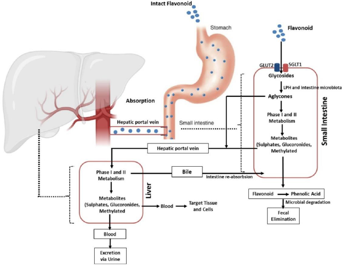
Absorption of flavonoids in the intestine can occur through several mechanisms, namely active transport, passive diffusion, or both. Some flavonoids, such as quercetin, are absorbed completely in the small intestine by involving sodium-dependent glucose transporters 1 (SGLT1). These transporters are located in the apical membrane of intestinal epithelial cells that utilize Na+/K+-ATPase pumps to transport this flavonoid. In addition, the absorption of this compound is also carried out by glucose transporter 2 (GLUT2) located on the basolateral membrane of the intestine. However, the exact mechanism used by GLUT2 in transporting flavonoids is still unclear [63-65]. Further, quercetin is hydrolysed by the intracellular enzymes called lactase-phloridzin hydrolase (LPH) and cytosolic β-glucosidase (CBG) in the aglycone form so that it is easily absorbed through the small intestine. In addition to quercetin, these enzymes can work on other flavonoid compounds. However, the effectiveness of these enzymes depends on the glycosides present in the flavonoid structure [41,64,66,67]. It should be noted that each compound grouped as flavonoids have different maximum absorption times. A study that was conducted in rats revealed that the absorption of some flavonoid derivatives such as apigenin, luteolin, and glucosides generally occurred rapidly with a maximum absorption time (tmax) of about 1 hour for maximum plasma concentration (Cmax) of 1 to 100 μmol/L and it will change depends on co-consumed food [41,68].

Distribution

In circulation, flavonoids can bind to several blood proteins [69]. Gecibesler & Aydin [70] demonstrated that flavonoids could bind strongly to human serum albumin (HSA). HSA is an essential protein in blood plasma found abundantly in the body [71]. This soluble protein can bind and transport various metabolites and organic compounds such as flavonoids, unesterified fatty acids, hormones, and metal ions to various tissues throughout the body [72,73].

Research by Boer et al. [74] revealed that flavonoid compounds, in particular quercetin and its derivatives, are widely distributed in rat tissues with the highest levels in the lungs, testes, and kidneys, while the brain, white fat, and spleen showed the lowest levels. This group also reported that this compound was deposited in several other organs in the rat, including the thymus, heart, liver, brown fat, bone, and muscle. Meanwhile, in pigs, the deposition of quercetin was more abundant in the liver and kidney tissue after a quercetin diet of 500 mg/kg with a lesser extent in the brain, heart, and spleen .

A similar finding was reported by Beiger et al. [75] demonstrating that the distribution of quercetin and some of its derivatives was found to be highest deposited in the liver, kidney, jejunum, lung, and muscle (longissimus dorsi) after a single dietary quercetin dose (25 mg/kg) in pigs. Furthermore, quercetin deposition was more distributed when administered at a dose of 50 mg/kg per day for four weeks, including colon (higher), kidney, jejunum, liver, lymph node, mesentery, lung, white adipose tissue, muscle (diaphragm), muscle (longissimus dorsi), and brain (as the smaller deposition).

Metabolism

After ingestion, approximately 10 % of flavonoid glycosides are absorbed in the upper gastrointestinal tract, while the remaining 90 % pass through the small intestine to reach the large intestine as unmetabolized and unabsorbed flavonoids. The unabsorbed flavonoids undergo a further enzymatic transformation in the small intestine, such as oxidation, reduction, and decarburization, as the preparatory steps before entering the large intestine. Once in the large intestine, they are hydrolysed and cleaved to remove glycosides and produce flavonoid aglycones, which are further metabolized to ring fission products. This process is carried out by the involvement of the lactase-phlorizin hydrolase (LPH) and colonic enzymes produced by the intestinal microbiota [41,76-78].

Flavonoids then extensively undergo two metabolic phases, namely phases 1 and 2, which occur in enterocytes and hepatocytes [79,80]. In the first phase, which takes place in hepatocytes, flavonoids will be oxidized by the cytochrome P450 (CYP450) enzyme to produce a minor contribution to flavonoid clearance. Different isoforms of the CYP450 enzyme play the metabolic process of several flavonoid-derived compounds in this phase. Isoform CYP2C9 is the most efficient enzyme for the metabolism of galangin followed by CYP1A3 and CYP1A1. Meanwhile, most of the oxidation of galangin is determined by the action of CYP1A2 and followed by CYP2C9 and CYP1A1 [81]. Another investigation reported that apigenin was metabolized by CYP1A1, CYP2B, and CYP2E to form three monohydroxylated derivatives [82].

In contrast to the first phase, phase II metabolism occurs in enterohepatic and enteric and is predominant in the direction of flavonoid disposition. In this stage, flavonoids undergo two main processes, namely glucuronidation and sulfation. While the former is mediated by UDP glucuronosyltransferases (e.g., UGT1A1, UGT1A8, and UGT1A9), the latter is catalysed by sulfotransferases (e.g., SULT1A1 and SULT1E1). Methylation can also occur in this phase. The process is mediated by a group of enzymes known as methyltransferases mostly found in many tissues, including the liver and intestines. The most common methylation reaction associated with flavonoid metabolism is O-methylation which occurs in the liver and is catalysed by catechol-O-methyltransferase (COMT) [41,64,69,79,83,84]. The products of these metabolic processes will then be circulated and deposited in various body components to carry out their actions, including their anti-inflammatory activities, as elaborated in the section “Flavonoid derivates as anti-inflammatory agents and their mechanisms of action”.

Excretion

Flavonoids are mainly excreted through the urine. A study reported that about 60-80 % of the metabolites of anthocyanins are excreted in the urine [85,86]. The excretion pathway of flavonoids depends on the conjugate form and the site of elimination. Glucuronide formed in the intestine tends to enter the systemic circulation directly and is excreted in the urine, while the products formed in the liver are mainly eliminated through the bile. This indicates that glucuronidation in enterocytes Favors the loss of flavonoids from the enterohepatic circulation (EHC) into the systemic circulation.

EHC-undergoing flavonoid compounds may also be further sulphated in the intestine or hepatocytes, leading to urinary excretion (without bypassing biliary excretion). Another study in isolated human faecal colonic bacteria found the presence of flavonoids such as quercetin, indicating the importance of the faecal route in facilitating the excretion of these compounds [76,86,87]. To sum up, the biological activities of flavonoids are diminished by the fact that these compounds have low aqueous solubility and poor bioavailability, as depicted in their pharmacokinetic profile described above. This leads to the development of various strategies to increase their solubility and oral bioavailability, including the development of nanoparticle-based delivery systems, which will be discussed in the section “Application of nanomedicine as delivery system of flavonoid derivates”.

Flavonoid derivates as anti-inflammatory agents and their mechanisms of action

Various studies of in silico, in vitro, in vivo, and clinical trials have demonstrated that flavonoids have potential activities in mitigating inflammation [11,88]. They can modulate various key points in regulating inflammation in the early and late stages. As displayed inTable 1, these compounds consist of several subclasses, and they can exert their anti-inflammatory properties via various mechanisms, including inhibition of the various inflammatory signaling pathways such as NF-κB [88], MAPK [89], blockage of pro-inflammatory enzymes (COX-1, COX-2, and 5-LOX) [11], inhibition of proinflammatory cytokines release (TNFα, IL-1β, and IL-6), and suppression of other inflammatory proteins [62]. The summary of the putative anti-inflammatory mechanisms of flavonoids is presented inTable 2 andFigure 3.

Flavonoid inhibit eicosanoids metabolism and function

Prostaglandins and leukotrienes are potent lipid mediators derived from phospholipase-released arachidonic acid that play a critical role in inflammation [160]. They are produced from locally damaged cell membranes with the help of several enzymes such as phospholipase A2 (PLA2), cyclooxygenase, and lipoxygenases as previously explained (in section Lipid-based signaling: arachidonic acid-derived eicosanoids). Several approved drugs such as corticosteroid-including drugs (such as methylpredisolone etc.) and nonsteroidal anti-inflammatory drugs (NSAIDs; e.g., mefenamic acid, indomethacin, ibuprofen) have targeted these enzymes for the treatment of several inflammation-related diseases such as pain, injury, asthma, allergies, and inflammation. Elevated COX-2 levels have indeed shown adverse effects on inflamematory conditions and even on some other inflammation-related diseases such as tumor development, so their suppression could significantly suppress worsening prognosis [161].

Different studies demonstrated that flavonoid-derived compounds can inhibit enzymes, including phospholipase A2, cyclooxygenase, and lipoxygenases, improving inflammatory disorders [52,160,162,163]. Several pieces of evidence have proposed their effect to block the pathway and mediate improvement in an intestinal inflammatory response. A study by López-Posadas [162] reported that flavonoids could inhibit the formation of prostaglandins from the AA pathway by suppressing epithelial COX-2 expression during inflammation in Intestinal epithelial cells. This finding is supported by another study reported by Serra and colleagues that advocated C3G to provide strong inhibitory activity against COX-2 and lead downregulation of several inflammatory mediators in the colonic carcinoma cell [164].

Apart from intestinal inflammation, several flavonoid derivative compounds have also been reported to be effective in inhibiting eicosanoid production in other inflammatory cases. A study by Husain et al. suggested that epigallocatechin-3-gallate (EGCG) can downregulate COX-2 in human prostate carcinoma LNCaP and PC-3 cells in a dose-dependent fashion [165]. Similar findings demonstrated the efficacy of EGCG could downmodulation of COX-2 were further lead reduction of PGE2 levels [166,167]. In another finding, giving EGCG results in the inhibition of NF-κB that in order leads to the inhibition of COX-2 promoter activity [168]. Several other flavonoid derivative compounds, such as luteolin, kaempferol, hesperetin, and naringin, have been shown to have inhibitory effects on this COX-2 enzyme [161,169]. Topical pre-treatment of kaempferol could attenuate UVB-induced COX-2 expression in mouse skin. This effect is hypothesized to arise through the modulation of Src kinase activity in the mouse skin [161]. Another promotion study demonstrated by He and colleagues [170], isolated and elucidated flavonoid compounds from Hosta plantagine and found twelve 12 different ones that showed significant inhibitory effects to both enzyme isoforms from 53.00 to 80.55 % (in COX-1) and 52.19 to 66.29 % (to COX-2) in concentration 50 μM.

The increase in eicosanoids is also mediated by the COX-1 isoform, especially in some cases of neuroinflammation. Elevated COX-1 levels in microglia may explain PGE2 levels in the cerebrospinal fluid of Creutzfeldt-Jacob disease (CJD) patients associated with shorter survival [171]. Regarding that, it has been reported that quercetin and tomentodiplacone-O could strongly inhibit COX-1 [11]. Similar to this finding, Kaempferol has been advocated to have an inhibitory effect on COX-1 in vitro. In addition to the cyclooxygenase pathway, several researchers have reported that this compound can inhibit the expression of lipooxygenase (LOX) enzymes which play an important role in the metabolism of arachidonic acid into leukotrienes, which is involved in various inflammatory-related diseases including asthmatics, rheumatoid arthritis, psoriasis, inflammatory bowel disease etc. [51,172]. Administration of several other flavonoid derivative compounds, such as quercetin and luteolin has also been shown to have a strong inhibitory effect on these enzymes to lead to improvement in symptoms of inflammation-related leukotrienes expression [169].

The effectiveness of flavonoids in inhibiting the formation of eicosanoids in the AA-related pathway has also been proposed through the PLA2 enzyme inhibition mechanism. This enzyme plays an important role in the process of converting membrane phospholipids into product intermediates, namely arachidonic acid, before being further converted into lipid mediators. Many recent studies have reported the activity of various flavonoid derivative compounds having good activity in inhibiting this enzyme and treating inflammation [173,174]. A study conducted by Enechi and reported colleagues [175] that flavonoid-rich extract from Peltophorum pterocarpum stem-bark provide a strong inhibitory effect in PLA2 and generate downmodulate of the inflamematory symptoms of paw oedema induced by egg albumin, in comparison with prednisolone as well approve anti-inflammatory drugs that act in PLA2. In accordance, several other studies have also demonstrated compounds such as quercetin, kaempferol, and galangin showing promising inhibitory activity against PLA2 [173,174,176].

Flavonoid inhibit MAPK and NF-κβ signalling pathways

Various evidence has suggested the crucial role of Nuclear Factor Kappa Beta (NF-κβ) and Mitogen-activated protein kinases (MAPKs) as two main signaling pathways that are interrelated in underlying inflamemation [177]. NF-κβ accumulation has been implicated with increased transcription and expression of various inflammatory mediators. Meanwhile, MAPKs, consisting of p38 kinase, ERK, and c-Jun NH2-terminal kinase (JNK), have been advocated to regulate the transcription of COX-2, iNOS, and inflammatory cytokines in inflammation. Therefore, increasing evidence has studied and targeted these signaling pathways in the development of anti-inflammatory drugs [177-179]. A study conducted by Hour and colleagues [177] reported that dihydromyricetin, a flavonoid isolated from Ampelopsis grossedentata has been proposed to have good and promising inflamematory potential by inhibiting the phosphorylation of NF-κB and IκBα as well as p38 and JNK. This suppression simultaneously leads to a downmodulation effect on various proinflammatory cytokines such as TNF-α, IL-1β, and interleukin-6, as well as increasing levels of IL-10 as an anti-inflammatory cytokine. Furthermore, inhibition of these two signaling pathways also reduces the levels of iNOS, TNF-α, and COX-2.

In another finding, an in vivo study conducted by Ichikawa and colleagues [180] had proposed epigallocatechin gallate (EGCG) to have a significant effect of reducing IL-6 expression, IKK activation, IκBα degradation, and activation of NF-κB which in turn inhibited inflammatory events that mediated by this pathway. The inhibition also reduces myocardial damage after ischemia and reperfusion in rats [181]. Furthermore, these compounds show promising effects in inhibiting IκBα degradation and the action of NF-κB in binding to DNA that leads to suppressing IL-12p40 and iNOS expression in LPS-activated macrophages [180]. In addition to EGCG, quercetin has been reported to have good anti-inflammatory activity through the same mechanism as EGCG, inhibiting IκBα phosphorylation and downmodulating the NF-κB pathway [182].

Growing evidence has been demonstrated that compounds that can inhibit JNK, p38, and IKKβ generally exhibit anti-inflammatory effects and suggest a relationship between MAPK and NFκB in inflamematory events. Flavonoid derivatives have been reported to affect the MAPK signalling pathway at various stages. A study has proven that EGCG is able to influence this pathway through the inhibition of ERK1/2, p-JNK, and p-p38 in human RA synovial fibroblasts (RASFs) [183]. Another study reported that kaempferol, luteolin, chrysin, and apigenin exert antiinflammatory activity by inhibiting MAPK signalling pathways such as JNK, p38, and ERK [181].

Application of nanomedicine as delivery system of flavonoid derivates

Herbs have been widely used to promote human health since ancient times. Advances in phytochemistry and pharmacology have made it possible to determine the composition and bioactivity of many medicinal products. It has been proved that the effectiveness of many herbal medicines depends on the delivery of the active compounds. However, their success in clinical trials has been less impressive, partly due to the compound's poor bioavailability [184,185]. Furthermore, the efficacy of many drugs obtained from natural sources, including flavonoids in their micro/macro formulations, has also been shown to have poor bioavailability and pharmacokinetics following their oral administration, leading to their less effectivity [184,186]. Researchers have developed other drug delivery methods while avoiding potential causes of decreased bioavailability, such as first-pass metabolism and drug malabsorption in the digestive tract [187,188].

Recently, the use of nanotechnology has shown tremendous success in the field of drug delivery, including natural compound-based drugs for the treatment of inflammation. This technique formulates the natural compounds in the form of nanosized particles (1 to 100 nm) [189,190]. It has been suggested that the encapsulation of flavonoids into nanocarriers is a beneficial technique to protect drug molecules from changes in their size, shape, and surface characteristics that might be caused by environmental factors. It has been proved that this method can increase the bioavailability of flavonoids and improve the targeted and controlled release profile of the natural products, which in turn increases the drug's effectiveness [185]. Furthermore, nano-delivery systems could protect the therapeutic agents from being enzymatically metabolized, resulting in their increased stability and circulation time [191-195]. The smaller size of the nanoparticles has also been proven to increase the penetration of the compounds compared to conventional topical formulations [196]. Recently, the most common types of nanoparticles used for drug delivery are polymer nanoparticles, including solid lipid nanoparticles, liposomes, micelles, crystalline nanoparticles, and dendrimers [185,197,198].

Solid lipid nanoparticles

Solid lipid nanoparticles (SLNs) are nanometer colloidal carriers composed of the solid particle lipid core in which active substances are trapped and stabilized by surfactants [199]. Their matrix is typically composed of solid lipids such as glycerides, sterols, fatty acids, and waxes. In particular, SLN has recently been proposed for oral and dermal administration of phenolic compounds to protect them from chemical degradation [200]. Tan et al. [201] demonstrated that the formulation of total flavonoid-based solid lipid nanoparticles (TF-SLN) could produce spherical particles in shape with a uniform size distribution of 104.83 nm and a zeta potential of 28.7 mV. Furthermore, this formulation was proved to have a better myocardial protection effect than flavonoids alone. Another study showed that the formulation of quercetin-loaded solid lipid nanoparticles (QSLNs) exhibited an increase in their bioavailability by 3.5-fold compared to quercetin alone in rats [12].

Polymeric nanoparticles

Natural or synthetic biodegradable polymer nanoparticles have become prominent in the field of nanomedicine for targeted drug delivery to improve biocompatibility, bioavailability, safety, increased permeability, preferable retention time, and less toxicity [186,202]. Drugs that are conjugated to macromolecules such as synthetic polymers, natural polysaccharides, or proteins through covalent bonds provide a direct and efficient approach to modifying pharmacokinetic performance, promoting the stability of drugs, and preventing drug leakage or explosive release during transport [203].

Chitosan is the most frequently used polymer for the formulation of nano-based drugs for their delivery to their target of action. Wang et.al., [204] reported that the administration of the inhaled baicalein encapsulated with chitosan-nanoparticle could mediate delivery of this drug and lead to the reduction of IL-5 level, enhancement of IL-12 and controlled inflammation-related asthma in mice. In addition, polymer nanoparticles are also effective for mediating drug delivery given topically. A study by Nan et.al. [205] revealed that the formulation of a topical preparation of quercetin-loaded chitosan nanoparticles led to the increase of percutaneous absorption and retention of quercetin in the skin and improve its effects against ultraviolet B radiation.

Nanoliposomes

Nanoliposomes are phospholipid vesicles containing one or more bilayers surrounding an aqueous core. Nanoliposome-based drugs are effective for the development of transdermal drugs, topical drugs, drugs used to target hair follicles and other uses. Nanoliposomes usually consist of phospholipids with cholesterol often added to improve the stability of the liposome bilayer by filling the gaps caused by incomplete packaging. Some of the literature report that the use of nanoliposomes in natural product-based drug, especially flavonoids, is effective in delivering drugs well to the targeted sites leading to the generation of desired effects. A study by Wang et al. [206] explained that the formulation of nanoliposome of quercetin could induce type III-programmed cell death in C6 glioma cells. Furthermore, another study by Jin et al. [207] exhibited that treatment of apigenin-loaded D-alpha-tocopheryl polyethylene glycol 1000 succinate (TPGS) liposomes could deliver apigenin well and exert inhibitory effects on tumor growth in A549 cell-bearing mice.

Nanoemulsions

Nanoemulsions are formed by combining two mechanically shear immiscible liquid phases with a surfactant having a droplet size of less than 100 nm. The formulation of natural product-based drugs with this method is able to overcome the weakness of flavonoid compounds that have problems with their bioavailability by improving their pharmacokinetic profile. A recent study by Fuior et al. [208] demonstrated that the formulation of flavonoid-loaded lipid nanoemulsions (FLNs) generated good characteristics (size of l200 nm, negative zeta potential, a high encapsulation efficiency >80 %, good in vitro stability, and steady release). Further, this formulation facilitated the action of the flavonoid in reducing endothelial inflammation. This finding is supported by a study conducted by Zain et al. [209] investigating the relationship between the delivery of nanoemulsion flavonoid-enriched oil palm and its wound-healing activity.

Self-nano-emulsifying drug delivery system (SNEEDs)

SNEDDS is a nanoemulsion pre-concentrate in which the drug is encapsulated in an oil phase in the presence of a surfactant/cosurfactant, capable of forming very small nanoscale droplets/nanoemulsion when gently mixed with an aqueous medium. Self-emulsifying drug delivery systems are isotropic mixtures of oils, surfactants, solvents, and cosolvents/surfactants which are developed to improve the physicochemical as well as pharmacological properties of highly lipophilic drugs, including flavonoids [210-212]. It has been demonstrated that the loading of quercetin into SNEDDs could effectively enhance the hepatoprotective activity of quercetin in mice suffering from hepatotoxicity [213]. Another study performed in Wistar rats confirmed that the formulation of apigenin into SNEDDs enhanced the oral bioavailability of the compound [214].

Concluding remarks

Flavonoids are secondary metabolites found abundantly in various plants. They exist in several subclasses, including flavanols, flavanones, flavonols, flavones, isoflavones, anthocyanins, and flavanonols. It has been demonstrated that flavonoids possess potential pharmacological activities, including their potency in mitigating inflammation. A number of studies conducted in silico, in vitro, in vivo, and clinical trials have deciphered that the anti-inflammatory activities of flavonoids are closely linked to their ability to modulate various biochemical mediators, enzymes, and signalling pathways involved in the inflammatory processes. However, these anti-inflammatory activities have been facing challenges as flavonoids have poor solubility and bioavailability. To tackle these challenges, the application of nanotechnology in developing better pharmaceutical dosage forms has brought promising results. Various nanotechnology platforms, including solid nanoparticles, polymeric nanoparticles, nanoliposomes, nanoemulsion, self-nano emulsifying drug delivery systems, have been reported to have better action in delivering flavonoids to their targeted sites. These platforms can potentially increase the solubility, bioavailability, and pharmacological activity of flavonoids and reduce the toxic effects of these compounds.

Notes

[1] Conflicts of interest Conflict of interest: There is no conflict of Interest associated of this article as well as no significant support that influenced the content.

Acknowledgements

The author gratefully thank to Ysrafil, to conductiing this projects, apt. Firzan Nainu, M. Biomed., Sc., Ph.D and Talha Bin Emran, Ph.D for supervised and grammar/structural editing of this manuscript.

References

[1] 

Ginwala R.; Bhavsar R.; Chigbu D.I.; Jain P.; Khan Z.K.. Potential Role of Flavonoids in Treating Chronic Inflammatory Diseases with a Special Focus on the Anti-Inflammatory Activity of Apigenin. Antioxidants 8(2) (2019). https://doi.org/10.3390/antiox8020035. https://doi.org/10.3390/antiox8020035

[2] 

Maleki S.J.; Crespo J.F.; Cabanillas B.. Anti-inflammatory effects of flavonoids. Food Chemistry 299 (2019) 125124. https://doi.org/10.1016/j.foodchem.2019.125124. https://doi.org/10.1016/j.foodchem.2019.125124

[3] 

Nguyen T.Y.; To D.C.; Tran M.H.; Lee J.S.; Lee J.H.; Kim J.A.; Woo M.H.; Min B.S.. Anti-inflammatory Flavonoids Isolated from Passiflora foetida. Natural Product Communications 10(6) (2015) 929-931.

[4] 

Herwald H.; Egesten A.. On PAMPs and DAMPs. Journal of Innate Immunity 8(5) (2016) 427-428. https://doi.org/10.1159/000448437. https://doi.org/10.1159/000448437

[5] 

Ma K.C.; Schenck E.J.; Pabon M.A.; Choi A.M.K.. The Role of Danger Signals in the Pathogenesis and Perpetuation of Critical Illness. American Journal of Respiratory and Critical Care Medicine 197(3) (2018) 300-309. https://doi.org/10.1164/rccm.201612-2460PP. https://doi.org/10.1164/rccm.201612-2460PP

[6] 

Netea M.G.; Balkwill F.; Chonchol M.; Cominelli F.; Donath M.Y.; Giamarellos-Bourboulis E.J.; Golenbock D.; Gresnigt M.S.; Heneka M.T.; Hoffman H.M.; Hotchkiss R.; Joosten L.A.B.; Kastner D.L.; Korte M.; Latz E.; Libby P.; Mandrup-Poulsen T.; Mantovani A.; Mills K.H.G.; Nowak K.L.; O'Neill L.A.; Pickkers P.; van der Poll T.; Ridker P.M.; Schalkwijk J.; Schwartz D.A.; Siegmund B.; Steer C.J.; Tilg H.; van der Meer J.W.M.; van de Veerdonk F.L.; Dinarello C.A.. A guiding map for inflammation. Nature Immunology 18(8) (2017) 826-831. https://doi.org/10.1038/ni.3790. https://doi.org/10.1038/ni.3790

[7] 

Schmid T.; Brüne B.. Prostanoids and Resolution of Inflammation - Beyond the Lipid-Mediator Class Switch. Frontiers in Immunology 12 (2021) 714042. https://doi.org/10.3389/fimmu.2021.714042. https://doi.org/10.3389/fimmu.2021.714042

[8] 

Choy K.W.; Murugan D.; Leong X.F.; Abas R.; Alias A.; Mustafa M.R.. Flavonoids as Natural Anti-Inflammatory Agents Targeting Nuclear Factor-Kappa B (NFκB) Signaling in Cardiovascular Diseases: A Mini Review. Frontiers in Pharmacology 10 (2019) 1295. https://doi.org/10.3389/fphar.2019.01295. https://doi.org/10.3389/fphar.2019.01295

[9] 

Peng Y.; Ao M.; Dong B.; Jiang Y.; Yu L.; Chen Z.; Hu C.; Xu R.. Anti-Inflammatory Effects of Curcumin in the Inflammatory Diseases: Status, Limitations and Countermeasures. Drug Design, Development and Therapy 15 (2021) 4503-4525. https://doi.org/10.2147/dddt.S327378. https://doi.org/10.2147/dddt.S327378

[10] 

Fürst R.; Zündorf I.. Plant-derived anti-inflammatory compounds: hopes and disappointments regarding the translation of preclinical knowledge into clinical progress. Mediators of Inflammation 2014 (2014) 146832. https://doi.org/10.1155/2014/146832. https://doi.org/10.1155/2014/146832

[11] 

Hanáková Z.; Hošek J.; Kutil Z.; Temml V.; Landa P.; Vaněk T.; Schuster D.; Dall'Acqua S.; Cvačka J.; Polanský O.; Šmejkal K.. Anti-inflammatory Activity of Natural Geranylated Flavonoids: Cyclooxygenase and Lipoxygenase Inhibitory Properties and Proteomic Analysis. Journal of Natural Products 80(4) (2017) 999-1006. https://doi.org/10.1021/acs.jnatprod.6b01011. https://doi.org/10.1021/acs.jnatprod.6b01011

[12] 

Ahmad N.; Banala V.T.; Kushwaha P.; Karvande A.; Sharma S.; Tripathi A.K.; Verma A.; Trivedi R.; Mishra P.R.. Quercetin-loaded solid lipid nanoparticles improve osteoprotective activity in an ovariectomized rat model: a preventive strategy for post-menopausal osteoporosis. RSC Advances 6(100) (2016) 97613-97628. https://doi.org/10.1039/C6RA17141A. https://doi.org/10.1039/C6RA17141A

[13] 

Batiha G.E.; Beshbishy A.M.; Ikram M.; Mulla Z.S.; El-Hack M.E.A.; Taha A.E.; Algammal A.M.; Elewa Y.H.A.. The Pharmacological Activity, Biochemical Properties, and Pharmacokinetics of the Major Natural Polyphenolic Flavonoid: Quercetin. Foods 9(3) (2020). https://doi.org/10.3390/foods9030374. https://doi.org/10.3390/foods9030374

[14] 

Tasneem S.; Liu B.; Li B.; Choudhary M.I.; Wang W.. Molecular pharmacology of inflammation: Medicinal plants as anti-inflammatory agents. Pharmacological Research 139 (2019) 126-140. https://doi.org/10.1016/j.phrs.2018.11.001. https://doi.org/10.1016/j.phrs.2018.11.001

[15] 

Ullah H.M.A.; Kwon T.H.; Park S.; Kim S.D.; Rhee M.H.. Isoleucilactucin Ameliorates Coal Fly Ash-Induced Inflammation through the NF-κB and MAPK Pathways in MH-S Cells. International Journal of Molecular Sciences 22(17) (2021). https://doi.org/10.3390/ijms22179506. https://doi.org/10.3390/ijms22179506

[16] 

Souto A.L.; Tavares J.F.; da Silva M.S.; Diniz Mde F.; de Athayde-Filho P.F.; Barbosa Filho J.M.. Anti-inflammatory activity of alkaloids: an update from 2000 to 2010. Molecules 16(10) (2011) 8515-8534. https://doi.org/10.3390/molecules16108515. https://doi.org/10.3390/molecules16108515

[17] 

Ito T.. PAMPs and DAMPs as triggers for DIC. The Journal of Clinical Investigation 2(1) (2014) 67. https://doi.org/10.1186/s40560-014-0065-0. https://doi.org/10.1186/s40560-014-0065-0

[18] 

Watanabe S.; Alexander M.; Misharin A.V.; Budinger G.R.S.. The role of macrophages in the resolution of inflammation. Journal of Intensive Care 129(7) (2019) 2619-2628. https://doi.org/10.1172/jci124615. https://doi.org/10.1172/jci124615

[19] 

Roh J.S.; Sohn D.H.. Damage-Associated Molecular Patterns in Inflammatory Diseases. Immune Network 18(4) (2018) e27. https://doi.org/10.4110/in.2018.18.e27. https://doi.org/10.4110/in.2018.18.e27

[20] 

Su Y.; Gao J.; Kaur P.; Wang Z.. Neutrophils and Macrophages as Targets for Development of Nanotherapeutics in Inflammatory Diseases. Pharmaceutics 12(12) (2020). https://doi.org/10.3390/harmaceutics12121222. https://doi.org/10.3390/harmaceutics12121222

[21] 

Chen L.; Deng H.; Cui H.; Fang J.; Zuo Z.; Deng J.; Li Y.; Wang X.; Zhao L.. Inflammatory responses and inflammation-associated diseases in organs. Oncotarget 9(6) (2018) 7204-7218. https://doi.org/0.18632/oncotarget.23208. https://doi.org/10.18632/oncotarget.23208

[22] 

Kawasaki T.; Kawai T.. Toll-like receptor signaling pathways. Frontiers in Immunology 5 (2014) 461. https://doi.org/10.3389/fimmu.2014.00461. https://doi.org/10.3389/fimmu.2014.00461

[23] 

Takeuchi O.; Akira S.. Pattern recognition receptors and inflammation. Cell 140(6) (2010) 805-820. https://doi.org/10.1016/j.cell.2010.01.022. https://doi.org/10.1016/j.cell.2010.01.022

[24] 

Murakami M.; Hirano T.. The molecular mechanisms of chronic inflammation development. Frontiers in Immunology 3 (2012) 323. https://doi.org/10.3389/fimmu.2012.00323. https://doi.org/10.3389/fimmu.2012.00323

[25] 

Sheppe A.E.F.; Edelmann M.J.. Roles of Eicosanoids in Regulating Inflammation and Neutrophil Migration as an Innate Host Response to Bacterial Infections. Infection and Immunity 89(8) (2021) e0009521. https://doi.org/10.1128/iai.00095-21. https://doi.org/10.1128/iai.00095-21

[26] 

Calder P.C.. Omega-3 polyunsaturated fatty acids and inflammatory processes: nutrition or pharmacology? British Journal of Clinical Pharmacology 75(3) (2013) 645-662. https://doi.org/10.1111/j.1365-2125.2012.04374.x. https://doi.org/10.1111/j.1365-2125.2012.04374.x

[27] 

Das U.N.. Essential Fatty Acids and Their Metabolites in the Pathobiology of Inflammation and Its Resolution. Biomolecules 11(12) (2021). https://doi.org/10.3390/biom11121873. https://doi.org/10.3390/biom11121873

[28] 

Jang Y.; Kim M.; Hwang S.W.. Molecular mechanisms underlying the actions of arachidonic acid-derived prostaglandins on peripheral nociception. Journal of Neuroinflammation 17(1) (2020) 30. https://doi.org/10.1186/s12974-020-1703-1. https://doi.org/10.1186/s12974-020-1703-1

[29] 

Wang B.; Wu L.; Chen J.; Dong L.; Chen C.; Wen Z.; Hu J.; Fleming I.; Wang D.W.. Metabolism pathways of arachidonic acids: mechanisms and potential therapeutic targets. Signal Transduction and Targeted Therapy 6(1) (2021) 94. https://doi.org/10.1038/s41392-020-00443-w. https://doi.org/10.1038/s41392-020-00443-w

[30] 

Ramesh G.; MacLean A.G.; Philipp M.T.. Cytokines and chemokines at the crossroads of neuroinflammation, neurodegeneration, and neuropathic pain. Mediators of Inflammation 2013 (2013) 480739. https://doi.org/10.1155/2013/480739. https://doi.org/10.1155/2013/480739

[31] 

Zhang J.M.; An J.. Cytokines, inflammation, and pain. International Anesthesiology Clinics 45(2) (2007) 27-37. https://doi.org/10.1097/AIA.0b013e318034194e. https://doi.org/10.1097/AIA.0b013e318034194e

[32] 

Turner M.D.; Nedjai B.; Hurst T.; Pennington D.J.. Cytokines and chemokines: At the crossroads of cell signalling and inflammatory disease. Biochimica et Biophysica Acta 1843(11) (2014) 2563-2582. https://doi.org/10.1016/j.bbamcr.2014.05.014. https://doi.org/10.1016/j.bbamcr.2014.05.014

[33] 

Shachar I.; Karin N.. The dual roles of inflammatory cytokines and chemokines in the regulation of autoimmune diseases and their clinical implications. Journal of Leukocyte Biology 93(1) (2013) 51-61. https://doi.org/10.1189/jlb.0612293. https://doi.org/10.1189/jlb.0612293

[34] 

Kaminska B.. MAPK signalling pathways as molecular targets for anti-inflammatory therapy-from molecular mechanisms to therapeutic benefits. Biochimica et Biophysica Acta 1754(1-2) (2005) 253-262. https://doi.org/10.1016/j.bbapap.2005.08.017. https://doi.org/10.1016/j.bbapap.2005.08.017

[35] 

Yang Y.L.; Liu M.; Cheng X.; Li W.H.; Zhang S.S.; Wang Y.H.; Du G.H.. Myricitrin blocks activation of NF-κB and MAPK signaling pathways to protect nigrostriatum neuron in LPS-stimulated mice. Journal of Neuroimmunology 337 (2019) 577049. https://doi.org/10.1016/j.jneuroim.2019.577049. https://doi.org/10.1016/j.jneuroim.2019.577049

[36] 

Yu H.; Lin L.; Zhang Z.; Zhang H.; Hu H.. Targeting NF-κB pathway for the therapy of diseases: mechanism and clinical study. Signal Transduction and Targeted Therapy 5(1) (2020) 209. https://doi.org/10.1038/s41392-020-00312-6. https://doi.org/10.1038/s41392-020-00312-6

[37] 

Wang T.Y.; Li Q.; Bi K.S.. Bioactive flavonoids in medicinal plants: Structure, activity and biological fate. Asian Journal of Pharmaceutical Sciences 13(1) (2018) 12-23. https://doi.org/10.1016/j.ajps.2017.08.004. https://doi.org/10.1016/j.ajps.2017.08.004

[38] 

Di Lorenzo C.; Colombo F.; Biella S.; Stockley C.; Restani P.. Polyphenols and Human Health: The Role of Bioavailability. Nutrients 13(1) (2021). https://doi.org/10.3390/nu13010273. https://doi.org/10.3390/nu13010273

[39] 

Kumar S.; Pandey A.K.. Chemistry and biological activities of flavonoids: an overview. Scientific World Journal 2013 (2013) 162750. https://doi.org/10.1155/2013/162750. https://doi.org/10.1155/2013/162750

[40] 

Dajas F.; Andrés A.C.; Florencia A.; Carolina E.; Felicia R.M.. Neuroprotective actions of flavones and flavonols: mechanisms and relationship to flavonoid structural features. Central Nervous System Agents in Medicinal Chemistry 13(1) (2013) 30-35. https://doi.org/10.2174/1871524911313010005. https://doi.org/10.2174/1871524911313010005

[41] 

Hostetler G.L.; Ralston R.A.; Schwartz S.J.. Flavones: Food Sources, Bioavailability, Metabolism, and Bioactivity. Advances in Nutrition 8(3) (2017) 423-435. https://doi.org/10.3945/an.116.012948. https://doi.org/10.3945/an.116.012948

[42] 

Zhang N.; Bi F.; Xu F.; Yong H.; Bao Y.; Jin C.; Liu J.. Structure and functional properties of active packaging films prepared by incorporating different flavonols into chitosan based matrix. International Journal of Biological Macromolecules 165(Pt A) (2020) 625-634. https://doi.org/10.1016/j.ijbiomac.2020.09.209. https://doi.org/10.1016/j.ijbiomac.2020.09.209

[43] 

Barreca D.; Gattuso G.; Bellocco E.; Calderaro A.; Trombetta D.; Smeriglio A.; Laganà G.; Daglia M.; Meneghini S.; Nabavi S.M.. Flavanones: Citrus phytochemical with health-promoting properties. Biofactors 43(4) (2017) 495-506. https://doi.org/10.1002/biof.1363. https://doi.org/10.1002/biof.1363

[44] 

Singla R.K.; Dubey A.K.; Garg A.; Sharma R.K.; Fiorino M.; Ameen S.M.; Haddad M.A.; Al-Hiary M.. Natural Polyphenols: Chemical Classification, Definition of Classes, Subcategories, and Structures. Journal of AOAC International 102(5) (2019) 1397-1400. https://doi.org/10.5740/jaoacint.19-0133. https://doi.org/10.5740/jaoacint.19-0133

[45] 

Ko K.P.. Isoflavones: chemistry, analysis, functions and effects on health and cancer. Asian Pacific Journal of Cancer Prevention 15(17) (2014) 7001-7010. https://doi.org/10.7314/apjcp.2014.15.17.7001. https://doi.org/10.7314/apjcp.2014.15.17.7001

[46] 

Aziz N.; Kim M.Y.; Cho J.Y.. Anti-inflammatory effects of luteolin: A review of in vitro, in vivo, and in silico studies. Journal of Ethnopharmacology (2018) 342-358. https://doi.org/10.1016/j.jep.2018.05.019. https://doi.org/10.1016/j.jep.2018.05.019

[47] 

Barreca D.; Mandalari G.; Calderaro A.; Smeriglio A.; Trombetta D.; Felice M.R.; Gattuso G.. Citrus Flavones: An Update on Sources, Biological Functions, and Health Promoting Properties. Plants 9(3) (2020) 288. https://doi.org/10.3390/plants9030288. https://doi.org/10.3390/plants9030288

[48] 

Nabavi S.F.; Braidy N.; Gortzi O.; Sobarzo-Sanchez E.; Daglia M.; Skalicka-Woźniak K.; Nabavi S.M.. Luteolin as an anti-inflammatory and neuroprotective agent: A brief review. Brain Research Bulletin 119(Pt A) (2015) 1-11. https://doi.org/10.1016/j.brainresbull.2015.09.002. https://doi.org/10.1016/j.brainresbull.2015.09.002

[49] 

Salehi B.; Venditti A.; Sharifi-Rad M.; Kręgiel D.; Sharifi-Rad J.; Durazzo A.; Lucarini M.; Santini A.; Souto E.B.; Novellino E.; Antolak H.; Azzini E.; Setzer W.N.; Martins N.. The Therapeutic Potential of Apigenin. International Journal of Molecular Medicine 20(6) (2019) 1305. https://doi.org/10.3390/ijms20061305. https://doi.org/10.3390/ijms20061305

[50] 

Singh S.; Gupta P.; Meena A.; Luqman S.. Acacetin, a flavone with diverse therapeutic potential in cancer, inflammation, infections and other metabolic disorders. Food and Chemical Toxicology 145 (2020) 111708. https://doi.org/10.1016/j.fct.2020.111708. https://doi.org/10.1016/j.fct.2020.111708

[51] 

Alam W.; Khan H.; Shah M.A.; Cauli O.; Saso L.. Kaempferol as a Dietary Anti-Inflammatory Agent: Current Therapeutic Standing. Molecules 25(18) (2020). https://doi.org/10.3390/molecules25184073. https://doi.org/10.3390/molecules25184073

[52] 

Devi K.P.; Malar D.S.; Nabavi S.F.; Sureda A.; Xiao J.; Nabavi S.M.; Daglia M.. Kaempferol and inflammation: From chemistry to medicine. Pharmacological Research 99 (2015) 1-10. https://doi.org/10.1016/j.phrs.2015.05.002. https://doi.org/10.1016/j.phrs.2015.05.002

[53] 

Lee H.N.; Shin S.A.; Choo G.S.; Kim H.J.; Park Y.S.; Kim B.S.; Kim S.K.; Cho S.D.; Nam J.S.; Choi C.S.; Che J.H.; Park B.K.; Jung J.Y.. Anti-inflammatory effect of quercetin and galangin in LPS-stimulated RAW264.7 macrophages and DNCB-induced atopic dermatitis animal models. International Journal of Molecular Medicine 41(2) (2018) 888-898. https://doi.org/10.3892/ijmm.2017.3296. https://doi.org/10.3892/ijmm.2017.3296

[54] 

Magar R.T.; Sohng J.K.. A Review on Structure, Modifications and Structure-Activity Relation of Quercetin and Its Derivatives. Journal of Microbiology and Biotechnology 30(1) (2020) 11-20. https://doi.org/10.4014/jmb.1907.07003. https://doi.org/10.4014/jmb.1907.07003

[55] 

Song X.; Tan L.; Wang M.; Ren C.; Guo C.; Yang B.; Ren Y.; Cao Z.; Li Y.; Pei J.. Myricetin: A review of the most recent research. Biomedicine & Pharmacotherapy 134 (2021) 111017. https://doi.org/10.1016/j.biopha.2020.111017. https://doi.org/10.1016/j.biopha.2020.111017

[56] 

Song Z.; Shanmugam M.K.; Yu H.; Sethi G.. Butein and Its Role in Chronic Diseases. Advances in Experimental Medicine and Biology 928(2016) 419-433. https://doi.org/10.1007/978-3-319-41334-1_17. https://doi.org/10.1007/978-3-319-41334-1_17

[57] 

Ohishi T.; Goto S.; Monira P.; Isemura M.; Nakamura Y.. Anti-inflammatory Action of Green Tea. Anti-Inflammatory & Anti-Allergy Agents in Medicinal Chemistry 15(2) (2016) 74-90. https://doi.org/10.2174/1871523015666160915154443. https://doi.org/10.2174/1871523015666160915154443

[58] 

Wen R.; Lv H.; Jiang Y.; Tu P.. Anti-inflammatory Flavanones and Flavanols from the Roots of Pongamia pinnata. Planta Medica 84(16) (2018) 1174-1182. https://doi.org/10.1055/a-0626-7356. https://doi.org/10.1055/a-0626-7356

[59] 

Křížová L.; Dadáková K.; Kašparovská J.; Kašparovský T.. Isoflavones. Molecules 24(6) (2019). https://doi.org/10.3390/molecules24061076. https://doi.org/10.3390/molecules24061076

[60] 

Lee Y.M.; Yoon Y.; Yoon H.; Park H.M.; Song S.; Yeum K.J.. Dietary Anthocyanins against Obesity and Inflammation. Nutrients 9(10) (2017). https://doi.org/10.3390/nu9101089. https://doi.org/10.3390/nu9101089

[61] 

Thilakarathna S.H.; Rupasinghe H.P.. Flavonoid bioavailability and attempts for bioavailability enhancement. Nutrients 5(9) (2013) 3367-3387. https://doi.org/10.3390/nu5093367. https://doi.org/10.3390/nu5093367

[62] 

Yang H.L.; Chen S.C.; Senthil Kumar K.J.; Yu K.N.; Lee Chao P.D.; Tsai S.Y.; Hou Y.C.; Hseu Y.C.. Antioxidant and anti-inflammatory potential of hesperetin metabolites obtained from hesperetin-administered rat serum: an ex vivo approach. Journal of Agricultural and Food Chemistry 60(1) (2012) 522-532. https://doi.org/10.1021/jf2040675. https://doi.org/10.1021/jf2040675

[63] 

Fang Y.; Cao W.; Xia M.; Pan S.; Xu X.. Study of Structure and Permeability Relationship of Flavonoids in Caco-2 Cells. Nutrients 9(12) (2017). https://doi.org/10.3390/nu9121301. https://doi.org/10.3390/nu9121301

[64] 

Li S.; Liu J.; Li Z.; Wang L.; Gao W.; Zhang Z.; Guo C.. Sodium-dependent glucose transporter 1 and glucose transporter 2 mediate intestinal transport of quercetrin in Caco-2 cells. Food & Nutrition Research 64 (2020). https://doi.org/10.29219/fnr.v64.3745. https://doi.org/10.29219/fnr.v64.3745

[65] 

Röder P.V.; Geillinger K.E.; Zietek T.S.; Thorens B.; Koepsell H.; Daniel H.. The role of SGLT1 and GLUT2 in intestinal glucose transport and sensing. PLoS One 9(2) (2014) e89977. https://doi.org/10.1371/journal.pone.0089977. https://doi.org/10.1371/journal.pone.0089977

[66] 

Hollman P.C.H.. Absorption, Bioavailability, and Metabolism of Flavonoids. Pharmaceutical Biology 42(sup1) (2009) 74-83. https://doi.org/10.3109/13880200490893492. https://doi.org/10.3109/13880200490893492

[67] 

Zhang Y.; Zeng G.; Pan H.; Li C.; Hu Y.; Chu K.; Han W.; Chen Z.; Tang R.; Yin W.; Chen X.; Hu Y.; Liu X.; Jiang C.; Li J.; Yang M.; Song Y.; Wang X.; Gao Q.; Zhu F.. Safety, tolerability, and immunogenicity of an inactivated SARS-CoV-2 vaccine in healthy adults aged 18-59 years: a randomised, double-blind, placebo-controlled, phase 1/2 clinical trial. The Lancet Infectious Diseases 21(2) (2021) 181-192. https://doi.org/10.1016/s1473-3099(20)30843-4. https://doi.org/10.1016/s1473-3099(20)30843-4

[68] 

Cao J.; Zhang Y.; Chen W.; Zhao X.. The relationship between fasting plasma concentrations of selected flavonoids and their ordinary dietary intake. The British Journal of Nutrition 103(2) (2010) 249-255. https://doi.org/10.1017/s000711450999170x. https://doi.org/10.1017/s000711450999170x

[69] 

Gonzales G.B.; Smagghe G.; Grootaert C.; Zotti M.; Raes K.; Van Camp J.. Flavonoid interactions during digestion, absorption, distribution and metabolism: a sequential structure-activity/property relationship-based approach in the study of bioavailability and bioactivity. Drug Metabolism Reviews 47(2) (2015) 175-190. https://doi.org/10.3109/03602532.2014.1003649. https://doi.org/10.3109/03602532.2014.1003649

[70] 

Gecibesler I.H.; Aydin M.. Plasma Protein Binding of Herbal-Flavonoids to Human Serum Albumin and Their Anti-proliferative Activities. Biochimica et Biophysica Acta General Subjects 92(1) (2020) e20190819. https://doi.org/10.1590/0001-3765202020190819. https://doi.org/10.1590/0001-3765202020190819

[71] 

Barreca D.; Laganà G.; Toscano G.; Calandra P.; Kiselev M.A.; Lombardo D.; Bellocco E.. The interaction and binding of flavonoids to human serum albumin modify its conformation, stability and resistance against aggregation and oxidative injuries. Biochimica et Biophysica Acta - General Subjects 1861(1B) (2017) 3531-3539. https://doi.org/10.1016/j.bbagen.2016.03.014. https://doi.org/10.1016/j.bbagen.2016.03.014

[72] 

Lin C.Z.; Hu M.; Wu A.Z.; Zhu C.C.. Investigation on the differences of four flavonoids with similar structure binding to human serum albumin. Journal of Pharmaceutical Analysis 4(6) (2014) 392-398. https://doi.org/10.1016/j.jpha.2014.04.001. https://doi.org/10.1016/j.jpha.2014.04.001

[73] 

Wang B.; Qin Q.; Chang M.; Li S.; Shi X.; Xu G.. Molecular interaction study of flavonoids with human serum albumin using native mass spectrometry and molecular modeling. Analytical and Bioanalytical Chemistry 410(3) (2018) 827-837. https://doi.org/10.1007/s00216-017-0564-7. https://doi.org/10.1007/s00216-017-0564-7

[74] 

de Boer V.C.; Dihal A.A.; van der Woude H.; Arts I.C.; Wolffram S.; Alink G.M.; Rietjens I.M.; Keijer J.; Hollman P.C.. Tissue distribution of quercetin in rats and pigs. The Journal of Nutrition 135(7) (2005) 1718-1725. https://doi.org/10.1093/jn/135.7.1718. https://doi.org/10.1093/jn/135.7.1718

[75] 

Bieger J.; Cermak R.; Blank R.; de Boer V.C.; Hollman P.C.; Kamphues J.; Wolffram S.. Tissue distribution of quercetin in pigs after long-term dietary supplementation. The Journal of Nutrition 138(8) (2008) 1417-1420. https://doi.org/10.1093/jn/138.8.1417. https://doi.org/10.1093/jn/138.8.1417

[76] 

Al-Ishaq R.K.; Liskova A.; Kubatka P.; Büsselberg D.. Enzymatic Metabolism of Flavonoids by Gut Microbiota and Its Impact on Gastrointestinal Cancer. Cancers 13(16) (2021) 3934. https://doi.org/10.3390/cancers13163934. https://doi.org/10.3390/cancers13163934

[77] 

Murota K.; Nakamura Y.; Uehara M.. Flavonoid metabolism: the interaction of metabolites and gut microbiota. Bioscience, Biotechnology, and Biochemistry 82(4) (2018) 600-610. https://doi.org/10.1080/09168451.2018.1444467. https://doi.org/10.1080/09168451.2018.1444467

[78] 

Pei R.; Liu X.; Bolling B.. Flavonoids and gut health. Current Opinion in Biotechnology 61 (2020) 153-159. https://doi.org/10.1016/j.copbio.2019.12.018. https://doi.org/10.1016/j.copbio.2019.12.018

[79] 

Chen Z.; Chen M.; Pan H.; Sun S.; Li L.; Zeng S.; Jiang H.. Role of catechol-O-methyltransferase in the disposition of luteolin in rats. Drug Metabolism and Disposition: the Biological Fate of Chemicals 39(4) (2011) 667-674. https://doi.org/10.1124/dmd.110.037333. https://doi.org/10.1124/dmd.110.037333

[80] 

Chen Z.; Zheng S.; Li L.; Jiang H.. Metabolism of flavonoids in human: a comprehensive review. Current Drug Metabolism 15(1) (2014) 48-61. https://doi.org/10.2174/138920021501140218125020. https://doi.org/10.2174/138920021501140218125020

[81] 

Gradolatto A.; Canivenc-Lavier M.C.; Basly J.P.; Siess M.H.; Teyssier C.. Metabolism of apigenin by rat liver phase I and phase ii enzymes and by isolated perfused rat liver. Drug Metabolism and Disposition: the Biological Fate of Chemicals 32(1) (2004) 58-65. https://doi.org/10.1124/dmd.32.1.58. https://doi.org/10.1124/dmd.32.1.58

[82] 

Jiang W.; Hu M.. Mutual interactions between flavonoids and enzymatic and transporter elements responsible for flavonoid disposition via phase II metabolic pathways. RSC advances 2(21) (2012) 7948-7963. https://doi.org/10.1039/c2ra01369j. https://doi.org/10.1039/c2ra01369j

[83] 

Akazawa T.; Uchida Y.; Miyauchi E.; Tachikawa M.; Ohtsuki S.; Terasaki T.. High Expression of UGT1A1/1A6 in Monkey Small Intestine: Comparison of Protein Expression Levels of Cytochromes P450, UDP-Glucuronosyltransferases, and Transporters in Small Intestine of Cynomolgus Monkey and Human. Molecular Pharmaceutics 15(1) (2018) 127-140. https://doi.org/10.1021/acs.molpharmaceut.7b00772. https://doi.org/10.1021/acs.molpharmaceut.7b00772

[84] 

Lv X.; Hou J.; Xia Y.L.; Ning J.; He G.Y.; Wang P.; Ge G.B.; Xiu Z.L.; Yang L.. Glucuronidation of bavachinin by human tissues and expressed UGT enzymes: Identification of UGT1A1 and UGT1A8 as the major contributing enzymes. Drug Metabolism and Pharmacokinetics 30(5) (2015) 358-365. https://doi.org/10.1016/j.dmpk.2015.07.001. https://doi.org/10.1016/j.dmpk.2015.07.001

[85] 

Bredsdorff L.; Obel T.; Dethlefsen C.; Tjønneland A.; Schmidt E.B.; Rasmussen S.E.; Overvad K.. Urinary flavonoid excretion and risk of acute coronary syndrome in a nested case-control study. The American Journal of Clinical Nutrition 98(1) (2013) 209-216. https://doi.org/10.3945/ajcn.112.046169. https://doi.org/10.3945/ajcn.112.046169

[86] 

Kay C.D.. Aspects of anthocyanin absorption, metabolism and pharmacokinetics in humans. Nutrition Research Reviews 19(1) (2006) 137-146. https://doi.org/10.1079/nrr2005116. https://doi.org/10.1079/nrr2005116

[87] 

Spencer J.P.; Abd-el-Mohsen M.M.; Rice-Evans C.. Cellular uptake and metabolism of flavonoids and their metabolites: implications for their bioactivity. Archives of Biochemistry and Biophysics 423(1) (2004) 148-161. https://doi.org/10.1016/j.abb.2003.11.010. https://doi.org/10.1016/j.abb.2003.11.010

[88] 

Giorgi V.S.; Peracoli M.T.; Peracoli J.C.; Witkin S.S.; Bannwart-Castro C.F.. Silibinin modulates the NF-κb pathway and pro-inflammatory cytokine production by mononuclear cells from preeclamptic women. Journal of Reproductive Immunology 95(1-2) (2012) 67-72. https://doi.org/10.1016/j.jri.2012.06.004. https://doi.org/10.1016/j.jri.2012.06.004

[89] 

Syed Hussein S.S.; Kamarudin M.N.; Kadir H.A.. (+)-Catechin Attenuates NF-κB Activation Through Regulation of Akt, MAPK, and AMPK Signaling Pathways in LPS-Induced BV-2 Microglial Cells. The American Journal of Chinese Medicine 43(5) (2015) 927-952. https://doi.org/10.1142/s0192415x15500548. https://doi.org/10.1142/s0192415x15500548

[90] 

Fan F.Y.; Sang L.X.; Jiang M.. Catechins and Their Therapeutic Benefits to Inflammatory Bowel Disease. Molecules 22(3) (2017) 484. https://doi.org/10.3390/molecules22030484. https://doi.org/10.3390/molecules22030484

[91] 

Lagha A.B.; Grenier D.. Tea polyphenols inhibit the activation of NF-κB and the secretion of cytokines and matrix metalloproteinases by macrophages stimulated with Fusobacterium nucleatum. Scientific Reports 6 (2016) 34520. https://doi.org/10.1038/srep34520. https://doi.org/10.1038/srep34520

[92] 

Nakanishi T.; Mukai K.; Yumoto H.; Hirao K.; Hosokawa Y.; Matsuo T.. Anti-inflammatory effect of catechin on cultured human dental pulp cells affected by bacteria-derived factors. European Journal of Oral Sciences 118(2) (2010) 145-150. https://doi.org/10.1111/j.1600-0722.2010.00714.x. https://doi.org/10.1111/j.1600-0722.2010.00714.x

[93] 

Nakano E.; Kamei D.; Murase R.; Taki I.; Karasawa K.; Fukuhara K.; Iwai S.. Anti-inflammatory effects of new catechin derivatives in a hapten-induced mouse contact dermatitis model. European Journal of Pharmacology 845 (2019) 40-47. https://doi.org/10.1016/j.ejphar.2018.12.036. https://doi.org/10.1016/j.ejphar.2018.12.036

[94] 

Khan A.; Ikram M.; Hahm J.R.; Kim M.O.. Antioxidant and Anti-Inflammatory Effects of Citrus Flavonoid Hesperetin: Special Focus on Neurological Disorders. Antioxidants 9(7) (2020). https://doi.org/10.3390/antiox9070609. https://doi.org/10.3390/antiox9070609

[95] 

Manchope M.F.; Casagrande R.; Verri W.A. Jr.. Naringenin: an analgesic and anti-inflammatory citrus flavanone. Oncotarget 8(3) (2017) 3766-3767. https://doi.org/10.18632/oncotarget.14084. https://doi.org/10.18632/oncotarget.14084

[96] 

Salehi B.; Fokou P.V.T.; Sharifi-Rad M.; Zucca P.; Pezzani R.; Martins N.; Sharifi-Rad J.. The Therapeutic Potential of Naringenin: A Review of Clinical Trials. Pharmaceuticals 12(1) (2019) 11. https://doi.org/10.3390/ph12010011. https://doi.org/10.3390/ph12010011

[97] 

Tsai S.J.; Huang C.S.; Mong M.C.; Kam W.Y.; Huang H.Y.; Yin M.C.. Anti-inflammatory and antifibrotic effects of naringenin in diabetic mice. Journal of Agricultural and Food Chemistry 60(1) (2012) 514-521. https://doi.org/10.1021/jf203259h. https://doi.org/10.1021/jf203259h

[98] 

Amaro M.I.; Rocha J.; Vila-Real H.; Eduardo-Figueira M.; Mota-Filipe H.; Sepodes B.; Ribeiro M.H.. Anti-inflammatory activity of naringin and the biosynthesised naringenin by naringinase immobilized in microstructured materials in a model of DSS-induced colitis in mice. Food Research International 42(8) (2009) 1010-1017. https://doi.org/10.1016/j.foodres.2009.04.016. https://doi.org/10.1016/j.foodres.2009.04.016

[99] 

Hassan R.A.; Hozayen W.G.; Abo Sree H.T.; Al-Muzafar H.M.; Amin K.A.; Ahmed O.M.. Naringin and Hesperidin Counteract Diclofenac-Induced Hepatotoxicity in Male Wistar Rats via Their Antioxidant, Anti-Inflammatory, and Antiapoptotic Activities. Oxidative Medicine and Cellular Longevity 2021 (2021) 9990091. https://doi.org/10.1155/2021/9990091. https://doi.org/10.1155/2021/9990091

[100] 

Kandhare A.D.; Alam J.; Patil M.V.; Sinha A.; Bodhankar S.L.. Wound healing potential of naringin ointment formulation via regulating the expression of inflammatory, apoptotic and growth mediators in experimental rats. Pharmaceutical Biology 54(3) (2016) 419-432. https://doi.org/10.3109/13880209.2015.1038755. https://doi.org/10.3109/13880209.2015.1038755

[101] 

Kandhare A.D.; Ghosh P.; Bodhankar S.L.. Naringin, a flavanone glycoside, promotes angiogenesis and inhibits endothelial apoptosis through modulation of inflammatory and growth factor expression in diabetic foot ulcer in rats. Chemico-Biological Interactions 219 (2014) 101-112. https://doi.org/10.1016/j.cbi.2014.05.012. https://doi.org/10.1016/j.cbi.2014.05.012

[102] 

Mohanty S.; Sahoo A.K.; Konkimalla V.B.; Pal A.; Si S.C.. Correction to Naringin in Combination with Isothiocyanates as Liposomal Formulations Potentiates the Anti-inflammatory Activity in Different Acute and Chronic Animal Models of Rheumatoid Arthritis. ACS Omega 6(4) (2021) 3434. https://doi.org/10.1021/acsomega.0c06294. https://doi.org/10.1021/acsomega.0c06294

[103] 

Abuelsaad A.S.; Allam G.; Al-Solumani A.A.. Hesperidin inhibits inflammatory response induced by Aeromonas hydrophila infection and alters CD4+/CD8+ T cell ratio. Mediators of Inflammation 2014 (2014) 393217. https://doi.org/10.1155/2014/393217. https://doi.org/10.1155/2014/393217

[104] 

Adefegha S.A.; Rosa Leal D.B.; Olabiyi A.A.; Oboh G.; Castilhos L.G.. Hesperidin attenuates inflammation and oxidative damage in pleural exudates and liver of rat model of pleurisy. Redox Report 22(6) (2017) 563-571. https://doi.org/10.1080/13510002.2017.1344013. https://doi.org/10.1080/13510002.2017.1344013

[105] 

Meng C.; Guo Z.; Li D.; Li H.; He J.; Wen D.; Luo B.. Preventive effect of hesperidin modulates inflammatory responses and antioxidant status following acute myocardial infarction through the expression of PPAR-γ and Bcl-2 in model mice. Molecular Medicine Reports 17(1) (2018) 1261-1268. https://doi.org/10.3892/mmr.2017.7981. https://doi.org/10.3892/mmr.2017.7981

[106] 

Parhiz H.; Roohbakhsh A.; Soltani F.; Rezaee R.; Iranshahi M.. Antioxidant and anti-inflammatory properties of the citrus flavonoids hesperidin and hesperetin: an updated review of their molecular mechanisms and experimental models. Phytotherapy Research 29(3) (2015) 323-331. https://doi.org/10.1002/ptr.5256. https://doi.org/10.1002/ptr.5256

[107] 

Tamilselvam K.; Nataraj J.; Janakiraman U.; Manivasagam T.; Essa M.M.. Antioxidant and anti-inflammatory potential of hesperidin against 1-methyl-4-phenyl-1, 2, 3, 6-tetrahydropyridine-induced experimental Parkinson's disease in mice. International Journal of Nutrition Pharmacology Neurological Diseases 3(3) (2013) 294. https://doi.org/10.4103/2231-0738.114875. https://doi.org/10.4103/2231-0738.114875

[108] 

Tejada S.; Pinya S.; Martorell M.; Capó X.; Tur J.A.; Pons A.; Sureda A.. Potential Anti-inflammatory Effects of Hesperidin from the Genus Citrus. International Journal of Nutrition Pharmacology Neurological Diseases 25(37) (2018) 4929-4945. https://doi.org/10.2174/0929867324666170718104412. https://doi.org/10.2174/0929867324666170718104412

[109] 

Tsai Y.-F.; Chen Y.-R.; Chen J.-P.; Tang Y.; Yang K.-C.. Effect of hesperidin on anti-inflammation and cellular antioxidant capacity in hydrogen peroxide-stimulated human articular chondrocytes. Process Biochemistry 85 (2019) 175-184. https://doi.org/10.1016/j.procbio.2019.07.014. https://doi.org/10.1016/j.procbio.2019.07.014

[110] 

Xiao S.; Liu W.; Bi J.; Liu S.; Zhao H.; Gong N.; Xing D.; Gao H.; Gong M.. Anti-inflammatory effect of hesperidin enhances chondrogenesis of human mesenchymal stem cells for cartilage tissue repair. Journal of Inflammation 15 (2018) 14. https://doi.org/10.1186/s12950-018-0190-y. https://doi.org/10.1186/s12950-018-0190-y

[111] 

Deng Z.; Hassan S.; Rafiq M.; Li H.; He Y.; Cai Y.; Kang X.; Liu Z.; Yan T.. Pharmacological Activity of Eriodictyol: The Major Natural Polyphenolic Flavanone. Evidence-Based Complementary and Alternative Medicine 2020 (2020) 6681352. https://doi.org/10.1155/2020/6681352. https://doi.org/10.1155/2020/6681352

[112] 

Lee J.K.. Anti-inflammatory effects of eriodictyol in lipopolysaccharide-stimulated raw 264.7 murine macrophages. Archives of Pharmacal Research 34(4) (2011) 671-679. https://doi.org/10.1007/s12272-011-0418-3. https://doi.org/10.1007/s12272-011-0418-3

[113] 

Wang Y.; Chen Y.; Chen Y.; Zhou B.; Shan X.; Yang G.. Eriodictyol inhibits IL-1β-induced inflammatory response in human osteoarthritis chondrocytes. Biomedicine & Pharmacotherapy 107 (2018) 1128-1134. https://doi.org/10.1016/j.biopha.2018.08.103. https://doi.org/10.1016/j.biopha.2018.08.103

[114] 

Zhu G.F.; Guo H.J.; Huang Y.; Wu C.T.; Zhang X.F.. Eriodictyol, a plant flavonoid, attenuates LPS-induced acute lung injury through its antioxidative and anti-inflammatory activity. Experimental and Therapeutic Medicine 10(6) (2015) 2259-2266. https://doi.org/10.3892/etm.2015.2827. https://doi.org/10.3892/etm.2015.2827

[115] 

Trappoliere M.; Caligiuri A.; Schmid M.; Bertolani C.; Failli P.; Vizzutti F.; Novo E.; di Manzano C.; Marra F.; Loguercio C.; Pinzani M.. Silybin, a component of sylimarin, exerts anti-inflammatory and anti-fibrogenic effects on human hepatic stellate cells. Journal of Hepatology 50(6) (2009) 1102-1111. https://doi.org/10.1016/j.jhep.2009.02.023. https://doi.org/10.1016/j.jhep.2009.02.023

[116] 

Yang H.L.; Shi X.W.. Silybin Alleviates Experimental Autoimmune Encephalomyelitis by Suppressing Dendritic Cell Activation and Th17 Cell Differentiation. Frontiers in Neurology 12 (2021) 659678. https://doi.org/10.3389/fneur.2021.659678. https://doi.org/10.3389/fneur.2021.659678

[117] 

Huang W.; Li M.L.; Xia M.Y.; Shao J.Y.. Fisetin-treatment alleviates airway inflammation through inhbition of MyD88/NF-κB signaling pathway. International Journal of Molecular Medicine 42(1) (2018) 208-218. https://doi.org/10.3892/ijmm.2018.3582. https://doi.org/10.3892/ijmm.2018.3582

[118] 

Molagoda I.M.N.; Jayasingha J.; Choi Y.H.; Jayasooriya R.; Kang C.H.; Kim G.Y.. Fisetin inhibits lipopolysaccharide-induced inflammatory response by activating β-catenin, leading to a decrease in endotoxic shock. Scientific Reports 11(1) (2021) 8377. https://doi.org/10.1038/s41598-021-87257-0. https://doi.org/10.1038/s41598-021-87257-0

[119] 

Hosseinpour-Niazi S.; Mirmiran P.; Fallah-Ghohroudi A.; Azizi F.. Non-soya legume-based therapeutic lifestyle change diet reduces inflammatory status in diabetic patients: a randomised cross-over clinical trial. The British Journal of Nutrition 114(2) (2015) 213-219. https://doi.org/10.1017/s0007114515001725. https://doi.org/10.1017/s0007114515001725

[120] 

Kadioglu O.; Nass J.; Saeed M.E.; Schuler B.; Efferth T.. Kaempferol Is an Anti-Inflammatory Compound with Activity towards NF-κB Pathway Proteins. Anticancer Research 35(5) (2015) 2645-2650. https://pubmed.ncbi.nlm.nih.gov/25964540/.

[121] 

Khazdair M.R.; Anaeigoudari A.; Agbor G.A.. Anti-viral and anti-inflammatory effects of kaempferol and quercetin and COVID-2019: A scoping review. Asian Pacific Journal of Tropical Biomedicine 11(8) (2021) 327-334. https://doi.org/10.4103/2221-1691.319567. https://doi.org/10.4103/2221-1691.319567

[122] 

Kim S.H.; Park J.G.; Lee J.; Yang W.S.; Park G.W.; Kim H.G.; Yi Y.S.; Baek K.S.; Sung N.Y.; Hossen M.J.; Lee M.N.; Kim J.H.; Cho J.Y.. The dietary flavonoid Kaempferol mediates anti-inflammatory responses via the Src, Syk, IRAK1, and IRAK4 molecular targets. Mediators of inflammation 2015 (2015) 904142. https://doi.org/10.1155/2015/904142. https://doi.org/10.1155/2015/904142

[123] 

Saw C.L.; Guo Y.; Yang A.Y.; Paredes-Gonzalez X.; Ramirez C.; Pung D.; Kong A.N.. The berry constituents quercetin, kaempferol, and pterostilbene synergistically attenuate reactive oxygen species: involvement of the Nrf2-ARE signaling pathway. Food and Chemical Toxicology 72 (2014) 303-311. https://doi.org/10.1016/j.fct.2014.07.038. https://doi.org/10.1016/j.fct.2014.07.038

[124] 

Tang X.L.; Liu J.X.; Dong W.; Li P.; Li L.; Hou J.C.; Zheng Y.Q.; Lin C.R.; Ren J.G.. Protective effect of kaempferol on LPS plus ATP-induced inflammatory response in cardiac fibroblasts. Inflammation 38(1) (2015) 94-101. https://doi.org/10.1007/s10753-014-0011-2. https://doi.org/10.1007/s10753-014-0011-2

[125] 

Cho B.O.; Yin H.H.; Park S.H.; Byun E.B.; Ha H.Y.; Jang S.I.. Anti-inflammatory activity of myricetin from Diospyros lotus through suppression of NF-κB and STAT1 activation and Nrf2-mediated HO-1 induction in lipopolysaccharide-stimulated RAW264.7 macrophages. Bioscience, Biotechnology, and Biochemistry 80(8) (2016) 1520-1530. https://doi.org/10.1080/09168451.2016.1171697. https://doi.org/10.1080/09168451.2016.1171697

[126] 

Lee da H.; Lee C.S.. Flavonoid myricetin inhibits TNF-α-stimulated production of inflammatory mediators by suppressing the Akt, mTOR and NF-κB pathways in human keratinocytes. European Journal of Pharmacology 784 (2016) 164-172. https://doi.org/10.1016/j.ejphar.2016.05.025. https://doi.org/10.1016/j.ejphar.2016.05.025

[127] 

Wang B.; Hao D.; Zhang Z.; Gao W.; Pan H.; Xiao Y.; He B.; Kong L.. Inhibition effects of a natural inhibitor on RANKL downstream cellular signalling cascades cross-talking. Journal of Cellular and Molecular Medicine 22(9) (2018) 4236-4242. https://doi.org/10.1111/jcmm.13703. https://doi.org/10.1111/jcmm.13703

[128] 

Dower J.I.; Geleijnse J.M.; Gijsbers L.; Schalkwijk C.; Kromhout D.; Hollman P.C.. Supplementation of the Pure Flavonoids Epicatechin and Quercetin Affects Some Biomarkers of Endothelial Dysfunction and Inflammation in (Pre)Hypertensive Adults: A Randomized Double-Blind, Placebo-Controlled, Crossover Trial. The Journal of Nutrition 145(7) (2015) 1459-1463. https://doi.org/10.3945/jn.115.211888. https://doi.org/10.3945/jn.115.211888

[129] 

Forney L.A.; Lenard N.R.; Stewart L.K.; Henagan T.M.. Dietary Quercetin Attenuates Adipose Tissue Expansion and Inflammation and Alters Adipocyte Morphology in a Tissue-Specific Manner. International Journal of Molecular Sciences 19(3) (2018) 895. https://doi.org/10.3390/ijms19030895. https://doi.org/10.3390/ijms19030895

[130] 

Javadi F.; Ahmadzadeh A.; Eghtesadi S.; Aryaeian N.; Zabihiyeganeh M.; Rahimi Foroushani A.; Jazayeri S.. The Effect of Quercetin on Inflammatory Factors and Clinical Symptoms in Women with Rheumatoid Arthritis: A Double-Blind, Randomized Controlled Trial. Journal of the American College of Nutrition 36(1) (2017) 9-15. https://doi.org/10.1080/07315724.2016.1140093. https://doi.org/10.1080/07315724.2016.1140093

[131] 

Lin W.; Wang W.; Wang D.; Ling W.. Quercetin protects against atherosclerosis by inhibiting dendritic cell activation. Molecular Nutrition & Food Research 61(9) (2017) 1700031. https://doi.org/10.1002/mnfr.201700031. https://doi.org/10.1002/mnfr.201700031

[132] 

Overman A.; Chuang C.C.; McIntosh M.. Quercetin attenuates inflammation in human macrophages and adipocytes exposed to macrophage-conditioned media. International Journal of Obesity 35(9) (2011) 1165-1172. https://doi.org/10.1038/ijo.2010.272. https://doi.org/10.1038/ijo.2010.272

[133] 

Sato S.; Mukai Y.. Modulation of Chronic Inflammation by Quercetin: The Beneficial Effects on Obesity. Journal of Inflammation Research 13 (2020) 421-431. https://doi.org/10.2147/jir.S228361. https://doi.org/10.2147/jir.S228361

[134] 

Kim A.; Lee C.S.. Apigenin reduces the Toll-like receptor-4-dependent activation of NF-κB by suppressing the Akt, mTOR, JNK, and p38-MAPK. Naunyn-Schmiedeberg's Archives of Pharmacology 391(3) (2018) 271-283. https://doi.org/10.1007/s00210-017-1454-4. https://doi.org/10.1007/s00210-017-1454-4

[135] 

Patil R.H.; Babu R.L.; Naveen Kumar M.; Kiran Kumar K.M.; Hegde S.M.; Nagesh R.; Ramesh G.T.; Sharma S.C.. Anti-Inflammatory Effect of Apigenin on LPS-Induced Pro-Inflammatory Mediators and AP-1 Factors in Human Lung Epithelial Cells. Inflammation 39(1) (2016) 138-147. https://doi.org/10.1007/s10753-015-0232-z. https://doi.org/10.1007/s10753-015-0232-z

[136] 

Soares R.; Azevedo I.. Apigenin: is it a pro- or anti-inflammatory agent? The American Journal of Pathology 168(5) (2006) 1762-1763. https://doi.org/10.2353/ajpath.2006.060087. https://doi.org/10.2353/ajpath.2006.060087

[137] 

Wang J.; Liu Y.T.; Xiao L.; Zhu L.; Wang Q.; Yan T.. Anti-inflammatory effects of apigenin in lipopolysaccharide-induced inflammatory in acute lung injury by suppressing COX-2 and NF-kB pathway. Inflammation 37(6) (2014) 2085-2090. https://doi.org/10.1007/s10753-014-9942-x. https://doi.org/10.1007/s10753-014-9942-x

[138] 

Wang Y.C.; Huang K.M.. In vitro anti-inflammatory effect of apigenin in the Helicobacter pylori-infected gastric adenocarcinoma cells. Food and Chemical Toxicology 53 (2013) 376-383. https://doi.org/10.1016/j.fct.2012.12.018. https://doi.org/10.1016/j.fct.2012.12.018

[139] 

Zhou Q.; Xu H.; Yu W.; Li E.; Wang M.. Anti-Inflammatory Effect of an Apigenin-Maillard Reaction Product in Macrophages and Macrophage-Endothelial Cocultures. Oxidative Medicine and Cellular Longevity 2019 (2019) 9026456. https://doi.org/10.1155/2019/9026456. https://doi.org/10.1155/2019/9026456

[140] 

Ahad A.; Ganai A.A.; Mujeeb M.; Siddiqui W.A.. Chrysin, an anti-inflammatory molecule, abrogates renal dysfunction in type 2 diabetic rats. Toxicology and Applied Pharmacology 279(1) (2014) 1-7. https://doi.org/10.1016/j.taap.2014.05.007. https://doi.org/10.1016/j.taap.2014.05.007

[141] 

Chang Y.H.; Chiang Y.F.; Chen H.Y.; Huang Y.J.; Wang K.L.; Hong Y.H.; Ali M.; Shieh T.M.; Hsia S.M.. Anti-Inflammatory and Anti-Hyperuricemic Effects of Chrysin on a High Fructose Corn Syrup-Induced Hyperuricemia Rat Model via the Amelioration of Urate Transporters and Inhibition of NLRP3 Inflammasome Signaling Pathway. Antioxidants 10(4) (2021) 564. https://doi.org/10.3390/antiox10040564. https://doi.org/10.3390/antiox10040564

[142] 

Ramírez-Espinosa J.J.; Saldaña-Ríos J.; García-Jiménez S.; Villalobos-Molina R.; Ávila-Villarreal G.; Rodríguez-Ocampo A.N.; Bernal-Fernández G.; Estrada-Soto S.. Chrysin Induces Antidiabetic, Antidyslipidemic and Anti-Inflammatory Effects in Athymic Nude Diabetic Mice. Molecules 23(1) (2017) 67. https://doi.org/10.3390/molecules23010067. https://doi.org/10.3390/molecules23010067

[143] 

Choi E.Y.; Jin J.Y.; Lee J.Y.; Choi J.I.; Choi I.S.; Kim S.J.. Anti-inflammatory effects and the underlying mechanisms of action of daidzein in murine macrophages stimulated with Prevotella intermedia lipopolysaccharide. Journal of Periodontal Research 47(2) (2012) 204-211. https://doi.org/10.1111/j.1600-0765.2011.01422.x. https://doi.org/10.1111/j.1600-0765.2011.01422.x

[144] 

Liu M.H.; Lin Y.S.; Sheu S.Y.; Sun J.S.. Anti-inflammatory effects of daidzein on primary astroglial cell culture. Nutritional Neuroscience 12(3) (2009) 123-134. https://doi.org/10.1179/147683009x423274. https://doi.org/10.1179/147683009x423274

[145] 

Peng Y.; Shi Y.; Zhang H.; Mine Y.; Tsao R.. Anti-inflammatory and anti-oxidative activities of daidzein and its sulfonic acid ester derivatives. Journal of Functional Foods 35 (2017) 635-640. https://doi.org/10.1016/j.jff.2017.06.027. https://doi.org/10.1016/j.jff.2017.06.027

[146] 

Sakamoto Y.; Kanatsu J.; Toh M.; Naka A.; Kondo K.; Iida K.. The Dietary Isoflavone Daidzein Reduces Expression of Pro-Inflammatory Genes through PPARα/γ and JNK Pathways in Adipocyte and Macrophage Co-Cultures. PLoS One 11(2) (2016) e0149676. https://doi.org/10.1371/journal.pone.0149676. https://doi.org/10.1371/journal.pone.0149676

[147] 

Buathong N.; Poonyachoti S.; Deachapunya C.. Anti-inflammatory Effect of Genistein in Human Endometrial Cell Line Treatment with Endotoxin Lipopolysaccharide. Journal of the Medical Association of Thailand 99(Suppl 8) (2016) S134-s141.

[148] 

Goh Y.X.; Jalil J.; Lam K.W.; Husain K.; Premakumar C.M.. Genistein: A Review on its Anti-Inflammatory Properties. Frontiers in Pharmacology 13 (2022) 820969. https://doi.org/10.3389/fphar.2022.820969. https://doi.org/10.3389/fphar.2022.820969

[149] 

Jung H.; Kwak H.-K.; Hwang K.T.. Antioxidant and antiinflammatory activities of cyanidin-3-glucoside and cyanidin-3-rutinoside in hydrogen peroxide and lipopolysaccharide-treated RAW264. 7 cells. Food Science and Biotechnology 23(6) (2014) 2053-2062. https://doi.org/10.1096/fasebj.28.1_supplement.830.23. https://doi.org/10.1096/fasebj.28.1_supplement.830.23

[150] 

Huang W.Y.; Liu Y.M.; Wang J.; Wang X.N.; Li C.Y.. Anti-inflammatory effect of the blueberry anthocyanins malvidin-3-glucoside and malvidin-3-galactoside in endothelial cells. Molecules 19(8) (2014) 12827-12841. https://doi.org/10.3390/molecules190812827. https://doi.org/10.3390/molecules190812827

[151] 

Sogo T.; Terahara N.; Hisanaga A.; Kumamoto T.; Yamashiro T.; Wu S.; Sakao K.; Hou D.X.. Anti-inflammatory activity and molecular mechanism of delphinidin 3-sambubioside, a Hibiscus anthocyanin. Biofactors 41(1) (2015) 58-65. https://doi.org/10.1002/biof.1201. https://doi.org/10.1002/biof.1201

[152] 

Min S.W.; Ryu S.N.; Kim D.H.. Anti-inflammatory effects of black rice, cyanidin-3-O-beta-D-glycoside, and its metabolites, cyanidin and protocatechuic acid. International Immunopharmacology 10(8) (2010) 959-966. https://doi.org/10.1016/j.intimp.2010.05.009. https://doi.org/10.1016/j.intimp.2010.05.009

[153] 

Masheta D.Q.; Al-Azzawi S.K., IOP Conference Series: Materials Science and Engineering, 2018, P. 012061 1757-899X. https://doi.org/10.1088/1757-899X/454/1/012061. https://doi.org/10.1088/1757-899X/454/1/012061

[154] 

Bastin A.; Sadeghi A.; Nematollahi M.H.; Abolhassani M.; Mohammadi A.; Akbari H.. The effects of malvidin on oxidative stress parameters and inflammatory cytokines in LPS-induced human THP-1 cells. Journal of Cellular Physiology 236(4) (2021) 2790-2799. https://doi.org/10.1002/jcp.30049. https://doi.org/10.1002/jcp.30049

[155] 

Tong W.W.; Zhang C.; Hong T.; Liu D.H.; Wang C.; Li J.; He X.K.; Xu W.D.. Silibinin alleviates inflammation and induces apoptosis in human rheumatoid arthritis fibroblast-like synoviocytes and has a therapeutic effect on arthritis in rats. Scientific Reports 8(1) (2018) 3241. https://doi.org/10.1038/s41598-018-21674-6. https://doi.org/10.1038/s41598-018-21674-6

[156] 

Lim R.; Morwood C.J.; Barker G.; Lappas M.. Effect of silibinin in reducing inflammatory pathways in in vitro and in vivo models of infection-induced preterm birth. PLoS One 9(3) (2014) e92505. https://doi.org/10.1371/journal.pone.0092505. https://doi.org/10.1371/journal.pone.0092505

[157] 

Chen J.; Li D.L.; Xie L.N.; Ma Y.R.; Wu P.P.; Li C.; Liu W.F.; Zhang K.; Zhou R.P.; Xu X.T.; Zheng X.; Liu X.. Synergistic anti-inflammatory effects of silibinin and thymol combination on LPS-induced RAW264.7 cells by inhibition of NF-κB and MAPK activation. Phytomedicine 78 (2020) 153309. https://doi.org/10.1016/j.phymed.2020.153309. https://doi.org/10.1016/j.phymed.2020.153309

[158] 

Raina K.; Agarwal C.; Agarwal R.. Effect of silibinin in human colorectal cancer cells: targeting the activation of NF-κB signaling. Molecular Carcinogenesis 52(3) (2013) 195-206. https://doi.org/10.1002/mc.21843. https://doi.org/10.1002/mc.21843

[159] 

Zheng W.; Feng Z.; Lou Y.; Chen C.; Zhang C.; Tao Z.; Li H.; Cheng L.; Ying X.. Silibinin protects against osteoarthritis through inhibiting the inflammatory response and cartilage matrix degradation in vitro and in vivo. Oncotarget 8(59) (2017) 99649. https://doi.org/10.18632/oncotarget.20587. https://doi.org/10.18632/oncotarget.20587

[160] 

Funk C.D.. Prostaglandins and leukotrienes: advances in eicosanoid biology. Science 294(5548) (2001) 1871-1875. https://doi.org/10.1126/science.294.5548.1871. https://doi.org/10.1126/science.294.5548.1871

[161] 

Lee K.M.; Lee K.W.; Jung S.K.; Lee E.J.; Heo Y.S.; Bode A.M.; Lubet R.A.; Lee H.J.; Dong Z.. Kaempferol inhibits UVB-induced COX-2 expression by suppressing Src kinase activity. Biochemical Pharmacology 80(12) (2010) 2042-2049. https://doi.org/10.1016/j.bcp.2010.06.042. https://doi.org/10.1016/j.bcp.2010.06.042

[162] 

López-Posadas R.; Ballester I.; Mascaraque C.; Suárez M.D.; Zarzuelo A.; Martínez-Augustin O.; Sánchez de Medina F.. Flavonoids exert distinct modulatory actions on cyclooxygenase 2 and NF-kappaB in an intestinal epithelial cell line (IEC18). British Journal of Pharmacology 160(7) (2010) 1714-1726. https://doi.org/10.1111/j.1476-5381.2010.00827.x. https://doi.org/10.1111/j.1476-5381.2010.00827.x

[163] 

Vezza T.; Rodríguez-Nogales A.; Algieri F.; Utrilla M.P.; Rodriguez-Cabezas M.E.; Galvez J.. Flavonoids in Inflammatory Bowel Disease: A Review. Nutrients 8(4) (2016) 211. https://doi.org/10.3390/nu8040211. https://doi.org/10.3390/nu8040211

[164] 

Serra D.; Paixão J.; Nunes C.; Dinis T.C.; Almeida L.M.. Cyanidin-3-glucoside suppresses cytokine-induced inflammatory response in human intestinal cells: comparison with 5-aminosalicylic acid. PLoS One 8(9) (2013) e73001. https://doi.org/10.1371/journal.pone.0073001. https://doi.org/10.1371/journal.pone.0073001

[165] 

Hussain T.; Gupta S.; Adhami V.M.; Mukhtar H.. Green tea constituent epigallocatechin-3-gallate selectively inhibits COX-2 without affecting COX-1 expression in human prostate carcinoma cells. International Journal of Cancer 113(4) (2005) 660-669. https://doi.org/10.1002/ijc.20629. https://doi.org/10.1002/ijc.20629

[166] 

Hong J.; Smith T.J.; Ho C.T.; August D.A.; Yang C.S.. Effects of purified green and black tea polyphenols on cyclooxygenase- and lipoxygenase-dependent metabolism of arachidonic acid in human colon mucosa and colon tumor tissues. Biochemical Pharmacology 62(9) (2001) 1175-1183. https://doi.org/10.1016/s0006-2952(01)00767-5. https://doi.org/10.1016/s0006-2952(01)00767-5

[167] 

Kundu J.K.; Na H.K.; Chun K.S.; Kim Y.K.; Lee S.J.; Lee S.S.; Lee O.S.; Sim Y.C.; Surh Y.J.. Inhibition of phorbol ester-induced COX-2 expression by epigallocatechin gallate in mouse skin and cultured human mammary epithelial cells. The Journal of Nutrition 133(11 Suppl 1) (2003) 3805s-3810s. https://doi.org/10.1093/jn/133.11.3805S. https://doi.org/10.1093/jn/133.11.3805S

[168] 

Peng G.; Dixon D.A.; Muga S.J.; Smith T.J.; Wargovich M.J.. Green tea polyphenol (-)-epigallocatechin-3-gallate inhibits cyclooxygenase-2 expression in colon carcinogenesis. Molecular Carcinogenesis 45(5) (2006) 309-319. https://doi.org/10.1002/mc.20166. https://doi.org/10.1002/mc.20166

[169] 

Lee J.H.; Kim G.H.. Evaluation of antioxidant and inhibitory activities for different subclasses flavonoids on enzymes for rheumatoid arthritis. Journal of Food Science 75(7) (2010) H212-217. https://doi.org/10.1111/j.1750-3841.2010.01755.x. https://doi.org/10.1111/j.1750-3841.2010.01755.x

[170] 

He J.W.; Yang L.; Mu Z.Q.; Zhu Y.Y.; Zhong G.Y.; Liu Z.Y.; Zhou Q.G.; Cheng F.. Anti-inflammatory and antioxidant activities of flavonoids from the flowers of Hosta plantaginea. RSC advances 8(32) (2018) 18175-18179. https://doi.org/10.1039/c8ra00443a. https://doi.org/10.1039/c8ra00443a

[171] 

Choi S.H.; Aid S.; Bosetti F.. The distinct roles of cyclooxygenase-1 and -2 in neuroinflammation: implications for translational research. Trends in Pharmacological Sciences 30(4) (2009) 174-181. https://doi.org/10.1016/j.tips.2009.01.002. https://doi.org/10.1016/j.tips.2009.01.002

[172] 

Brain S.D.; Williams T.J.. Leukotrienes and inflammation. Pharmacology & Therapeutics 46(1) (1990) 57-66. https://doi.org/10.1016/0163-7258(90)90035-z. https://doi.org/10.1016/0163-7258(90)90035-z

[173] 

Lättig J.; Böhl M.; Fischer P.; Tischer S.; Tietböhl C.; Menschikowski M.; Gutzeit H.O.; Metz P.; Pisabarro M.T.. Mechanism of inhibition of human secretory phospholipase A2 by flavonoids: rationale for lead design. Journal of Computer-Aided Molecular Design 21(8) (2007) 473-483. https://doi.org/10.1007/s10822-007-9129-8. https://doi.org/10.1007/s10822-007-9129-8

[174] 

Thibane V.; Ndhlala A.; Finnie J.; Van Staden J.J.S.A.J.o.B.. Modulation of the enzyme activity of secretory phospholipase A2, lipoxygenase and cyclooxygenase involved in inflammation and disease by extracts from some medicinal plants used for skincare and beauty. South African Journal of Botany 120 (2019) 198-203. https://doi.org/10.1016/j.sajb.2018.06.001. https://doi.org/10.1016/j.sajb.2018.06.001

[175] 

Enechi O.C.; Okeke E.S.; Awoh O.E.; Okoye C.O.; Odo C.K.. Inhibition of phospholipase A2, platelet aggregation and egg albumin induced rat paw oedema as anti-inflammatory effect of Peltophorun pterocarpus stem-bark. Clinical Phytoscience 7(1) (2021) 75. https://doi.org/10.1186/s40816-021-00310-3. https://doi.org/10.1186/s40816-021-00310-3

[176] 

Cotrim C.A.; de Oliveira S.C.; Diz Filho E.B.; Fonseca F.V.; Baldissera L. Jr.; Antunes E.; Ximenes R.M.; Monteiro H.S.; Rabello M.M.; Hernandes M.Z.; de Oliveira Toyama D.; Toyama M.H.. Quercetin as an inhibitor of snake venom secretory phospholipase A2. Chemico-Biological Interactions 189(1-2) (2011) 9-16. https://doi.org/10.1016/j.cbi.2010.10.016. https://doi.org/10.1016/j.cbi.2010.10.016

[177] 

Hou X.L.; Tong Q.; Wang W.Q.; Shi C.Y.; Xiong W.; Chen J.; Liu X.; Fang J.G.. Suppression of Inflammatory Responses by Dihydromyricetin, a Flavonoid from Ampelopsis grossedentata, via Inhibiting the Activation of NF-κB and MAPK Signaling Pathways. Journal of Natural Products 78(7) (2015) 1689-1696. https://doi.org/10.1021/acs.jnatprod.5b00275. https://doi.org/10.1021/acs.jnatprod.5b00275

[178] 

Johnson G.L.; Lapadat R.. Mitogen-activated protein kinase pathways mediated by ERK, JNK, and p38 protein kinases. Science 298(5600) (2002) 1911-1912. https://doi.org/10.1126/science.1072682. https://doi.org/10.1126/science.1072682

[179] 

Xagorari A.; Roussos C.; Papapetropoulos A.. Inhibition of LPS-stimulated pathways in macrophages by the flavonoid luteolin. Br J Pharmacol 136(7) (2002) 1058-1064. https://doi.org/10.1038/sj.bjp.0704803. https://doi.org/10.1038/sj.bjp.0704803

[180] 

Ichikawa D.; Matsui A.; Imai M.; Sonoda Y.; Kasahara T.. Effect of various catechins on the IL-12p40 production by murine peritoneal macrophages and a macrophage cell line, J774.1. Biological & Pharmaceutical Bulletin 27(9) (2004) 1353-1358. https://doi.org/10.1248/bpb.27.1353. https://doi.org/10.1248/bpb.27.1353

[181] 

Behl T.; Rana T.; Alotaibi G.H.; Shamsuzzaman M.; Naqvi M.; Sehgal A.; Singh S.; Sharma N.; Almoshari Y.; Abdellatif A.A.H.; Iqbal M.S.; Bhatia S.; Al-Harrasi A.; Bungau S.. Polyphenols inhibiting MAPK signalling pathway mediated oxidative stress and inflammation in depression. Biomedicine & Pharmacotherapy 146 (2022) 112545. https://doi.org/10.1016/j.biopha.2021.112545. https://doi.org/10.1016/j.biopha.2021.112545

[182] 

Comalada M.; Camuesco D.; Sierra S.; Ballester I.; Xaus J.; Gálvez J.; Zarzuelo A.. In vivo quercitrin anti-inflammatory effect involves release of quercetin, which inhibits inflammation through down-regulation of the NF-kappaB pathway. European Journal of Immunology 35(2) (2005) 584-592. https://doi.org/10.1002/eji.200425778. https://doi.org/10.1002/eji.200425778

[183] 

Behl T.; Upadhyay T.; Singh S.; Chigurupati S.; Alsubayiel A.M.; Mani V.; Vargas-De-La-Cruz C.; Uivarosan D.; Bustea C.; Sava C.; Stoicescu M.; Radu A.F.; Bungau S.G.. Polyphenols Targeting MAPK Mediated Oxidative Stress and Inflammation in Rheumatoid Arthritis. Molecules 26(21) (2021). https://doi.org/10.3390/molecules26216570. https://doi.org/10.3390/molecules26216570

[184] 

Bonifácio B.V.; Silva P.B.; Ramos M.A.; Negri K.M.; Bauab T.M.; Chorilli M.. Nanotechnology-based drug delivery systems and herbal medicines: a review. Int J Nanomedicine 9 (2014) 1-15. https://doi.org/10.2147/ijn.S52634. https://doi.org/10.2147/ijn.S52634

[185] 

Watkins R.; Wu L.; Zhang C.; Davis R.M.; Xu B.. Natural product-based nanomedicine: recent advances and issues. International Journal of Nanomedicine 10 (2015) 6055-6074. https://doi.org/10.2147/ijn.S92162. https://doi.org/10.2147/ijn.S92162

[186] 

George A.; Shah P.A.; Shrivastav P.S.. Natural biodegradable polymers based nano-formulations for drug delivery: A review. International Journal of Pharmaceutics 561 (2019) 244-264. https://doi.org/10.1016/j.ijpharm.2019.03.011. https://doi.org/10.1016/j.ijpharm.2019.03.011

[187] 

Cherukuri S.; Batchu U.R.; Mandava K.; Cherukuri V.; Ganapuram K.R.. Formulation and evaluation of transdermal drug delivery of topiramate. International Journal of Pharmaceutical Investigation 7(1) (2017) 10-17. https://doi.org/10.4103/jphi.JPHI_35_16. https://doi.org/10.4103/jphi.JPHI_35_16

[188] 

Shaker D.S.; Ishak R.A.H.; Ghoneim A.; Elhuoni M.A.. Nanoemulsion: A Review on Mechanisms for the Transdermal Delivery of Hydrophobic and Hydrophilic Drugs. Scientia Pharmaceutica 87(3) (2019) 17. https://doi.org/10.3390/scipharm87030017. https://doi.org/10.3390/scipharm87030017

[189] 

Auffan M.; Rose J.; Bottero J.Y.; Lowry G.V.; Jolivet J.P.; Wiesner M.R.. Towards a definition of inorganic nanoparticles from an environmental, health and safety perspective. Nature Nanotechnology 4(10) (2009) 634-641. https://doi.org/10.1038/nnano.2009.242. https://doi.org/10.1038/nnano.2009.242

[190] 

Ysrafil Y.; Astuti I.. Chitosan nanoparticle-mediated effect of antimiRNA-324-5p on decreasing the ovarian cancer cell proliferation by regulation of GLI1 expression. Bioimpacts 12(3) (2022) 195-202. https://doi.org/10.34172/bi.2021.22119. https://doi.org/10.34172/bi.2021.22119

[191] 

Jeetah R.; Bhaw-Luximon A.; Jhurry D.. Nanopharmaceutics: phytochemical-based controlled or sustained drug-delivery systems for cancer treatment. Journal of Biomedical Nanotechnology 10(9) (2014) 1810-1840. https://doi.org/10.1166/jbn.2014.1884. https://doi.org/10.1166/jbn.2014.1884

[192] 

Mukerjee A.; Vishwanatha J.K.. Formulation, characterization and evaluation of curcumin-loaded PLGA nanospheres for cancer therapy. Anticancer Research 29(10) (2009) 3867-3875. https://ar.iiarjournals.org/content/29/10/3867.

[193] 

Mulik R.S.; Mönkkönen J.; Juvonen R.O.; Mahadik K.R.; Paradkar A.R.. Apoptosis-induced anticancer effect of transferrin-conjugated solid lipid nanoparticles of curcumin. Cancer Nanotechnology 3(1-6) (2012) 65-81. https://doi.org/10.1007/s12645-012-0031-2. https://doi.org/10.1007/s12645-012-0031-2

[194] 

Nam J.S.; Sharma A.R.; Nguyen L.T.; Chakraborty C.; Sharma G.; Lee S.S.. Application of Bioactive Quercetin in Oncotherapy: From Nutrition to Nanomedicine. Molecules 21(1) (2016) E108. https://doi.org/10.3390/molecules21010108. https://doi.org/10.3390/molecules21010108

[195] 

Sharma A.R.; Kundu S.K.; Nam J.S.; Sharma G.; Priya Doss C.G.; Lee S.S.; Chakraborty C.. Next generation delivery system for proteins and genes of therapeutic purpose: why and how? BioMed Research International 2014 (2014) 327950. https://doi.org/10.1155/2014/327950. https://doi.org/10.1155/2014/327950

[196] 

Cheng Y.C.; Li T.S.; Su H.L.; Lee P.C.; Wang H.D.. Transdermal Delivery Systems of Natural Products Applied to Skin Therapy and Care. Molecules 25(21) (2020). https://doi.org/10.3390/molecules25215051. https://doi.org/10.3390/molecules25215051

[197] 

Rizkita L.D.; Ysrafil Y.; Martien R.; Astuti I.. Chitosan Nanoparticles Mediated Delivery of miR-106b-5b to Breast Cancer Cell Lines MCF-7 and T47D. International Journal of Applied Pharmaceutics 13(1) (2021) 129-134. https://doi.org/10.22159/ijap.2021v13i1.39749. https://doi.org/10.22159/ijap.2021v13i1.39749

[198] 

Suardi R.B.; Ysrafil Y.; Sesotyosari S.L.; Martien R.; Wardana T.; Astuti I.; Haryana S.M.. The Effects of Combination of Mimic miR-155-5p and Antagonist miR-324-5p Encapsulated Chitosan in Ovarian Cancer SKOV3. Asian Pacific Journal of Cancer Prevention 21(9) (2020) 2603-2608. https://doi.org/10.31557/apjcp.2020.21.9.2603. https://doi.org/10.31557/apjcp.2020.21.9.2603

[199] 

Pardeike J.; Hommoss A.; Müller R.H.. Lipid nanoparticles (SLN, NLC) in cosmetic and pharmaceutical dermal products. International Journal of Pharmaceutics 366(1-2) (2009) 170-184. https://doi.org/10.1016/j.ijpharm.2008.10.003. https://doi.org/10.1016/j.ijpharm.2008.10.003

[200] 

Hallan S.S.; Sguizzato M.; Drechsler M.; Mariani P.; Montesi L.; Cortesi R.; Björklund S.; Ruzgas T.; Esposito E.. The Potential of Caffeic Acid Lipid Nanoparticulate Systems for Skin Application: In Vitro Assays to Assess Delivery and Antioxidant Effect. Nanomaterials 11(1) (2021). https://doi.org/10.3390/nano11010171. https://doi.org/10.3390/nano11010171

[201] 

Tan M.E.; He C.H.; Jiang W.; Zeng C.; Yu N.; Huang W.; Gao Z.G.; Xing J.G.. Development of solid lipid nanoparticles containing total flavonoid extract from Dracocephalum moldavica L. and their therapeutic effect against myocardial ischemia-reperfusion injury in rats. International Journal of Nanomedicine 12 (2017) 3253-3265. https://doi.org/10.2147/ijn.S131893. https://doi.org/10.2147/ijn.S131893

[202] 

Ysrafil Y.; Astuti I.; Anwar S.L.; Martien R.; Sumadi F.A.N.; Wardhana T.; Haryana S.M.. MicroRNA-155-5p Diminishes in Vitro Ovarian Cancer Cell Viability by Targeting HIF1α Expression. Advanced Pharmaceutical Bulletin 10(4) (2020) 630-637. https://doi.org/10.34172/apb.2020.076. https://doi.org/10.34172/apb.2020.076

[203] 

Wang Q.; Qin X.; Fang J.; Sun X.. Nanomedicines for the treatment of rheumatoid arthritis: State of art and potential therapeutic strategies. Acta Pharmaceutica Sinica B 11(5) (2021) 1158-1174. https://doi.org/10.1016/j.apsb.2021.03.013. https://doi.org/10.1016/j.apsb.2021.03.013

[204] 

Wang D.; Mehrabi Nasab E.; Athari S.S.. Study effect of Baicalein encapsulated/loaded Chitosan-nanoparticle on allergic Asthma pathology in mouse model. Saudi Journal of Biological Sciences 28(8) (2021) 4311-4317. https://doi.org/10.1016/j.sjbs.2021.04.009. https://doi.org/10.1016/j.sjbs.2021.04.009

[205] 

Nan W.; Ding L.; Chen H.; Khan F.U.; Yu L.; Sui X.; Shi X.. Topical Use of Quercetin-Loaded Chitosan Nanoparticles Against Ultraviolet B Radiation. Frontiers in Pharmacology 9 (2018) 826. https://doi.org/10.3389/fphar.2018.00826. https://doi.org/10.3389/fphar.2018.00826

[206] 

Wang G.; Wang J.J.; Yang G.Y.; Du S.M.; Zeng N.; Li D.S.; Li R.M.; Chen J.Y.; Feng J.B.; Yuan S.H.; Ye F.. Effects of quercetin nanoliposomes on C6 glioma cells through induction of type III programmed cell death. International Journal of Nanomedicine 7 (2012) 271-280. https://doi.org/10.2147/ijn.S26935. https://doi.org/10.2147/ijn.S26935

[207] 

Jin X.; Yang Q.; Zhang Y.. Synergistic apoptotic effects of apigenin TPGS liposomes and tyroservatide: implications for effective treatment of lung cancer. International Journal of Nanomedicine 12 (2017) 5109-5118. https://doi.org/10.2147/ijn.S140096. https://doi.org/10.2147/ijn.S140096

[208] 

Fuior E.V.; Deleanu M.; Constantinescu C.A.; Rebleanu D.; Voicu G.; Simionescu M.; Calin M.. Functional Role of VCAM-1 Targeted Flavonoid-Loaded Lipid Nanoemulsions in Reducing Endothelium Inflammation. Pharmaceutics 11(8) (2019) 391. https://doi.org/10.3390/pharmaceutics11080391. https://doi.org/10.3390/pharmaceutics11080391

[209] 

Zain M.S.C.; Edirisinghe S.L.; Kim C.-H.; De Zoysa M.; Shaari K.. Nanoemulsion of flavonoid-enriched oil palm (Elaeis guineensis Jacq.) leaf extract enhances wound healing in zebrafish. Phytomedicine Plus 1(4) (2021) 100124. https://doi.org/10.1016/j.phyplu.2021.100124. https://doi.org/10.1016/j.phyplu.2021.100124

[210] 

Nagaraja S.; Basavarajappa G.M.; Attimarad M.; Pund S.. Topical Nanoemulgel for the Treatment of Skin Cancer: Proof-of-Technology. Pharmaceutics 13(6) (2021) 902. https://doi.org/10.3390/pharmaceutics13060902. https://doi.org/10.3390/pharmaceutics13060902

[211] 

Ponto T.; Latter G.; Luna G.; Leite-Silva V.R.; Wright A.; Benson H.A.E.. Novel Self-Nano-Emulsifying Drug Delivery Systems Containing Astaxanthin for Topical Skin Delivery. Pharmaceutics 13(5) (2021) 649. https://doi.org/10.3390/pharmaceutics13050649. https://doi.org/10.3390/pharmaceutics13050649

[212] 

Sapiun Z.; Imran A.K.; Dewi S.T.R.; Pade D.F.M.; Ibrahim W.; Tungadi R.; Abdulkadir W.S.; Banne Y.; Rifai Y.; Sartini S.. Formulation and Characterization of Self Nano-Emulsifying Drug Delivery System (SNEDDS) Fraction of N-Hexane: Ethyl Acetate from Sesewanua Leaf (Clerodendrum Fragrans Wild.). International Journal of Applied Pharmaceutics 15(2) (2023) 72-77. https://doi.org/10.22159/ijap.2023v15i2.46365. https://doi.org/10.22159/ijap.2023v15i2.46365

[213] 

Ahmed O.A.; Badr-Eldin S.M.; Tawfik M.K.; Ahmed T.A.; El-Say K.M.; Badr J.M.. Design and optimization of self-nanoemulsifying delivery system to enhance quercetin hepatoprotective activity in paracetamol-induced hepatotoxicity. Journal of Pharmaceutical Sciences 103(2) (2014) 602-612. https://doi.org/10.1002/jps.23834. https://doi.org/10.1002/jps.23834

[214] 

Kazi M.; Alhajri A.; Alshehri S.M.; Elzayat E.M.; Al Meanazel O.T.; Shakeel F.; Noman O.; Altamimi M.A.; Alanazi F.K.. Enhancing Oral Bioavailability of Apigenin Using a Bioactive Self-Nanoemulsifying Drug Delivery System (Bio-SNEDDS): In Vitro, In Vivo and Stability Evaluations. Pharmaceutics 12(8) (2020). https://doi.org/10.3390/pharmaceutics12080749. https://doi.org/10.3390/pharmaceutics12080749

Floating objects

Figure 1. The basic structure of flavonoids.
ADMET-11-1918-g001.jpg
Figure 2. Pharmacokinetics of flavonoid.
ADMET-11-1918-g002.jpg
Figure 3. Mechanism of action of flavonoids as an anti-inflammatory agent.
ADMET-11-1918-g003.jpg
Table 1. Various causes that can trigger the activation of the inflammatory cascade.
Infectious causesNon-infectious causes
Bacterial
Flagellin, meso-diaminopimelic acid, lipopeptides, muramyl dipeptide, lipopolysaccharides, peptidoglycans, microbial DNA, toxoid.
Physical
Ionizing radiation, burnt, physical injury, the presence of foreign bodies, trauma, frostbite, blunt object.
Fungi
Zymosans
Chemical
Toxins, fatty acids, acidic environment, alcohol, and chemical irritants.
Viruses
Single or double-stranded RNA
Biological
Damaged or death cells
Allergen
Pollen, chemical compounds
Psychological
Excitement
Table 2. Subclasses of flavonoids
SubclassStructureTypeRef.
FlavonesADMET-11-1918-t001.jpgApigenin Luteolin
Acacetin Diosmetin
Chrysoeriol
[41,46-50]
FlavonolsADMET-11-1918-t002.jpgQuercetin Kaempferol
Galangin Myricetin
Fisetin
[51-56]
FlavanolsADMET-11-1918-t003.jpgCatechin Epicatechin
Gallocatechin
[57]
FlavanonesADMET-11-1918-t004.jpgNaringenin Naringin
Hesperidin Hesperetin
Eriodictyol Silybin
[43,58]
IsoflavonesADMET-11-1918-t005.jpgGenistein Daidzein
Glycitein Formononetin
Biochanin A
[59]
AnthocyaninsADMET-11-1918-t006.jpgCyanidin Malvidin
Peonidin Delphinidin
Pelargonidin Petunidin
[60]
Table 3. Anti-inflammatory activity of various flavonoids.
Class of flavonoidSourcesFlavonoid compoundStudyTarget moleculesReferences
FlavanolBlack and green tea, red wine, red grapes, bananas, apples, blueberries, peaches, and pearsCatechin, Epigallocatechin-3-galate, Epicatechin gallate.In vitro and in vivoNF-κB, IL-6, iNOS, COX-2, IL-1β, MAPK pathway, NOX, ROS, IKK, IL-8, JNK1/2, p38, JAK/STAT1 pathway, Nrf2, and PI3K/Akt[8,89-93]
FlavanonePaulownia tomentosa fruitsTomentodiplacone OIn silico, in vitro and In vivoCOX-1, COX-2, and 5-LOX[11]
Citrus fruitsHesperetinIn vitro, ex vivo and vivoTLR4, NF-κB, TNF-α, NO, PGE2, iNOS, I-κB, JNK1/2, p38, MAPK and COX-2[62,94]
Grapefruits, sour orange, tart cherries, tomatoes, Greek oregano.NaringeninIn vitro and in vivoTNF-α, IL-1β, IL-6, NF-κB, Nrf2, TRPV1, TRPM3, TRPM8, caspase-3, Bad, Bax, TLR4, iNOS, COX2, NOX2, MAPK, TGF-β1, hsp70, MMP-3, IL-33, and HO-1[95-97]
FlavanoneCitrus fruitsNaringinIn vitro, and in vivoTNF-α, IL-1β, IL-6, IL-8, NF-κB, smad-7, TGF-β, iNOS, VEGF, pol-γ, caspase-3, smad-3, and Bax[98-102]
Citrus fruitsHesperidinIn vitro and in vivoROS, CD14, NF- κβ, E-selectin, TNF-α, IL-1β, iNOS, IL-6, MCP-1, ICAM-1, COX-2, IFN-γ, IL-2, IL-4, IL-10, p65, Foxo1, Foxo3 and Nrf2 signaling pathway, MMP-3, MMP-9, IL-10, TIMP-1, and SOX9[103-110]
Citrus fruitsEriodictyolIn vitro and in vivoNF-κB, p38, MAPK, ERK1/2, JNK, TNF-α, IL-6, Nrf2 pathway, MIP-2, iNOS, IL-8, FOXO1, PI3K/Akt pathway, COX-2, PGE2, IκBα, p-p65, ERK1/2, JNK, NO and IL-1β[111-114]
Milk thistleSilybinIn vitro and in vivoERK, MEK, MCP-1, IL-5, IL-10, IFN-γ, IL-17A, GM-CSF, IL-1β, TGF-β, IL-6, Ik-Bα and Raf[115,116]
FlavonolBerries, grapefruit, onion, olive oil, and red wineFisetin,In vitro, and in vivoMyD88 and NF-κB signaling pathways, NO, PGE2, IL-6, TNF-α, iNOS, COX-2a, IL-1β, Ser9, β-catenin, IL-2, IL-4, IFN-γ, IL-18 and IL-5[117,118]
KaempferolIn silico, in vitro, in vivo, and clinical trialMyD88 and NF-κB signaling pathways, IL-6, IL-1β, IL-18, IL-8, TNF-α, Akt, Src, Syk, IRAK1, IRAK4, Nrf2, TLR4, iNOS, IL-10, IL-12, p70, LOX, ROS, ICAM-1, E-selectin, CRP, COX1 and COX2[51,119-124]
MyricetinIn vitro, and In vivoNO, iNOS, PGE2, COX-2, TNF-α, IL-6, NF-κB, IκBα, Nrf2, STAT1, I FN-β, IL-12, IL-1β, JAK/STAT1, NOX2/p47 phox, RANKL/RANK, MAPK, MALAT1, AKT, IKK, Akt, and mTOR.[55,125-127]
QuercetinIn vitro, in vivo and clinical trialTNF-α, IL-6, IL-8, IL-1β, IL-18, IL-12, NF-κB, JNK1/2, c-Jun, ERK1/2, MCP-1, NO, iNOS and COX-2, AMPK, Sirt1, CD80, CD86, Dabs, Src, PI3K, Akt, MHC-II.[128-133]
FlavoneMagnolia officinalis, fruit peels, red peppers, and tomato skinsApigeninIn vitro, and In vivoCOX-2, NF-κB, TLR4, TGF-β, TNF-α, IL-1β, IL-6, IL-2, IL-8, NO, iNOS, AP-1 (c-Jun, c-Fos, and JunB), ERK, MCP-1, IκBα, ICAM-1, ROS, Akt, mTOR, JNK, and p38-MAPK[134-139]
chrysinIn vitro, and In vivoIL-1β, IL-6, NLRP3, TGF-β, TNF-α, NF-κB[140-142]
luteolinIn silico in vitro, and In vivoNF-κB, MAPK, AP- 1, SOCS3, STAT3, IL-1β, IL-6, IL-2, IL-8, IL-12, IL-10, IL-17, TNF-α, IFN-β, CCL2, CXCL2, CXCL8, and CXCL9, NLRP3, TGF-β, NF-κB, iNOS, NO, COX– 2, PGE2, GM-CSF, MCP– 1. MMP– 2 and MMP– 9.[46]
IsoflavoneSoya bean and peanutsDaidzinIn vitro and In vivoTNF-α, IL6, IL-1β, IL-8, NO, iNOS, Cxcl2, PPARα/γ, p38, IκB-α, STAT1, Ccl2, and JNK[143-146]
genisteinIn vitro and In vivoIL-6, IL-1β, IL-8, IL-12, IL-20, IL-23, IFN-γ, TNF-α, VEGFA, CCL2, TNF-α, NF-κB, PGs, iNOS, ROS, AMPK, MAPK, JNK, TLR4, IκBα, IKK, COX-2, MCP-1, KC, ICAM-1 and VCAM-1[147,148]
AnthocyaninsCherry, Elsberry, blueberry, hibiscus plants, and strawberryCyanidin, delphinidin, malvidinIn vitro and in vivoNF-κB, MAPK, MEK1/2-ERK1/2, JNK, IL-1β, IL-6, TNF-α, COX-2, PGE2, iNOS, NO, MCP-1, ICAM-1, VCAM-1, and CINC-1.[149-154]
FlavanonolsMilk thistleSilibininIn vitro, in vivo, and clinical trialNF-κB, MAPK, TNF-α, IL-6, IL-8, IL-1β, IL-10, MMP-9, COX-2, PGE2, PGF2α, Th17, SIRT1, cyclin D1, Bcl-2, iNOS, NO, VEGF, MMP-1, MMP-3, MMP-13, PI3K/Akt[88,155-159]

This display is generated from NISO JATS XML with jats-html.xsl. The XSLT engine is libxslt.