Skoči na glavni sadržaj

Pregledni rad

https://doi.org/10.5599/admet.1999

Eco-friendly synthesis of chitosan and its medical application: from chitin extraction to nanoparticle preparation

Riyona Desvy Pratiwi
Sjaikhurrizal El Muttaqien
Nunik Gustini
Najla Salsabilla Difa
Gita Syahputra
A’liyatur Rosyidah


Puni tekst: engleski pdf 682 Kb

str. 435-455

preuzimanja: 72

citiraj

Preuzmi JATS datoteku


Sažetak

Background and Purpose
Chitosan, a chitin deacetylation product, has been applied in nanoparticle or nano-chitosan for medical applications. However, the chitin extraction from crustacean shells and other natural resources, chitin deacetylation, and crosslinking of the chitosan forming the nano-chitosan mostly involve hazardous chemical and physical processes. The risks of these processes to human health and the environment attract the attention of scientists to develop safer and greener techniques. This review aims to describe the progress of harmless chitosan synthesis.
Experimental Approach
All strongly related publications to each section, which were found on scientific search engines (Google Scholar, Scopus, and Pubmed), were studied, selected, and then used as references in writing this review. No limitation for the publication year was applied. The publications were searched from April 2022 - June 2023.
Key Results
Nano-chitosan could be synthesized in harmless techniques, including the preparation of the chitosan raw materials and crosslinking the chitosan polymer. Enzymatic processes in shell deproteination in the chitin extraction and deacetylation are preferable to reduce the negative effects of conventional chemical-physical processes. Mild alkalines and deep eutectic solvents also provide similar benefits. In the nano-chitosan synthesis, naturally derived compounds (carrageenan, genipin, and valinin) show potency as safer crosslinkers, besides tripolyphosphate, the most common safe crosslinker.
Conclusion
A list of eco-friendly and safer processes in the synthesis of nano-chitosan has been reported in recent years. These findings are suggested for the nano-chitosan synthesis on an industrial scale in the near future.

Ključne riječi

green synthesis; nano-chitosan; drug delivery

Hrčak ID:

309601

URI

https://hrcak.srce.hr/309601

Datum izdavanja:

18.5.2024.

Posjeta: 165 *


Introduction

Nanotechnology is a developing field of research for designing tools and systems with specific cellular, atomic, and molecular features involving nanomaterials of size 1-100 nm. Nowadays, the application of nanotechnology has been expanded in the field of biomedical research and clinical practice called nanomedicine [1]. Nanomaterials are used in imaging, drug delivery, and diagnostics in nanomedicine. Since they are small, have a high surface area, and are polydisperse, nanomaterials may have different properties from bulk materials. One of the main advantages of nanoparticles is their small size, which enables them to circulate within the body without obstructing blood flow and to avoid being cleared by the complement and renal systems [2].

Chitosan is a naturally occurring polysaccharide primarily made up of (1-4)-linked glucosamine units with a smidgen of N-acetylglucosamine units. Chitosan is a promising material for medicine and pharmacy due to its biocompatibility—biodegradability, low toxicity, and mucoadhesiveness [3]. Nano-chitosan exhibits various applications in medicine and health. The system is suitable for mucosal delivery, such as oral application, nasal drug, pulmonary, muco-adhesin, and other mucosal routes [4]. Like other largely polymeric nanoparticles, nano-chitosan is biodegradable, biocompatible, and applicable for a broad range of active pharmaceutical ingredients, either small or large molecules, for instance, proteins. The nano-sized chitosan offers numerous valuable chemical, physical, and biological properties for better therapy outcomes [5].

In terms of environmental issues, experts agreed that the final product of nanomedicine, such as nano-chitosan, is not necessarily considered to contribute to high ecological risk [6]. However, from past experiences, chitosan was prepared through a rough process, including irritative chemicals in high concentrations [7]. Moreover, in the earlier years, the chitosan particles were synthesized by threatening substances, for instance, glutaraldehyde [8]. This article aims to review a list of eco-friendly processes to prepare the nano-chitosan, from the extraction of chitin as the main raw material, chitosan synthesis by the chitin deacetylation, and selecting the greener and safer crosslinkers in order to form the nano-chitosan.

Conventional method for chitin and chitosan production

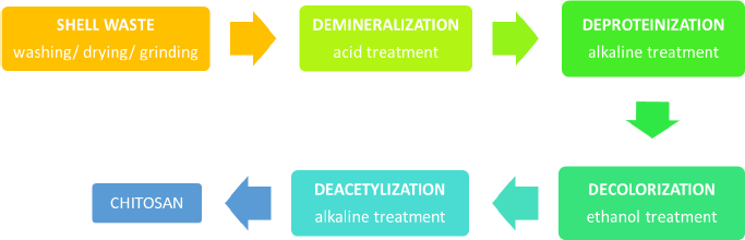
Chitin is an abundant polysaccharide in nature after cellulose in the fungal cell wall and exoskeleton of insects, arthropods, crustaceans, and other invertebrates [9,10]. It has been discovered and explored in several periods since the late eighteenth century [10]. The chemical structure of chitin and cellulose is similar, yet hydroxyl at cellulose C-2 is substituted with an acetamide group in chitin [11]. Chitin is a polymorphic compound with α, β, and γ configurations [12]. It is obtained by extraction from those natural sources, but mostly from shrimp and crab, as up to 80 % of these animals are food wastes [13]. In brief, there are several steps of industrial production of chitin: demineralization, deproteination, bleaching/discoloration, and deacetylation to obtain chitosan, as illustrated inFigure 1. The lengthy extraction process usually takes 17 to 72 h, which includes 1 to 24 h of treatment with hydrochloric acid and 16 to 48 h of sodium hydroxide processing, resulting in a high cost, high energy, and non-environmentally friendly process.

The synthesis is started by grinding the shrimp waste into smaller sizes, followed by the removal of the contained mineral, including calcium carbonate, calcite, and calcium phosphate (demineralization). In this demineralization step, excessive treatment using dilute acidic solutions such as sulfuric, hydrochloric, nitric, acetic, oxalic and formic acids is done, in which the use of hydrochloric acid during the extraction is preferable to obtain a better quality of chitin [14]. After stirring the pulverized dried shells in acidic solution at room temperature, the mineral such as calcium carbonate is transformed into precipitated calcium chloride and carbon dioxide. Following the demineralization step, the reaction mixture should be rinsed with distilled water to neutralize the pH of the mixture. As chitin occurs naturally in association with protein (chitinprotein), a deproteinization process is needed to disrupt the chemical bonds between chitin and proteins. It is done by treating it with aqueous solution of sodium hydroxide (1-10 %) at elevated temperature (65 to 100 °C) for 0.5 to 12 h. In this step, complete removal of the protein from chitin is preferred, particularly for the biomedical application of chitosan, as the remaining shellfish protein may induce allergic reactions in humans. These acid and alkaline treatments resulted in coloured chitin products. The next step is decolorization to remove the red or orange pigment-formed complexes with chitin. The process could be accomplished mainly by treating it with ethanol or acetone and bleaching it with sodium hypochlorite, yielding a white-coloured chitin powder.

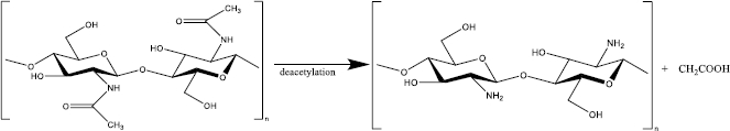
To convert chitin to chitosan, the resulting chitin should be deacetylated in the concentrated sodium or potassium hydroxide solution [15]. The acetyl group in the chitin structure causes low solubility and reactivity in many solvents; thus, it is not convenient to use [9]. In 1859, C. Rouget found that the solubilization of chitin in acid increased after being reacted with concentrated potassium hydroxide at high temperatures [16]. This process yields a derivate compound with better solubility and reactivity than its native substance because of the presence of primary and secondary hydroxyl groups [16-18]. The deacetylated chitin was then identified as chitosan by Felix Hoppe Seyler in 1894 [16,19]. After deacetylation, the typical characteristic of each chitin polymorph disappears and becomes amorphous chitosan [12].

Chitosan itself is a cationic polymer (one positive charge for each glucosamine residue) derived from partial (at minimum 50 %) or almost complete alkaline deacetylation (reaching 95 %) at C-2 chitin which presents -NH2 primary group (Figure 2) [20,21]. Among many chitosan derivates such as glycosamine, tosyl - and trityl chitin, chitosan is the most widely used in biomedical and nutraceutical fields [22]. Consisting of amine moiety, chitosan has beneficial biological properties such as biodegradable, highly biocompatible, non-toxic, and low allergenic [23]. Moreover, because of the amino groups, chitosan is a weak base with pKa 6.3 to 6.5, which is protonated below physiological pH, leading to application in a pH-responsive system [24]. Because of the D-glucosamine, chitosan is generally insoluble in water and organic solvents and and is soluble in acidic aqueous solutions, such as acetic acid. However, the solubility highly depends on the degree of deacetylation (DD) [24].

DD is a parameter to determine the chitosan characteristic and its biological activity. For instance, aqueous soluble chitosan requires at least 50 % DD, while enzyme or lysozyme non-degradable chitosan must be 95 % DD [25,26]. This parameter is calculated as a unit ratio of 2-acetamido-2-deoxy-glucopyranose (GIcNAc) to 2-amino-2-deoxy-D-glucopyranose (GIcN)) which are linked by a β (1-4) glycosidic bond [27]. DD of the chitosan is analysed by various methods, such as hydrogen bromide titrimetry, infrared spectrometry, UV-Vis spectrophotometry, and 1HNMR spectrometry [28-31].

Besides the excessive need for acid and basic chemicals for chitosan production, which affects the quality of the obtained biopolymer, the waste generated from this well-established process should also be carefully considered. The wastewater containing diluted proteins and huge amounts of chloride, calcium, and sodium could potentially induce marine and freshwater ecotoxicity as well as human carcinogenic toxicity [32,33]. Numerous proteins, peptides, and chitooligosaccharides were produced during the deproteinization step as impurities could not be utilized further due to the high level of alkali solvents contained [34]. Neutralization and detoxification of the discharged wastewater using large amounts of fresh water are necessary to avoid waste disposal problems and reduce the environmental impact, leading to the increased cost of the chitin-purification process. Therefore, more eco-friendly processes of chitosan production are needed as alternative methods.

Eco-friendly chitin and chitosan production

The eco-friendly method for chitosan production was introduced to resolve the shortcomings of the conventional counterparts. The green extraction technique could be done through numerous approaches, such as microbial fermentation, enzyme-assisted extraction, and microwave-assisted extraction. For example, the use of organic acid and protease-producing microorganisms like lactic acid bacteria could replace those chemicals in demineralization, deproteinization, and deacetylation steps without critically affecting the yields of chitin and chitosan [7,35,36]. The lists of enzymatic-producing bacteria used in the chitosan production method have been comprehensively reviewed [37]. In the demineralization step, the use of organic lactic acid and/or acetic acid could result in a relatively comparable effectivity to that obtained with hydrochloric acid [38]. Another benefit of using this biological extraction approach is that the obtained protein-rich by-product could be utilized for other applications, including feed or fertilizer, due to the absence of corrosive chemicals. The resulting wastes from this approach are less harmful to the environment while preserving the characteristics of the obtained chitin. Also, a strong base for neutralization is not needed in the demineralization step using organic acid, limiting the production of salt that could inhibit the enzymatic activity during the enzyme-based deproteinization process. Such a combination of greener routes of organic acid demineralization using citric acid and enzymatic deproteinization using papain or bromelain is beneficial and was reported recently by Pérez to produce chitosan from shrimp shells [39].

In the enzyme-assisted extraction approach, Younes and coworkers [40] used a non-commercial Bacillus mojavensis A21 crude enzyme for the deproteinization step of shrimp shells. They optimized several operating parameters, such as enzyme/substrate ratio, temperature and incubation time, by response surface methodology (RSM). The optimal conditions were an enzyme/substrate ratio of 7.75 U/mg, a temperature of 60 °C, and an incubation time of 6 h for obtaining a deproteinization degree of 88 ± 5 %. Valdez-Peña et al. explored the potency of commercial enzymes (Alcalase®, Flavorzyme®, Lysozyme, Inovapure 300, Trypsin VI, Papain DSM) on the deproteinization step of shell-shrimp powder [41]. From this research, a high weight loss of 61 % was achieved by using Alcalase®, while using other commercial enzymes resulted in 35 to 38 % weight losses. The use of Streptomyces griseus protease for enzymatic protein removal from a shrimp-shell powder of Litopenaeus vannamei was reported by Hongkulsup and coworkers [42]. This process achieved 91 % deproteinization at 37 °C, 3 h and 1 wt.% of the enzyme.

In the deacetylation step, the combination of the enzymatic method with a mild alkaline solution was advantageous for the quality of chitin and ecological compensation [43]. In the earlier, chitin deacetylation was only performed using highly concentrated sodium hydroxide reaching 60 % at high temperatures (65 to >100 °C) and long incubation time [44]. Different times, temperatures, and concentrations of the alkaline solution affect the number of DD and molecular weight [45,46]. Higher temperature and hydroxyl solution concentration resulted in higher DD, while after saturation, prolonging the incubation time did not significantly increase DD [47]. High DD (>90 %) was only reached in high hydroxyl solution at high temperature and long incubation. On the contrary, the extreme deacetylation condition damaged the polysaccharide chain, yielding low molecular weight (MW) chitosan [48]. According to the MW, chitosan is classified into three groups, i.e. low MW (<100 kDa); medium MW (100 to 1,000 kDa), and high MW (>1,000 kDa) [49]. High DD and low MW chitosan show antimicrobial and antioxidant activity, high DD and high MW chitosan are needed for mucoadhesive activity and application in regenerative medicine or drug delivery, meanwhile, medium MW chitosan has anticancer activity [50,51]. In recent years, modified chitin deacetylations were proposed in order to obtain high DD with lower side effects on the polysaccharide chain. As mentioned above, chemical chitin deacetylation, particularly on a manufacturing scale, inflicts environmental pollution [52]. It was reported that to produce one kilogram of 70 % deacetylated chitosan, at least six kilograms of hydroxy chloride, two kilograms of sodium hydroxide, and a large quantity of nitrogen, water, and organic solvents are customized [53]. Thus, biological processes involving deacetylase enzymes are preferable [54].

The first reported chitin deacetylase (CDA) was identified and purified from Mucor rouxii, a dimorphism (yeast-like and filamentous) fungi microbial. The enzyme is a 75 to 80 kDa glycoprotein with high mannose, approximately 30 % carbohydrate, and commonly expressed in the periplasmic space but also secreted to the extracellular medium [55,56]. The enzyme activity was optimum at pH 4.5, 50 °C for glycol chitin model substrate and catalysed at least four N-acetylglucosamine [56]. Afterwards, numerous CADs from other fungi were discovered, isolated, and studied on chitosan production [57]. The CADs have typical biochemical properties, including different molecular sizes from 12 to 150 kDa, acidic pI (2.7 to 4.8), and show optimum bioactivity at 30 to 60 °C in pH 4.5 to 12 [58]. The inefficient yield of the isolated CADs initiated the production of recombinant CADs [59]. Hitherto, several recombinant CADs genes from Colletotrichum lindemuthianum, Saccharomyces cerevisiae, and Mucor circinelloides have been expressed in different bacterial or yeast expression systems such as Escherichia coli or Pichia pastoris, respectively, and showed proper biological activity [59-65].

As microwave has a high potential to accelerate chemical reactions and increase the reaction yield, a chitin deacetylation method using microwave energy was also introduced to reduce incubation time in the hydroxyl solution from hours to less than twenty minutes [66]. The microwave exposure reduced the heating temperature as well as shortened the heating time due to its heating efficiency and homogeneous microwave field within the sample, generating uniform heating [67,68]. Also, the obtained chitosan products from chemical and microwave energy-assisted extraction displayed similar structures, morphologies and chemical compositions. The direct and quick energy transfer of the microwave irradiation to the substrate and catalyst may be responsible for increased reaction efficiency [69]. In this approach, various process parameters, including reaction time, solvent concentration, and solid-to-liquid ratio, should be optimized to obtain the designated degree of deacetylation and molecular mass of the obtained chitosan.

In addition, the concentration of hydroxyl solution could be decreased by combination with urea [70]. The deep eutectic solvents (DESs) approach proved more sustainable, biocompatible, inexpensive, and more convenient in preparation [71]. DESs are a mixture of at least one hydrogen-bond acceptor (HBA), such as halide salts, and one hydrogen-bond donor (HBD): urea, glycerol, benzamide, and citric acid [72]. The application of the DESs in chitosan production was not only effective in the deacetylation process but also in the chitin extraction, including deproteination and demineralization [73].

Aside from deacetylation products, natural chitosan has also been found in some micro and invertebrate organisms. From M. rouxii, the first natural chitosan was isolated from the cell wall using an acidic solution, hydrochloric acid, and formic acid [74]. Afterwards, the natural chitosan was also discovered from other microorganisms like bacteria (Serratia sp. and Bacillus sp.), yeast (Candida albicans and S. cerevisiae), and other fungal microorganisms (Rhizopus oryzae, Gongronella butleri, Cunninghamella elegans, and Phycomyces blakesleeanus [53]. Zhang and colleagues reported that natural chitosan was only found in budding spores of S. cerevisiae, not in the vegetative form [75]. Chitosan extracted from S. cerevisiae and Aspergillus niger had almost similar DD with chitosan processed from chitin’s shrimp [76]. Producing natural chitosan from those sources reduces chemical and toxic materials during the process, including chitin extraction and deacetylation [53].

Considering the risk and benefit of each process, chemically processed chitin-derivate chitosan is still widely produced on the industrial scale. The enzymatic deacetylation encompasses an eco-friendly method; even in economic value, the enzyme is relatively higher priced than the chemicals [57]. Likewise, the natural chitosan final yield is still relatively low compared with the chitin-derivate chitosan and highly depends on the culture medium and incubation condition [53,76]. Modified chemically processed chitosan has become interesting because of the rational cost and benefit of reducing harm by the conventional chemical method [57].

Applications of chitosan in nanomedicine

Chitosan has been used and reported in many fields, for instance, in the food and beverage industry; pharmacy and health, including biotechnology, biomedicine, dentistry, and veterinary; agriculture and aquaculture; textile; pulp and paper industry; chemistry and environmental chemistry [77]. In pharmaceutical and health, chitosan has been applied in drug and gene delivery systems, tissue engineering and artificial implants, bioimaging, protein binding, wound healing, and contact lenses [78].

As the concept of nanotechnology introduced by Richard Feynman in 1959, the application of nanotechnology in medicine has been also emerged [79,80]. Nanotechnology in medicine, terminologically defined as nanomedicine, is nano-scaled medicine obtained from an engineering process resulting in different physical, chemical, and biological features than its original material for diagnosis, monitoring, control, prevention, and treatment of diseases [81,82]. However, the exact definition of nanomedicine is still debated among scientists, regulatory agencies, and enterprisers related to size, organic/inorganic material, pharmacodynamic profile, pathology, and route of administration [83]. In general, nanomaterial should be between 1 to 100 nm in size. Still, for nanomedicine, the material, particularly organic nanomedicine such as liposome or micelle, which is more than 100 nm (i.e., 400 nm), is also considered as ‘nano,’ as long it shows significantly improved properties (i.e., higher solubility and bioavailability) than the bulk material [83]. On the other hand, iron oxide only exhibits altered characteristics below 20 nm [83]. In another literature, nanomedicine is determined in the 1 to 1000 nm range [84], but in the context of pathology and route of administration (i.e., enhanced permeability and retention for cytotoxic therapy), the effect could not be observed from >200 nm particles [85].

According to the types of material, nanomedicine is classified into seven groups: oxide/metal nanoparticles; polymer-based nanoparticles; lipid-based nanoparticles; micelles, liposomes, protein-based nanoparticles, and unspecified others [86,87]. Chitosan-based nanoparticle or nano-chitosan is categorized in polymer-based nanoparticles [88]. In the field of medicine, nano-chitosan has been explored for small and large molecule delivery carrier, wound healing, and tissue regeneration, besides the potential as antimicrobial/anticancer/antioxidant given from the chitosan itself [88].

Eco-friendly crosslinkers for nano-chitosan preparations

Nano-chitosan (and micro-chitosan) is formed by several methods, i.e., ionic gelation crosslinking, reverse micelles, emulsification, precipitation, radical polymerization, and top-down method [8]. The nano-chitosan is characterized differently by each method [89]. Emulsification, reverse micelles, and precipitation involve aqueous chitosan, stabilizer, and organic phases. On the other hand, the organic solvent-free method is much preferred to those methods in which the usage of organic solvent is required. Thus, to date, ionic gelation is the most favourable method for nano-chitosan production. The particle is produced by ionic interaction between positively charged amine groups of chitosan with polyanions crosslinker [8,89].

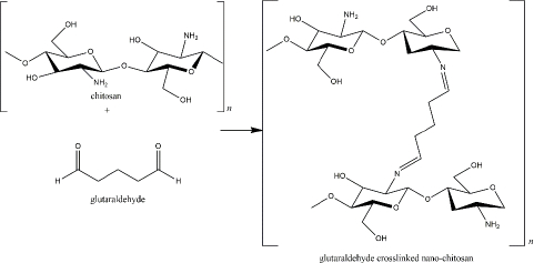
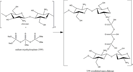
At the beginning of chitosan development, glutaraldehyde was used as the crosslinker [8]. The crosslinking is formed via imine bonds by the interaction of amine groups of the chitosan and an aldehydic group of the glutaraldehyde. The amount of glutaraldehyde affects the particle size as the higher concentration of glutaraldehyde reduces particle size [90]. In addition, the reaction of glutaraldehyde-chitosan depends on the pH solution, which determines the protonation of the amine groups (Figure 3) [91]. Glutaraldehyde is known as a stable crosslinker for chitosan. However, because of its high toxicity to human health, for instance, skin and eye irritation, respiratory tract problems, and environmental issues, it has been substituted with other safer crosslinkers, such as tripolyphosphate (TPP) [92,93]. As described inTable 1, TPP has been the most commonly used crosslinker for nano-chitosan production. Besides the TPP, some crosslinkers, which are natural-derived compounds, have been reported to exhibit remarkable advantages as harmless crosslinker in the nano-chitosan synthesis (Table 2).

TPP, also available in sodium TPP (Na5P3O10), reacts with the amine groups of the chitosan through the negatively charged P3O105- (Figure 4). Characteristics of nano-chitosan from the chitosan polymer and TPP depend on several experimental factors, such as the ratio of chitosan and TPP, pH solution, ionic strength, stirring type, and rate [131]. TPP was reviewed as the smallest and strongest nano-chitosan’s crosslinker among the others crosslinkers [132]. To overcome the environmental issues, a certain amount of innovative natural-based crosslinker for nano-chitosan has been explored, including carrageenan, genipin, citric acid, and vanillin [129].

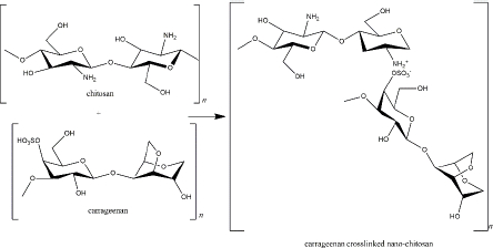
Carrageenan is a sulphated galactosan extracted from the Rhodophyceae family containing main sugars (o-galactose) and 3,6-anhydro-o-galactose; the main substituent (sulphate); carbohydrate residues (xylose, glucose, and uronic acids), and other substituents (methyl esters) [141]. It is an anionic polymer because of the high component of the ester-sulphate. In addition, because of variations in the sulphate content, carrageenan is classified into six types (κ, λ, ι, θ, β, ν, and μ carrageenan). Each type shows different physiochemical properties (such as solubility and gelling mechanism) and bioactivity [142].

As a negatively charged polymer, carrageenan is suitable as a crosslinker in the nano-chitosan preparation. Carrageenan (κ-carrageenan) and chitosan were used to form polyelectrolyte complexes (PEC) (Figure 5). Not only does it function as a drug delivery system, tissue regeneration, cell cultivation, and enzyme immobilization, but the PEC also exhibits biological activity as an anti-inflammatory via histamine activation [143].

Khaliq and colleagues reported that cefotaxime sodium loaded κ-carrageenan - chitosan hydrogel was applicable to dress diabetic wounds by releasing the drug in 24 h for effectively inhibiting bacterial propagation [144]. The drug release profile depends on pKa or pI and the extrinsic pH. For instance, diflunisal (pKa = 2.94) was released from a κ-carrageenan - chitosan nanocapsule in intestinal fluid and blood distribution (pH 7.4) in which the drug was found in anionic and highly soluble form. Otherwise, in lower pH, such as in acidic gastric fluid (pH 2-3), diflunisal was neutral and insoluble in an aqueous solution [145]. Along with TPP, the combination of these two marine polymers was also used for the delivery system of α-mangostin and showed cytotoxicity in the MCF-7 cell line and antibacterial activity [146,147].

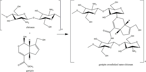
Genipin, another naturally derived crosslinker in nano-chitosan preparation, is found in Genipa americana fruit. It was soluble in aqueous solution, alcohol, and propylene glycol, less toxic than other synthetic crosslinkers (showed 0.01 % cytotoxicity compared with glutaraldehyde), and reported for several pharmacological activities [148]. This compound actively reacts with primary amine groups of chitosan by substituting oxygen at the dihydropyran ring and the aldehyde group, which depends on environmental pH and oxidation level (Figure 6) [149,150]. Most of the recent applications of genipin-crosslinker chitosan were proposed for tissue regeneration and wound healing. Heimbuck and team described the capabilities of genipin-crosslinked chitosan hydrogel to hamper bacterial growth and to neutralize environmental pH as a wound dressing [151]. A list of genipin-crosslinked chitosan for skeletal regeneration has been reviewed by Wang et al. [152].

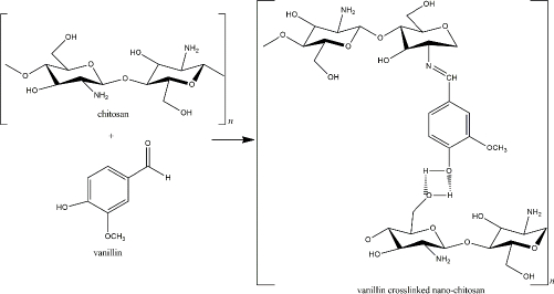
Another fascinating natural chitosan crosslinker is vanillin or 4-hydroxy-3-methoxy benzaldehyde, an aromatic compound extracted from Vanilla sp. beans and commonly used in food and beverage industries. The exorbitant cost of the natural vanilla extraction and harvest limitation caused the idea to chemically synthesize the vanillin compound for a more rational commercial value [153]. 4-hydroxybenzaldehyde was used to produce this artificial vanillin.

However, the precursor was then reported to harm the environment and human health [153,154]. Therefore, biotechnological-derived approaches, such as plant-based, enzyme-based, and microorganism-based methods, have been applied in vanillin production to obtain lower costs with decreased side effects [153]. The aldehyde group of the vanillin reacts with the chitosan’s primary amine, yielding an imine group (Schiff base). The reaction is reversible by the presence of a para-hydroxyl group (of the imine group) that forms a hydrogen bond with the adjacent hydroxyl of the amino group (Figure 7). Because the reversible hydrogen bond exists, the interaction of vanillin and chitosan relies on pH and temperature. The hydrogen bonds showed high stability at low temperatures [155].

Discussion and conclusion

The idea of an eco-friendly process, previously called green chemistry, appeared in 1991 to avoid excessive exposure to harmful chemicals to the environment and human health. The harmful chemicals are classified into three following groups: physical problem (explosive and flammable substances); toxicity to human health (causing cytotoxicity, mortality); environmental danger (causing climate change; ozone layer depletion; over-consumption of energy; or uncontrollable waste) [156]. Pharmaceutical industries were reported to contribute significantly to pollutant and waste by-products among chemistry industries. In the last decade, the pharmaceutical industries have applied green chemistry principles as reviewed by Mishra et al., including reducing waste and preventing pollution, using safer chemical or renewable materials, and increasing energy efficiency [157].

Production of nanomaterial for nanomedicine applications, such as nano-chitosan, is also known to involve hazardous substances and waste for the environment and health. Therefore, eco-friendly or green synthesis is requested not only during the nanoparticle assembly but must be applied during the raw materials preparations. In short, two crucial eco-friendly concepts in the nano-chitosan preparation, which are using safer or reducing dangerous chemicals in the chitosan synthesis (including chitin extraction and deacetylation) and determining harmless crosslinkers for the nano-chitosan preparation, have been discussed. Biotechnology processes, particularly enzymatic and microbiological methods, have also been applied in this field, even so, up to now, in terms of industrial capacity and cost, it gives less benefit than the other green methods, for instance, by using sustainable reagents such as DESs. Above all, the natural properties of chitin, chitosan, or nano-chitosan (hydrophilicity, biocompatibility, no organic solvent needed) are the main key to the appropriateness of the eco-friendly or green synthesis concept [158].

Acknowledgements

We would like to thank to Vaccine and Drug Funding Program 2023, Organization of Health, The National Innovation and Research Agency, Republic of Indonesia.

Notes

[1] Conflicts of interest Conflict of interest: The authors declare no conflicts of interest.

[2] Contributions: RDP contributed to conceptualization, writing and revising the manuscript; SEM wrote and improved the manuscript in the section on chitin and chitosan production; NG wrote the abstract; NSD & GS contributed in writing technical process; AR contributed to conceptualization and corrected the whole first manuscript. All authors read, corrected, and approved the final manuscript.

References

[1] 

Teli M.K.; Mutalik S.; Rajanikant G.K.. Nanotechnology and Nanomedicine: Going Small Means Aiming Big; Current Pharmaceutical Design 16 (2010) 1882-1892. https://doi.org/10.2174/138161210791208992 https://doi.org/10.2174/138161210791208992

[2] 

Foulkes R.; Man E.; Thind J.; Yeung S.; Joy A.; Hoskins C.. The Regulation of Nanomaterials and Nanomedicines for Clinical Application: Current and Future Perspectives. Biomaterial Science 8 (2020) 4653-4664. https://doi.org/10.1039/d0bm00558d https://doi.org/10.1039/d0bm00558d

[3] 

Peniche H.; Peniche C.. Chitosan Nanoparticles: A Contribution to Nanomedicine. Polymer International 60 (2011) 883-889. https://doi.org/10.1002/pi.3056 https://doi.org/10.1002/pi.3056

[4] 

Mohammed M.A.; Syeda J.T.M.; Wasan K.M.; Wasan E.K.. An Overview of Chitosan Nanoparticles and Its Application in Non-Parenteral Drug Delivery. Pharmaceutics 9 (2017) 53. https://doi.org/10.3390/pharmaceutics9040053 https://doi.org/10.3390/pharmaceutics9040053

[5] 

Chenthamara D.; Subramaniam S.; Ramakrishnan S.G.; Krishnaswamy S.; Essa M.M.; Lin F.H.; Qoronfleh M.W.. Therapeutic Efficacy of Nanoparticles and Routes of Administration. Biomaterial Research 23 (2019) 20. https://doi.org/10.1186/s40824-019-0166-x https://doi.org/10.1186/s40824-019-0166-x

[6] 

Mahapatra I.; Clark J.R.A.; Dobson P.J.; Owen R.; Lynch I.; Lead J.R.. Expert Perspectives on Potential Environmental Risks from Nanomedicines and Adequacy of the Current Guideline on Environmental Risk Assessment. Environmental Science Nano 5 (2018) 1873-1889. https://doi.org/10.1039/c8en00053k https://doi.org/10.1039/c8en00053k

[7] 

Beaney P.; Lizardi-Mendoza J.; Healy M.. Comparison of Chitins Produced by Chemical and Bioprocessing Methods. Journal of Chemical Technology and Biotechnology 80 (2005) 145-150. https://doi.org/10.1002/jctb.1164 https://doi.org/10.1002/jctb.1164

[8] 

Yanat M.; Schroën K.. Preparation Methods and Applications of Chitosan Nanoparticles; with an Outlook toward Reinforcement of Biodegradable Packaging. Reactive and Functional Polymer 161 (2021) 104849. https://doi.org/10.1016/J.REACTFUNCTPOLYM.2021.104849 https://doi.org/10.1016/J.REACTFUNCTPOLYM.2021.104849

[9] 

Iber B.T.; Kasan N.A.; Torsabo D.; Omuwa J.W.. A Review of Various Sources of Chitin and Chitosan in Nature. Journal of Renewable Materials 10 (2022) 1097-1123. https://doi.org/10.32604/JRM.2022.018142 https://doi.org/10.32604/JRM.2022.018142

[10] 

Crini G.. Historical Review on Chitin and Chitosan Biopolymers. Environmental Chemistry Letter 17 (2019) 1623-1643. https://doi.org/10.1007/s10311-019-00901-0 https://doi.org/10.1007/s10311-019-00901-0

[11] 

Chawla S.P.; Kanatt S.R.; Sharma A.K.. Chitosan, in Polysaccharides Ramawat K.; Mérillon J.M. (Eds)., Springer Cham, 2015, 219-246. https://doi.org/10.1007/978-3-319-16298-0_13 https://doi.org/10.1007/978-3-319-16298-0_13

[12] 

Tsurkan M.V.; Voronkina A.; Khrunyk Y.; Wysokowski M M.; Petrenko I.; Ehrlich H.. Progress in Chitin Analytics. Carbohydrate Polymers 252 (2021) 117204. https://doi.org/10.1016/J.CARBPOL.2020.117204 https://doi.org/10.1016/J.CARBPOL.2020.117204

[13] 

Hossin M.A.; Al Shaqsi N.H.K.; Al Touby S.J.; Al Sibani M.A.. Review of Polymeric Chitin Extraction, Characterization, and Applications. Arabian Journal of Geosciences 14 (2021) 1870. https://doi.org/10.1007/s12517-021-08239-0 https://doi.org/10.1007/s12517-021-08239-0

[14] 

Younes I.; Rinaudo M.. Chitin and chitosan preparation from marines sources. Structure, properties and applications. Marine Drugs 13 (2015) 1133-1174. https://doi.org/10.3390/md13031133 https://doi.org/10.3390/md13031133

[15] 

Kumar M.N.R.. A review of chitin and chitosan applications. Reactive and Functional Polymers 46 (2000) 1-27. https://doi.org/10.1016/S1381-5148(00)00038-9 https://doi.org/10.1016/S1381-5148(00)00038-9

[16] 

Periayah M.H.; Halim A.S.; Saad A.Z.M.. Chitosan: A Promising Marine Polysaccharide for Biomedical Research. Pharmacognosy Review 10 (2016) 39-42. https://doi.org/10.4103/0973-7847.176545 https://doi.org/10.4103/0973-7847.176545

[17] 

Islam S.; Bhuiyan M.A.R.; Islam M.N.. Chitin and Chitosan: Structure, Properties and Applications in Biomedical Engineering. Journal of Polymers and The Environment 25 (2017) 854-866. https://doi.org/10.1007/s10924-016-0865-5 https://doi.org/10.1007/s10924-016-0865-5

[18] 

Ravindra R.; Krovvidi K.R.; Khan A.A.. Solubility Parameter of Chitin and Chitosan. Carbohydrate Polymers 36 (1998) 121-127. https://doi.org/10.1016/S0144-8617(98)00020-4 https://doi.org/10.1016/S0144-8617(98)00020-4

[19] 

Nimbeni S.B.; Nimbeni B.S.; Divakar D.D.. Role of Chitosan in Remineralization of Enamel and Dentin: A Systematic Review. International Journal of Clinical Pediatric Dentistry 14 (2021) 562-568. https://doi.org/10.5005/jp-journals-10005-1971 https://doi.org/10.5005/jp-journals-10005-1971

[20] 

Gao F.; Zhang B.S.; Zhao J.H.; Huang J.F.; Jia P.S.; Wang S.; Zhang J.; Zhou J.M.; Guo H.S.. Deacetylation of Chitin Oligomers Increases Virulence in Soil-Borne Fungal Pathogens. Nature Plants 5 (2019) 1167-1176. https://doi.org/10.1038/s41477-019-0527-4 https://doi.org/10.1038/s41477-019-0527-4

[21] 

Pérez-Álvarez L.; Ruiz-Rubio L.; Vilas-Vilela J.L.. Determining the Deacetylation Degree of Chitosan: Opportunities to Learn Instrumental Techniques. Journal of Chemical Education 95 (2018) 1022-1028. https://doi.org/10.1021/acs.jchemed.7b00902 https://doi.org/10.1021/acs.jchemed.7b00902

[22] 

Akakuru O.U.; Louis H.; Amos P.I.; Akakuru O.C.; Nosike E.I.; Ogulewe E.F.. The Chemistry of Chitin and Chitosan Justifying Their Nanomedical Utilities. Biochemistry and Pharmacology 7 (2018) 241. https://doi.org/10.4172/2167-0501.1000241 https://doi.org/10.4172/2167-0501.1000241

[23] 

Forsythe J.; Rassu G.; Giunchedi P.; James Gilbert R.; Mateos-Díaz J.C.; Ojeda-Hernández D.D.; D.; Canales-Aguirre A.A.; Matias-Guiu J.; Gomez-Pinedo U.. Potential of Chitosan and Its Derivatives for Biomedical Applications in the Central Nervous System. Frontiers in Bioengineering and Biotechnology 8 (2020) 389. https://doi.org/10.3389/fbioe.2020.00389 https://doi.org/10.3389/fbioe.2020.00389

[24] 

Thakur V.K.; Thakur M.K.. Recent Advances in Graft Copolymerization and Applications of Chitosan: A Review. ACS Sustainable Chemistry and Engingeering 2 (2014) 2637-2652. https://doi.org/10.1021/sc500634p https://doi.org/10.1021/sc500634p

[25] 

Feng T.; Du Y.; Li J.; Wei Y. Y.; Yao P. Antioxidant Activity of Half N-Acetylated Water-Soluble Chitosan in Vitro. European Food Research and Technology 225 (2007) 133-138. https://doi.org/10.1007/s00217-006-0391-0 https://doi.org/10.1007/s00217-006-0391-0

[26] 

Deng Q.Y.; Zhou C.R.; Luo B.H.. Preparation and Characterization of Chitosan Nanoparticles Containing Lysozyme. Pharmceutical Biology 44 (2006) 336-342. https://doi.org/10.1080/13880200600746246 https://doi.org/10.1080/13880200600746246

[27] 

Sweidan K.; Abdel-Motalleb J.; Al-Jbour N.D.; Obaidat Jordan R.M.. Further investigation on the degree of deacetylation of chitosan determined by potentiometric titration. Journal of Excipients and Food Chemicals 2 (2011) 16-25.https://doaj.org/article/ad90534b4543471f998602bde4a1bf7f

[28] 

Khan T.A.; Peh K.K.; Ch’ng S.H.. Reporting Degree of Deacetylation Values of Chitosan: The Influence of Analytical Methods. Journal of Pharmacy and Pharmaceutical Science 5 (2002) 205-212. https://pubmed.ncbi.nlm.nih.gov/12553887/ PubMed: http://www.ncbi.nlm.nih.gov/pubmed/12553887

[29] 

Czechowska-Biskup R.; Jarosińska D.; Rokita B.; Ulański P.; Rosiak J.M.. Determination of Degree of Deacetylation of Chitosan - Comparison of Methods. Progress on Chemistry and Application of Chitin and Its Derivatives 17 (2012) 5-20.https://www.researchgate.net/publication/288104933_Determination_of_degree_of_deacetylation_of_chitosan_-_Comparision_of_methods

[30] 

Jiang Y.; Fu C.; Wu S.; Liu G.; Guo J.; Su Z.. Determination of the Deacetylation Degree of Chitooligosaccharides. Marine Drugs 15 (2017) 332. https://doi.org/10.3390/md15110332 https://doi.org/10.3390/md15110332

[31] 

Lavertu M.; Xia Z.; Serreqi A.N.; Berrada M.; Rodrigues A.; Wang D.; Buschmann M.D.; Gupta A.. A Validated 1H NMR Method for the Determination of the Degree of Deacetylation of Chitosan. Journal of Pharmaceutical and Biomedical Analysis 32 (2003) 1149-1158. https://doi.org/10.1016/S0731-7085(03)00155-9 https://doi.org/10.1016/S0731-7085(03)00155-9

[32] 

Riofrio A.; Alcivar T.; Baykara H.. Environmental and economic viability of chitosan production in Guayas-Ecuador: a robust investment and life cycle analysis. ACS Omega 6 (2021) 23038-23051. https://doi.org/10.1021/acsomega.1c01672 https://doi.org/10.1021/acsomega.1c01672

[33] 

Wang W.; Du Y.; Qiu Y.; Wang X.; Hu Y.; Yang J.; et al.. A new green technology for direct production of low molecular weight chitosan. Carbohydrate polymers 74 (2008) 127-132. https://doi.org/10.1016/j.carbpol.2008.01.25 https://doi.org/10.1016/j.carbpol.2008.01.25

[34] 

Younes I.; Hajj S.; Frachet V.; Rinaudo M.; Jellouli K.; Nasri M.. Chitin extraction from shrimp shell using enzymatic treatment. Antitumor, antioxidant and antimicrobial activities of chitosan. International Journal of Biological Macromolecules 69 (2014) 489-498. https://doi.org/10.1016/j.ijbiomac.2014.06.013 https://doi.org/10.1016/j.ijbiomac.2014.06.013

[35] 

Yu Y.; Liu X.; Miao J.; Leng K.. Chitin from Antarctic Krill shell: eco-preparation, detection, and characterization. International Journal of Biological Macromolecules 164 (2020) 4125-4137. https://doi.org/10.1016/j.ijbiomac.2020.08.244 https://doi.org/10.1016/j.ijbiomac.2020.08.244

[36] 

Zhang H.; Yun S.; Song L.; Zhang Y.; Zhao Y.. The preparation and characterization of chitin and chitosan under large-scale submerged fermentation level using shrimp by-products as substrate. International Journal of Biological Macromolecules 96 (2017) 334-339. https://doi.org/10.1016/j.ijbiomac.2016.12.017 https://doi.org/10.1016/j.ijbiomac.2016.12.017

[37] 

Mohan K.; Ganesan A.R.; Ezhilarasi P.N.; Kondamareddy K.K.; Rajan D.K.; Sathishkumar P.; Rajarajeswaran J.; Conterno L.. Green and Eco-Friendly Approaches for the Extraction of Chitin and Chitosan: A Review. Carbohydrate Polymers 287 (2022) 119349. https://doi.org/10.1016/j.carbpol.2022.119349 https://doi.org/10.1016/j.carbpol.2022.119349

[38] 

Mahmoud N.S.; Ghaly A.E.; Arab F.. Unconventional approach for demineralization of deproinated crustacean shells for chitin production. American Journal of Biochemistry and Biotechnology 3(1) (2007) 1-9. https://doi.org/10.3844/ajbbsp.2007.1.9 https://doi.org/10.3844/ajbbsp.2007.1.9

[39] 

Ameh A.O.; Isa M.T.; Abutu D.; Danlami A.. Kinetic modelling of the demineralization of shrimp exoskeleton using citric acid. Leonardo Electron Journal Practice and Technology 25 (2014) 99-108.http://lejpt.academicdirect.org/A25/099_108.pdf

[40] 

Younes I.; Ghorbel-Bellaj O.; Nasri R.; Chaabouni M.; Rinaudo M.; Nasri M.. Chitin and chitosan preparation from shrimp shells using optimized enzymatic deproteinization. Process Biochemistry 47 (2012) 2032-2039. https://doi.org/10.1016/j.procbio.2012.07.017 https://doi.org/10.1016/j.procbio.2012.07.017

[41] 

Valdez-Peña A.U.; Espinoza-Perez J.D.; Sandoval-Fabian G.C.; Balagurusamy N.; Hernandez-Rivera A.; De-la-Garza-Rodriguez I.M.; Contreras-Esquivel J.C., Screening of industrial enzymes for deproitenization of shrimp head for chitin recovery. Food Science and Biotechnology 19 (2010) 553-557. https://doi.org/10.1007/s10068-010-0077-z https://doi.org/10.1007/s10068-010-0077-z

[42] 

Hongkulsup C.; Khutoryanskiy V.V.; Niranjan K.. Enzyme assisted extraction of chitin from shrimp shells (Litopenaeus vannamei). Journal of Chemical Technology & Biotechnology 91 (2016) 1250-1256. https://doi.org/10.1002/jctb.4714 https://doi.org/10.1002/jctb.4714

[43] 

Marzieh M.N.; Zahra F.; Tahereh E.; Sara K.N.. Comparison of the physicochemical and structural characteristics of enzymatic produced chitin and commercial chitin. International Journal of Biological Macromolecules 139 (2019) 270-276. https://doi.org/10.1016/j.ijbiomac.2019.07.217 https://doi.org/10.1016/j.ijbiomac.2019.07.217

[44] 

Hossain M.; Iqbal A.. Production and Characterization of Chitosan from Shrimp Waste. Journal of the Bangladesh Agricultural University 12 (2014) 153-160. https://doi.org/10.3329/jbau.v12i1.21405 https://doi.org/10.3329/jbau.v12i1.21405

[45] 

Pires C.T.G.V.M.T.; Vilela J.A.P, Airoldi C.. The Effect of Chitin Alkaline Deacetylation at Different Condition on Particle Properties. Procedia Chemistry 9 (2014) 220-225. https://doi.org/10.1016/j.proche.2014.05.026 https://doi.org/10.1016/j.proche.2014.05.026

[46] 

Weska R.F.; Moura J.M.; Batista L.M.; Rizzi J.; Pinto L.A.A.. Optimization of Deacetylation in the Production of Chitosan from Shrimp Wastes: Use of Response Surface Methodology. Journal of Food Engineering 80 (2007) 749-753. https://doi.org/10.1016/J.JFOODENG.2006.02.006 https://doi.org/10.1016/J.JFOODENG.2006.02.006

[47] 

Galed G.; Diaz E.; Goycoolea F.M.; Heras A.. Influence of N-Deacetylation Conditions on Chitosan Production from α-Chitin. Natural Product Communications 3 (2008) 543-550. https://doi.org/10.1177/1934578X0800300414 https://doi.org/10.1177/1934578X0800300414

[48] 

Novikov V.Y.; Konovalova I.N.; Dolgopyatova N.; The N. Mechanisms of Chitin and Chitosan Deacetylation During Long-term Alkaline Treatment. Applied Biochemsitry and Microbiology 58 (2022) 273-279. https://doi.org/10.1134/S0003683822030097 https://doi.org/10.1134/S0003683822030097

[49] 

Santoso J.; Adiputra K.C.; Soerdirga L.C.; Tarman K.. Effect of Acetic Acid Hydrolysis on the Characteristics of Water-Soluble Chitosan. In Proceedings of the IOP Conference Series: Earth and Environmental Science 414 (2020) 012021. https://doi.org/10.1088/1755-1315/414/1/012021 https://doi.org/10.1088/1755-1315/414/1/012021

[50] 

Casadidio C.; Peregrina D.V.; Gigliobianco M.R.; Deng S.; Censi R.; di Martino P.. Chitin and Chitosans: Characteristics, Eco-Friendly Processes, and Applications in Cosmetic Science. Marine Drugs 17 (2019) 369. https://doi.org/10.3390/md17060369 https://doi.org/10.3390/md17060369

[51] 

Rasweefali M.K.; Sabu S.; Sunooj K.V.; Sasidharan A.; Xavier K.A.M.. Consequences of Chemical Deacetylation on Physicochemical, Structural and Functional Characteristics of Chitosan Extracted from Deep-Sea Mud Shrimp. Carbohydrate Polymer Technologies and Applications 2 (2021) 100032. https://doi.org/10.1016/j.carpta.2020.100032 https://doi.org/10.1016/j.carpta.2020.100032

[52] 

Pawaskar G.M.; Pangannaya S.; Raval K.; Trivedi D.R.; Raval R.. Screening of Chitin Deacetylase Producing Microbes from Marine Source Using a Novel Receptor on Agar Plate. International Journal of Biology Macromolecular 131 (2019) 716-720. https://doi.org/10.1016/j.ijbiomac.2019.03.118 https://doi.org/10.1016/j.ijbiomac.2019.03.118

[53] 

Sebastian J.; Rouissi T.; Brar S.K.. Fungal Chitosan: Prospects and Challenges. Handbook of Chitin and Chitosan Preparation and Properties 1 (2020) 419-452. https://doi.org/10.1016/B978-0-12-817970-3.00014-6 https://doi.org/10.1016/B978-0-12-817970-3.00014-6

[54] 

Hoell I.A.; Vaaje-Kolstad G.; Eijsink V.G.H.. Structure and Function of Enzymes Acting on Chitin and Chitosan. Biotechnology Genetic Engineering Review 27 (2010) 331-366. https://doi.org/10.1080/02648725.2010.10648156 https://doi.org/10.1080/02648725.2010.10648156

[55] 

Bartnicki-Garcia’ S.; Nickerson W.J.. Nutrition, Growth, and Morphogenesis of Mucor rouxii. Journal of Bacteriology 84 (1962) 841-867. https://doi.org/10.1128/jb.84.4.841-858.1962 https://doi.org/10.1128/jb.84.4.841-858.1962

[56] 

Kafetzopoulos D.; Martinou A.; Bouriotis V.. Bioconversion of Chitin to Chitosan: Purification and Characterization of Chitin Deacetylase from Mucor rouxii. Proceedings of the National Academy of Sciences of the United States of America 90 (1993) 2564-2568. https://doi.org/10.1073/pnas.90.7.2564 https://doi.org/10.1073/pnas.90.7.2564

[57] 

Kaczmarek M.B.; Struszczyk-Swita K.; Li X.; Szczęsna-Antczak Daroch M.; Enzymatic M. Modifications of Chitin, Chitosan, and Chitooligosaccharides. Frontiers Bioengineering and Biotechnolology 7 (2019) 243. https://doi.org/10.3389/fbioe.2019.00243 https://doi.org/10.3389/fbioe.2019.00243

[58] 

Grifoll-Romero L.; Pascual S.; Aragunde H.; Biarnés X.; Planas A.. Chitin Deacetylases: Structures, Specificities, and Biotech Applications. Polymers 10 (2018) 352. https://doi.org/10.3390/polym10040352 https://doi.org/10.3390/polym10040352

[59] 

Tokuyasu K.; Kaneko S.; Hayashi K.; Mori Y.. Production of a Recombinant Chitin Deacetylase in the Culture Medium of Escherichia coli Cells. FEBS Letters 458 (1999) 23-26. https://doi.org/10.1016/S0014-5793(99)01113-8 https://doi.org/10.1016/S0014-5793(99)01113-8

[60] 

Bhat P.; Pawaskar G.M.; Raval R.; Cord-Landwehr S.; Moerschbacher B.; B. Raval K. Expression of Bacillus Licheniformis Chitin Deacetylase in E. coli PLysS: Sustainable Production, Purification and Characterisation. International Journal of Biolology Macromolecule 131 (2019) 1008-1013. https://doi.org/10.1016/j.ijbiomac.2019.03.144 https://doi.org/10.1016/j.ijbiomac.2019.03.144

[61] 

Naqvi S.; Cord-Landwehr S.; Singh R.; Bernard F.; Kolkenbrock S.; Gueddari N.E.; Moerschbacher B.M.. A Recombinant Fungal Chitin Deacetylase Produces Fully Defined Chitosan Oligomers with Novel Patterns of Acetylation. Applied Environmental Microbiology 82 (2016) 6645-6655. https://doi.org/10.1128/AEM.01961-16 https://doi.org/10.1128/AEM.01961-16

[62] 

Shrestha B.; Blondeau K.; Stevens W.F.; Hegarat F.L.. Expression of Chitin Deacetylase from Colletotrichum Lindemuthianum in Pichia pastoris: Purification and Characterization. Protein Expression and Purification 38 (2004) 196-204. https://doi.org/10.1016/J.PEP.2004.08.012 https://doi.org/10.1016/J.PEP.2004.08.012

[63] 

Mishra C.; Semino C.E.; Mccreath K.J.; de La Vega H.; Jones B.J.; Specht C.A.; Robbins P.W.. Cloning and Expression of Two Chitin Deacetylase Genes of Saccharomyces cerevisiae. Yeast 13 (1997) 327-336. https://doi.org/10.1002/(SICI)1097-0061 https://doi.org/10.1002/(SICI)1097-0061

[64] 

Kaczmarek M.B.; Struszczyk-Swita K.; Xiao M.; Szczęsna-Antczak M.; Antczak T.; Gierszewska M.; Steinbüchel A.; Daroch M.. Polycistronic Expression System for Pichia pastoris Composed of Chitino- and Chitosanolytic Enzymes. Frontiers Bioengineering and Biotechnolology 9 (2021) 710922. https://doi.org/10.3389/fbioe.2021.710922. https://doi.org/10.3389/fbioe.2021.710922

[65] 

Martinou A.; Koutsioulis D.; Bouriotis V.. Cloning and Expression of a Chitin Deacetylase Gene (CDA2) from Saccharomyces cerevisiae in Escherichia coli: Purification and Characterization of the Cobalt-Dependent Recombinant Enzyme. Enzyme Microbiology Technology 32 (2003) 757-763. https://doi.org/10.1016/S0141-0229(03)00048-6 https://doi.org/10.1016/S0141-0229(03)00048-6

[66] 

el Knidri H.; Dahmani J.; Addaou A.; Laajeb A.; Lahsini A.. Rapid and Efficient Extraction of Chitin and Chitosan for Scale-up Production: Effect of Process Parameters on Deacetylation Degree and Molecular Weight. International Journal of Biological Macromolecules 139 (2019) 1092-1102. https://doi.org/10.1016/j.ijbiomac.2019.08.079 https://doi.org/10.1016/j.ijbiomac.2019.08.079

[67] 

Zaeni A.; Safitri E.; Fuadah B.; Sudiana I.N.. Microwave-assisted hydrolysis of chitosan from shrimp shell waster for glucosamine hydrochlorid production. The 5th International Conference on Theoretical and Applied Physics, IOP Publishing of Physics Conference Series. 846 (2015) 23-25. https://doi.org/10.1088/1742-6596/846/1/012011 https://doi.org/10.1088/1742-6596/846/1/012011

[68] 

Zhang J.; Feng M.; Lu X.; Shi C.; Li X.; Xin J.; et al. Base-free preparation of low molecular weight chitin from crab shell. Carbohydrate Polymers 190 (2018) 148-155. https://doi.org/10.1016/j.carbpol.2018.02.019 https://doi.org/10.1016/j.carbpol.2018.02.019

[69] 

Tsubaki S.; Azuma J.I.. Total fractional of green tea residue by microwave-assisted alkaline pretreatment and enzymatic hydrolysis. Biosensor Technology. 131 (2013) 485-491. https://doi.org/10.1016/j.biortech.2013.01.001 https://doi.org/10.1016/j.biortech.2013.01.001

[70] 

Li F.; You X.; Li Q.; Qin D.; Wang M.; Yuan S.; Chen X.; Bi S.. Homogeneous Deacetylation and Degradation of Chitin in NaOH/Urea Dissolution System. International Journal of Biological Macromolecules 189 (2021) 391-397. https://doi.org/10.1016/J.IJBIOMAC.2021.08.126 https://doi.org/10.1016/J.IJBIOMAC.2021.08.126

[71] 

Vicente F.A.; Huš M.; Likozar B.; Novak U.. Chitin Deacetylation Using Deep Eutectic Solvents: Ab Initio-Supported Process Optimization. ACS Sustainable Chemical Engineering 9 (2021) 3874-3886, https://doi.org/10.1021/acssuschemeng.0c08976 https://doi.org/10.1021/acssuschemeng.0c08976

[72] 

Smith E.L.; Abbott A.P.; Ryder K.S.. Deep Eutectic Solvents (DESs) and Their Applications. Chemical Reviews 114 (2014) 11060-11082. https://doi.org/10.1021/cr300162p https://doi.org/10.1021/cr300162p

[73] 

Zhao D.; Huang W.C.; Guo N.; Zhang S.; Xue C.; Mao X.. Two-Step Separation of Chitin from Shrimp Shells Using Citric Acid and Deep Eutectic Solvents with the Assistance of Microwave. Polymers 11 (2019) 409. https://doi.org/10.3390/polym11030409 https://doi.org/10.3390/polym11030409

[74] 

White S.A.; Farina-Peter R.; Fulton I.. Production and Isolation of Chitosan from Mucor rouxii. Applied and Environmental Microbiology 38 (1979) 323-326. https://doi.org/10.1128/aem.38.2.323-328.1979 https://doi.org/10.1128/aem.38.2.323-328.1979

[75] 

Zhang H.; Tachikawa H.; Gao X.D.; Nakanishi H.. Applied Usage of Yeast Spores as Chitosan Beads. Applied and Environmental Microbiology 80 (2014) 5098-5105. https://doi.org/10.1128/AEM.00677-14 https://doi.org/10.1128/AEM.00677-14

[76] 

Afroz M.; Nayeem M.; Kashem H.; Masirul Afroz M.; Kashem N.H.; Prottoy K.M.; Piash S.; Islam N.. Saccharomyces Cerevisiae as an Untapped Source of Fungal Chitosan for Antimicrobial Action. Applied Biochemistry and Biotechnology 193 (2021) 3765–3786. https://doi.org/10.21203/rs.3.rs-490748/v1 https://doi.org/10.21203/rs.3.rs-490748/v1

[77] 

Morin-Crini N.; Lichtfouse E.; Torri G.; Crini G.. Applications of Chitosan in Food, Pharmaceuticals, Medicine, Cosmetics, Agriculture, Textiles, Pulp and Paper, Biotechnology, and Environmental Chemistry. Environmental Chemistry Letters 17 (2019) 1667-1692. https://doi.org/10.1007/s10311-019-00904-x https://doi.org/10.1007/s10311-019-00904-x

[78] 

Shariatinia Z.. Pharmaceutical Applications of Chitosan. Advanced Colloid Interface Science 263 (2019) 131-194. https://doi.org/10.1016/J.CIS.2018.11.008 https://doi.org/10.1016/J.CIS.2018.11.008

[79] 

Bayda S.; Adeel M.; Tuccinardi T.; Cordani M.; Rizzolio F.; Baeza A.. Molecules The History of Nanoscience and Nanotechnology: From Chemical-Physical Applications to Nanomedicine. Molecules 25 (2020) 112. https://doi.org/10.3390/molecules25010112 https://doi.org/10.3390/molecules25010112

[80] 

Freitas R.A.. What Is Nanomedicine? Nanomedicine 1 (2005) 2-9. https://doi.org/10.1016/J.NANO.2004.11.003 https://doi.org/10.1016/J.NANO.2004.11.003

[81] 

Pelaz B.; Alexiou C.; Alvarez-Puebla R.A.; Alves F.; Andrews A.M.; Ashraf Φ.S.; Balogh L.P.; Ballerini L.; Bestetti A.; Brendel C.. Diverse Applications of Nanomedicine Nano Focus. ACS Nano 11 (2017) 2313-2381. https://doi.org/10.1021/acsnano.6b06040 https://doi.org/10.1021/acsnano.6b06040

[82] 

Catita J. Morais, Salas Valdez B.; Jorge R.; Vitorino C.; Soares S.; Sousa J.; Pais A.. Nanomedicine: Principles, Properties, and Regulatory Issues. Frontiers in Chemistry 1 (2018) 360. https://doi.org/10.3389/fchem.2018.00360 https://doi.org/10.3389/fchem.2018.00360

[83] 

Satalkar P.; Elger B.S.; Shaw D.M.. Defining Nano, Nanotechnology and Nanomedicine: Why Should It Matter? Science Engineering Ethics 22 (2016) 1255-1276. https://doi.org/10.1007/s11948-015-9705-6 https://doi.org/10.1007/s11948-015-9705-6

[84] 

Astruc D., Introduction to Nanomedicine. Molecules 21(1) (2016) 4. https://doi.org/10.3390/molecules21010004 https://doi.org/10.3390/molecules21010004

[85] 

Kobayashi H.; Watanabe R.; Choyke P.L.. Improving Conventional Enhanced Permeability and Retention (EPR) Effects; What Is the Appropriate Target? Theranostics 4 (2014) 81-89. https://doi.org/10.7150/thno.7193 https://doi.org/10.7150/thno.7193

[86] 

Germain M.; Caputo F.; Metcalfe S.; Tosi G.; Spring K.; Åslund A.K.O.; Pottier A.; Schiffelers R.; Ceccaldi A.; Schmid R.. Delivering the Power of Nanomedicine to Patients Today. Journal of Controlled Release 326 (2020) 164-171. https://doi.org/10.1016/J.JCONREL.2020.07.007 https://doi.org/10.1016/J.JCONREL.2020.07.007

[87] 

Ray P.; Haideri N.; Haque I.; Mohammed O.; Chakraborty S.; Banerjee S.; Quadir M.; Brinker A.E.; Banerjee S.K.. The Impact of Nanoparticles on the Immune System: A Gray Zone of Nanomedicine. The Journal of Immunological Science 5 (2021) 19-33. https://doi.org/10.29245/2578-3009/2021/1.1206 https://doi.org/10.29245/2578-3009/2021/1.1206

[88] 

Zhao D.; Yu S.; Sun B.; Gao S.; Guo S.; Zhao K.. Biomedical Applications of Chitosan and Its Derivative Nanoparticles. Polymers (Basel) 10 (2018) 462. https://doi.org/10.3390/polym10040462 https://doi.org/10.3390/polym10040462

[89] 

Ribeiro E.F.; de Barros-Alexandrino T.T.; Assis O.B.G.; Junior A.C.; Quiles A.; Hernando I.; Nicoletti V.R.. Chitosan and Crosslinked Chitosan Nanoparticles: Synthesis, Characterization and Their Role as Pickering Emulsifiers. Carbohydrate Polymers 250 (2020) 116878. https://doi.org/10.1016/J.CARBPOL.2020.116878 https://doi.org/10.1016/J.CARBPOL.2020.116878

[90] 

Monteiro O.A.C.; Airoldi C.. Some Studies of Crosslinking Chitosan-Glutaraldehyde Interaction in a Homogeneous System. International Journal of Biological Macromolecules 26 (1999) 119-128. https://doi.org/10.1016/S0141-8130(99)00068-9 https://doi.org/10.1016/S0141-8130(99)00068-9

[91] 

Kildeeva N.R.; Perminov P.A.; Vladimirov L.; Novikov V.; Mikhailov S.N.. About Mechanism of Chitosan Cross-Linking with Glutaraldehyde. Russian Journal of Bioorganic Chemsitry 35 (2009) 360-369. https://doi.org/10.1134/S106816200903011X https://doi.org/10.1134/S106816200903011X

[92] 

Song K.; Xu H.; Mu B.; Xie K.; Yang Y.. Non-Toxic and Clean Crosslinking System for Protein Materials: Effect of Extenders on Crosslinking Performance. Journal of Cleaner Production 150 (2017) 214-223. https://doi.org/10.1016/j.jclepro.2017.03.025 https://doi.org/10.1016/j.jclepro.2017.03.025

[93] 

Takigawa T.; Endo Y.. Effects of Glutaraldehyde Exposure on Human Health. Journal of Occupational Health 48 (2006) 75-87. https://doi.org/10.1539/joh.48.75 https://doi.org/10.1539/joh.48.75

[94] 

Zhu Y.; Marin L.M.; Xiao Y.; Gillies E.R.; Siqueira W.L.. pH-Sensitive Chitosan Nanoparticles for Salivary Protein Delivery. Nanomaterials 11 (2021) 1028. https://doi.org/10.3390/nano11041028 https://doi.org/10.3390/nano11041028

[95] 

Li L.; Jiang G.; Yu W.; Liu D.; Chen H.; Liu Y.; Tong Z.; Kong X.; Yao J.. Preparation of Chitosan-Based Multifunctional Nanocarriers Overcoming Multiple Barriers for Oral Delivery of Insulin. Materials Science and Engineering: C 70 (2017) 278-286. https://doi.org/10.1016/J.MSEC.2016.08.083 https://doi.org/10.1016/J.MSEC.2016.08.083

[96] 

Li L.; Yang L.; Li M.; Zhang L.. A Cell-Penetrating Peptide Mediated Chitosan Nanocarriers for Improving Intestinal Insulin Delivery. Carbohydrate Polymers 174 (2017) 182-189. https://doi.org/10.1016/J.CARBPOL.2017.06.061 https://doi.org/10.1016/J.CARBPOL.2017.06.061

[97] 

Batista P.; Castro P.; Madureira A.R.; Sarmento B.; Pintado M.. Development and Characterization of Chitosan Microparticles-in-Films for Buccal Delivery of Bioactive Peptides. Pharmaceutics 12 (2019) 32. https://doi.org/10.3390/ph12010032 https://doi.org/10.3390/ph12010032

[98] 

Lv Y.; Zhang J.; Wang C.. Self-Assembled Chitosan Nanoparticles for Intranasal Delivery of Recombinant Protein Interleukin-17 Receptor C (IL-17RC): Preparation and Evaluation in Asthma Mice. Bioengineered 12 (2021) 3029-3039. https://doi.org/10.1080/21655979.2021.1940622 https://doi.org/10.1080/21655979.2021.1940622

[99] 

Zohri M.; Javar H.A.; Gazori T.; Khoshayand M.R.; Hamid Aghaee-Bakhtiari S.; Ghahremani M.H.. Response Surface Methodology for Statistical Optimization of Chitosan/Alginate Nanoparticles as a Vehicle for Recombinant Human Bone Morphogenetic Protein-2 Delivery. International Journal of Nanomedicine 15 (2020) 8345-8356. https://doi.org/10.2147/IJN.S250630 https://doi.org/10.2147/IJN.S250630

[100] 

Ghosh R.; Susmita Mondal A.B.; Mukherjee D.; Adhikari A.; Ahmed S.A.; Reem Alsantali C.D.I.; Khder A.S.; Hatem Altass C.F.M.; Moussa Z.; Das R.; et al. Oral Drug Delivery Using a Polymeric Nanocarrier: Chitosan Nanoparticles in the Delivery of Rifampicin. Materials Advances 3 (2022), 4622-4628. https://doi.org/10.1039/d2ma00295g https://doi.org/10.1039/d2ma00295g

[101] 

Hosseini-Ashtiani N.; Tadjarodi A.; Zare-Dorabei R.. Low Molecular Weight Chitosan-Cyanocobalamin Nanoparticles for Controlled Delivery of Ciprofloxacin: Preparation and Evaluation. International Journal of Biological Macromolecules 176 (2021) 459-467. https://doi.org/10.1016/j.ijbiomac.2021.02.093 https://doi.org/10.1016/j.ijbiomac.2021.02.093

[102] 

Mazzotta E.; de Benedittis S.; Qualtieri A.; Muzzalupo R.. Pharmaceutics Actively Targeted and Redox Responsive Delivery of Anticancer Drug by Chitosan Nanoparticles. Pharmaceutics 12 (2020) 26. https://doi.org/10.3390/pharmaceutics12010026 https://doi.org/10.3390/pharmaceutics12010026

[103] 

Khairnar G.; Mokale V.; Mujumdar A.; Naik J.. Development of Nanoparticulate Sustained Release Oral Drug Delivery System for the Antihyperglycemic with Antihypertensive Drug. Materials Technology 34 (2019) 880-888. https://doi.org/10.1080/10667857.2019.1639019 https://doi.org/10.1080/10667857.2019.1639019

[104] 

Aminu N.; Chan S.Y.; Yam M.F.; Toh S.M.. A Dual-Action Chitosan-Based Nanogel System of Triclosan and Flurbiprofen for Localised Treatment of Periodontitis. International Journal of Pharmacy 570 (2019) 118659. https://doi.org/10.1016/j.ijpharm.2019.118659 https://doi.org/10.1016/j.ijpharm.2019.118659

[105] 

Tzeyung A.S.; Bhattamisra S.K.; Madheswaran T.; Alhakamy N.A.; Aldawsari H.M.; Radhakrishnan A.K.. Pharmaceutics Fabrication, Optimization, and Evaluation of Rotigotine-Loaded Chitosan Nanoparticles for Nose-To-Brain Delivery. Pharmaceutics 11 (2019) 26. https://doi.org/10.3390/pharmaceutics11010026 https://doi.org/10.3390/pharmaceutics11010026

[106] 

Shi G.N.; Zhang C.N.; Xu R.; Niu J.F.; Song H.J.; Zhang X.Y.; Wang W.W.; Wang Y.M.; Li C.; Wei X.Q.; et al. Enhanced Antitumor Immunity by Targeting Dendritic Cells with Tumor Cell Lysate-Loaded Chitosan Nanoparticles Vaccine. Biomaterials 113 (2017) 191-202. https://doi.org/10.1016/J.BIOMATERIALS.2016.10.047 https://doi.org/10.1016/J.BIOMATERIALS.2016.10.047

[107] 

Lopes P.D.; Okino C.H.; Fernando F.S.; Pavani C.; Casagrande V.M.; Lopez R.F.V.; Montassier M.; Montassier H.J.. Inactivated Infectious Bronchitis Virus Vaccine Encapsulated in Chitosan Nanoparticles Induces Mucosal Immune Responses and Effective Protection against Challenge. Vaccine 36 (2018) 2630-2636. https://doi.org/10.1016/J.VACCINE.2018.03.065 https://doi.org/10.1016/J.VACCINE.2018.03.065

[108] 

AbdelAllah N.H.; Gaber Y.; Rashed M.E.; Azmy A.F.; Abou-Taleb H.A.; Abdel Ghani S.. Alginate-Coated Chitosan Nanoparticles Act as Effective Adjuvant for Hepatitis A Vaccine in Mice. International Journal of Biological Macromolecules 152 (2020) 904-912. https://doi.org/10.1016/J.IJBIOMAC.2020.02.287 https://doi.org/10.1016/J.IJBIOMAC.2020.02.287

[109] 

Mehrabi M.; Dounighi N.M.; Sorkhabadi S.M.R.; Doroud D.; Amani A.; Khoobi M.; Ajdary S.; Pilehvar-Soltanahmadi Y.. Development and Physicochemical, Toxicity and Immunogenicity Assessments of Recombinant Hepatitis B Surface Antigen (RHBsAg) Entrapped in Chitosan and Mannosylated Chitosan Nanoparticles: As a Novel Vaccine Delivery System and Adjuvant. Artificial Cells Nanomedine and Biotechnology 46 (2018) 230-240. https://doi.org/10.1080/21691401.2017.1417868 https://doi.org/10.1080/21691401.2017.1417868

[110] 

Zhang S.; Huang S.; Lu L.; Song X.; Li P.; Wang F.. Curdlan Sulfate-O-Linked Quaternized Chitosan Nanoparticles: Potential Adjuvants to Improve the Immunogenicity of Exogenous Antigens via Intranasal Vaccination. International Journal of Nanomedicine 13 (2018) 2377-2394. https://doi.org/10.2147/IJN.S158536.132377-2394 https://doi.org/10.2147/IJN.S158536.132377-2394

[111] 

Zhang J.; Sun H.; Gao C.; Wang Y.; Cheng X.; Yang Y.; Gou Q.; Lei L.; Chen Y.; Wang X.; et al. Development of a Chitosan-modified PLGA Nanoparticle Vaccine for Protection against Escherichia coli K1 Caused Meningitis in Mice. Journal of Nanobiotechnology 19 (2021) 69. https://doi.org/10.1186/s12951-021-00812-9 https://doi.org/10.1186/s12951-021-00812-9

[112] 

Rodolfo C.; Eusébio D.; Ventura C.; Nunes R.; Florindo H.F.; Costa D.; Sousa A.; Valente J.; Panitch A.. Pharmaceutics Design of Experiments to Achieve an Efficient Chitosan-Based DNA Vaccine Delivery System. Pharmaceuitics 13 (2021) 1369. https://doi.org/10.3390/pharmaceutics13091369 https://doi.org/10.3390/pharmaceutics13091369

[113] 

Marandi B.H.G.; Zolfaghari M.R.; Kazemi R.; Motamedi M.J.; Amani J.. Immunization against Vibrio Cholerae, ETEC, and EHEC with Chitosan Nanoparticle Containing LSC Chimeric Protein. Microbial Pathogenesis 134 (2019) 103600. https://doi.org/10.1016/J.MICPATH.2019.103600 https://doi.org/10.1016/J.MICPATH.2019.103600

[114] 

Bagheri M.; Validi M.; Gholipour A.; Makvandi P.; Sharifi E.. Chitosan Nanofiber Biocomposites for Potential Wound Healing Applications: Antioxidant Activity with Synergic Antibacterial Effect. Bioengineering and Translational Medicine 7 (2021) 10254. https://doi.org/10.1002/btm2.10254 https://doi.org/10.1002/btm2.10254

[115] 

Abrica-González P.; Zamora-Justo A.; Sotelo-López A.; Vázquez-Martínez G.R.; Balderas-López J.A.; Muñoz-Diosdado A.; Ibáñez-Hernández M.. Gold Nanoparticles with Chitosan, N-Acylated Chitosan, and Chitosan Oligosaccharide as DNA Carriers. Nanoscale Research Letters 14 (2019) 258. https://doi.org/10.1186/s11671-019-3083-y https://doi.org/10.1186/s11671-019-3083-y

[116] 

Zhang Y.J.; Gao B.; Liu X.W.. Topical and Effective Hemostatic Medicines in the Battlefield. International Journal of Clinical Experimental Medicine 8 (2015) 10-19. https://www.ncbi.nlm.nih.gov/pmc/articles/PMC4358424/ PMC4358424

[117] 

Hu S.; Bi S.; Yan D.; Zhou Z.; Sun G.; Cheng X.; Chen X.. Preparation of Composite Hydroxybutyl Chitosan Sponge and Its Role in Promoting Wound Healing. Carbohydrate Polymers 184 (2018) 154-163. https://doi.org/10.1016/J.CARBPOL.2017.12.033 https://doi.org/10.1016/J.CARBPOL.2017.12.033

[118] 

Cui C.; Sun S.; Wu S.; Chen S.; Ma J.; Zhou F.. Electrospun Chitosan Nanofibers for Wound Healing Application. Engineered Regeneration 2 (2021) 82-90. https://doi.org/10.1016/J.ENGREG.2021.08.001 https://doi.org/10.1016/J.ENGREG.2021.08.001

[119] 

Du X.; Wu L.; Yan H.; Jiang Z.; Li S.; Li W.; Bai Y.; Wang, Cheng Z.; Kong D.; et al. Microchannelled Alkylated Chitosan Sponge to Treat Noncompressible Hemorrhages and Facilitate Wound Healing. Nature Communication 12 (2021) 4733. https://doi.org/10.1038/s41467-021-24972-2 https://doi.org/10.1038/s41467-021-24972-2

[120] 

Chi J.; Zhang X.; Chen C.; Shao C.; Zhao Y.; Wang Y.. Antibacterial and Angiogenic Chitosan Microneedle Array Patch for Promoting Wound Healing. Bioactive Materials 5 (2020) 253-259, https://doi.org/10.1016/J.BIOACTMAT.2020.02.004 https://doi.org/10.1016/J.BIOACTMAT.2020.02.004

[121] 

Venkataprasanna K.S.; Prakash J.; Vignesh S.; Bharath G.; Venkatesan M.; Banat F.; Sahabudeen S.; Ramachandran S.; Venkatasubbu G.D.. Fabrication of Chitosan/PVA/GO/CuO Patch for Potential Wound Healing Application. International Journal of Biological Macromolecules 143 (2020) 744-762. https://doi.org/10.1016/J.IJBIOMAC.2019.10.029 https://doi.org/10.1016/J.IJBIOMAC.2019.10.029

[122] 

Devi N.; Dutta J.. Preparation and Characterization of Chitosan-Bentonite Nanocomposite Films for Wound Healing Application. International Journal of Biological Macromolecules 104 (2017) 1897-1904. https://doi.org/10.1016/J.IJBIOMAC.2017.02.080 https://doi.org/10.1016/J.IJBIOMAC.2017.02.080

[123] 

Abdel-Mohsen A.M.; Frankova J.; Abdel-Rahman R.M.; Salem A.A.; Sahffie N.M.; Kubena I.; Jancar J.. Chitosan-Glucan Complex Hollow Fibers Reinforced Collagen Wound Dressing Embedded with Aloe Vera. II. Multifunctional Properties to Promote Cutaneous Wound Healing. International Journal of Pharmacy 582 (2020) 119349. https://doi.org/10.1016/J.IJPHARM.2020.119349 https://doi.org/10.1016/J.IJPHARM.2020.119349

[124] 

Bölgen N.; Demir D.; Yalçın M.S.; Özdemir S.. Development of Hypericum Perforatum Oil Incorporated Antimicrobial and Antioxidant Chitosan Cryogel as a Wound Dressing Material. International Journal of Biological Macromolecules 161 (2020) 1581-1590. https://doi.org/10.1016/J.IJBIOMAC.2020.08.056 https://doi.org/10.1016/J.IJBIOMAC.2020.08.056

[125] 

Koumentakou I.; Terzopoulou Z.; Michopoulou A.; Kalafatakis I.; Theodorakis K.; Tzetzis D.; Bikiaris D.. Chitosan Dressings Containing Inorganic Additives and Levofloxacin as Potential Wound Care Products with Enhanced Hemostatic Properties. International Journal of Biological Macromolecules 162 (2020) 693-703. https://doi.org/10.1016/J.IJBIOMAC.2020.06.187 https://doi.org/10.1016/J.IJBIOMAC.2020.06.187

[126] 

Si J.; Yang Y.; Xing X.; Yang F.; Shan P.. Controlled Degradable Chitosan/Collagen Composite Scaffolds for Application in Nerve Tissue Regeneration. Polymer Degradation and Stability 166 (2019) 73-85. https://doi.org/10.1016/J.POLYMDEGRADSTAB.2019.05.023 https://doi.org/10.1016/J.POLYMDEGRADSTAB.2019.05.023

[127] 

Fathi A.; Khanmohammadi M.; Goodarzi A.; Foroutani L.; Mobarakeh Z.T.; Saremi J.; Arabpour Z.; Ai J.. Fabrication of Chitosan-Polyvinyl Alcohol and Silk Electrospun Fiber Seeded with Differentiated Keratinocyte for Skin Tissue Regeneration in Animal Wound Model. Journal of Biological Engineering 14 (2020) 27. https://doi.org/10.1186/s13036-020-00249-y https://doi.org/10.1186/s13036-020-00249-y

[128] 

Dasgupta S.; Maji K.; Nandi S.K.. Investigating the Mechanical, Physiochemical and Osteogenic Properties in Gelatin-Chitosan-Bioactive Nanoceramic Composite Scaffolds for Bone Tissue Regeneration: In Vitro and in Vivo. Materials Science and Engineering C 94 (2019) 713-728. https://doi.org/10.1016/J.MSEC.2018.10.022 https://doi.org/10.1016/J.MSEC.2018.10.022

[129] 

Singh B.N.; Veeresh V.; Mallick S.P.; Jain Y.; Sinha S.; Rastogi A.; Srivastava P.. Design and Evaluation of Chitosan/Chondroitin Sulfate/Nano-Bioglass Based Composite Scaffold for Bone Tissue Engineering. International Journal of Biological Macromolecules 133 (2019) 817-830. https://doi.org/10.1016/J.IJBIOMAC.2019.04.107 https://doi.org/10.1016/J.IJBIOMAC.2019.04.107

[130] 

Mallick S.P.; Singh B.N.; Rastogi A.; Srivastava P.. Design and Evaluation of Chitosan/Poly(l-Lactide)/Pectin Based Composite Scaffolds for Cartilage Tissue Regeneration. International Journal Biological Macromolecules 112 (2018) 909-920. https://doi.org/10.1016/J.IJBIOMAC.2018.02.049 https://doi.org/10.1016/J.IJBIOMAC.2018.02.049

[131] 

dos Santos A.M.; Carvalho S.G.; Ferreira L.M.B.; Chorilli M.; Gremião M.P.D.. Understanding the Role of Electrostatic Interactions on the Association of 5-Fluorouracil to Chitosan-TPP Nanoparticles. Colloids Surface A Physicochemical Engingeering Aspects 640 (2022) 128417. https://doi.org/10.1016/j.colsurfa.2022.128417 https://doi.org/10.1016/j.colsurfa.2022.128417

[132] 

Warsito M.F.; Agustiani F.A.. A Review on Factors Affecting Chitosan Nanoparticles Formation. International Symposium on Applied Chemistry, IOP Conference Series: Materials Science and Engineering, Indonesia, 2020, 012027. https://doi.org/10.1088/1757-899X/1011/1/012027 https://doi.org/10.1088/1757-899X/1011/1/012027

[133] 

Hajji S.; Ktari N.; Ben Salah R.; Boufi S.; Debeaufort F.; Nasri M.. Development of Nanocomposite Films Based on Chitosan and Gelatin Loaded with Chitosan-Tripolyphosphate Nanoparticles: Antioxidant Potentials and Applications in Wound Healing. Journal of Polymers and the Environment 30 (2022) 833-854. https://doi.org/10.1007/s10924-021-02239-7 https://doi.org/10.1007/s10924-021-02239-7

[134] 

Bezerra J.M.N.A.; Oliveira A.C.J.; Silva-Filho E.C.; Severino P.; Souto S.B.; Souto E.B.; Soares M.F.L.R, Soares-Sobrinho J.L.. The Potential Role of Polyelectrolyte Complex Nanoparticles Based on Cashew Gum, Tripolyphosphate and Chitosan for the Loading of Insulin. Diabetology 2 (2021) 107-116. https://doi.org/10.3390/diabetology2020009 https://doi.org/10.3390/diabetology2020009

[135] 

Yew H.C.; Misran M.. Preparation and Characterization of pH Dependent κ-Carrageenan-Chitosan Nanoparticle as Potential Slow Released Delivery Carrier. Iranian Polymer Journal (English Edition) 25 (2016) 1037-1046. https://doi.org/10.1007/s13726-016-0489-6 https://doi.org/10.1007/s13726-016-0489-6

[136] 

Li C.; Hein S.; Wang K.. Chitosan-Carrageenan Polyelectrolyte Complex for the Delivery of Protein Drugs. ISRN Biomaterials 2013 (2013) 629807. https://doi.org/10.5402/2013/629807 https://doi.org/10.5402/2013/629807

[137] 

Vo N.T.N.; Huang L.; Lemos H.; Mellor A.L.; Novakovic K.. Genipin-Crosslinked Chitosan Hydrogels: Preliminary Evaluation of the in Vitro Biocompatibility and Biodegradation. Journal of Applied Polymer Science 138 (2021) 50848. https://doi.org/10.1002/app.50848 https://doi.org/10.1002/app.50848

[138] 

Razi M.A.; Wakabayashi R.; Tahara Y.; Goto M.; Kamiya N.. Genipin-Stabilized Caseinatehitosan Nanoparticles for Enhanced Stability and Anti-Cancer Activity of Curcumin. Colloids Surface B Biointerfaces 164 (2018) 308-315. https://doi.org/10.1016/j.colsurfb.2018.01.041 https://doi.org/10.1016/j.colsurfb.2018.01.041

[139] 

Huang C.; Liao H.; Liu X.; Xiao M.; Liao S.; Gong S.; Yang F.; Shu X.; Zhou X.. Preparation and Characterization of Vanillin-Chitosan Schiff Base Zinc Complex for a Novel Zn2+ Sustained Released System. International Journal of Biological Macromolecules 194 (2022) 611-618. https://doi.org/10.1016/j.ijbiomac.2021.11.104 https://doi.org/10.1016/j.ijbiomac.2021.11.104

[140] 

Li P.W.; Wang G.; Yang Z.M.; Duan W.; Peng Z.; Kong L.X.; Wang Q.H.. Development of Drug-Loaded Chitosan-Vanillin Nanoparticles and Its Cytotoxicity against HT-29 Cells. Drug Delivery 23 (2016) 30-35. https://doi.org/10.3109/10717544.2014.900590 https://doi.org/10.3109/10717544.2014.900590

[141] 

De Ruiter G.A.; Brian Rudolph. Carrageenan Biotechnology. Trends in Food Science & Technology 8 (1997) 389-395. https://doi.org/10.1016/S0924-2244(97)01091-1 https://doi.org/10.1016/S0924-2244(97)01091-1

[142] 

Pacheco-Quito E.M.; Ruiz-Caro R.; Veiga M.D.. Carrageenan: Drug Delivery Systems and Other Biomedical Applications. Marine Drugs 18 (2020) 583. https://doi.org/10.3390/md18110583 https://doi.org/10.3390/md18110583

[143] 

Davydova V.N.; Sorokina I.V.; Volod’ko A.V.; Sokolova E.V.; Borisova M.S.; Yermak I.M.. The Comparative Immunotropic Activity of Carrageenan, Chitosan and Their Complexes. Marine Drugs 18 (2020) 458. https://doi.org/10.3390/md18090458 https://doi.org/10.3390/md18090458

[144] 

Khaliq T.; Sohail M.; Minhas M.U.; Shah S.A.; Jabeen N.; Khan S.; Hussain Z.; Mahmood A.; Kousar M.; Rashid H.. Self-Crosslinked Chitosan/κ-Carrageenan-Based Biomimetic Membranes to Combat Diabetic Burn Wound Infections. International Journal of Biological Macromolecules 197 (2022) 157-168. https://doi.org/10.1016/j.ijbiomac.2021.12.100 https://doi.org/10.1016/j.ijbiomac.2021.12.100

[145] 

Rochín-Wong S.; Rosas-Durazo A.; Zavala-Rivera P.; Maldonado A.; Martínez-Barbosa M.E.; Vélaz I.; Tánori J.. Drug Release Properties of Diflunisal from Layer-by- Layer Self-Assembled k-Carrageenan/Chitosan Nanocapsules: Effect of Deposited Layers. Polymers (Basel) 10 (2018) 760. https://doi.org/10.3390/polym10070760 https://doi.org/10.3390/polym10070760

[146] 

Nguyen T.H.; Nguyen T.C.; Nguyen T.M.T.; Hoang D.H.; Tran D.M.T.; Tran D.T.; Hoang P.T.; Le V.T.; Tran T.K.N.; Thai H.. Characteristics and Bioactivities of Carrageenan/Chitosan Microparticles Loading α-Mangostin. Journal of Polymers and the Environmental 30 (2022) 631-643. https://doi.org/10.1007/s10924-021-02230-2 https://doi.org/10.1007/s10924-021-02230-2

[147] 

Wathoni N.; Meylina L.; Rusdin A.; Mohammed A.F.A.; Tirtamie D.; Herdiana Y.; Motoyama K.; Panatarani C.; Joni I.M.; Lesmana R.; et al. The Potential Cytotoxic Activity Enhancement of α-Mangostin in Chitosan-Kappa Carrageenan-Loaded Nanoparticle against Mcf-7 Cell Line. Polymers (Basel) 13 (2021) 1681. https://doi.org/10.3390/polym13111681 https://doi.org/10.3390/polym13111681

[148] 

Ramos-de-la-Peña A.M.; Renard C.M.G.C.; Montañez J.; de la Luz Reyes-Vega M.; Contreras-Esquivel J.C.. A Review through Recovery, Purification and Identification of Genipin. Phytochemistry Reviews 15 (2016) 37-49. https://doi.org/10.1007/s11101-014-9383-z https://doi.org/10.1007/s11101-014-9383-z

[149] 

Pizzolitto C.; Cok M.; Asaro F.; Scognamiglio F.; Marsich E.; Lopez F.; Donati I.; Sacco P.. On the Mechanism of Genipin Binding to Primary Amines in Lactose-Modified Chitosan at Neutral pH. International Journal of Molecular Science 21 (2020) 6831. https://doi.org/10.3390/ijms21186831 https://doi.org/10.3390/ijms21186831

[150] 

Muzzarelli R.A.A.. Genipin-Crosslinked Chitosan Hydrogels as Biomedical and Pharmaceutical Aids. Carbohydrate Polymers 77 (2009) 1-9. https://doi.org/10.1016/j.carbpol.2009.01.016 https://doi.org/10.1016/j.carbpol.2009.01.016

[151] 

Heimbuck A.M.; Priddy-Arrington T.R.; Padgett M.L.; Llamas C.B.; Barnett H.H.; Bun nell B.A.; Caldorera-Moore M.E.. Development of Responsive Chitosan-Genipin Hydrogels for the Treatment of Wounds. ACS Applied Bio Materials 2 (2019) 2879-2888. https://doi.org/10.1021/acsabm.9b00266 https://doi.org/10.1021/acsabm.9b00266

[152] 

Wang Z.; Liu H.; Luo W.; Cai T.; Li Z.; Liu Y.; Gao W.; Wan Q.; et al. Regeneration of Skeletal System with Genipin Crosslinked Biomaterials. Journal of Tissue Engineering 11 (2020) 1-24. https://doi.org/10.1177/2041731420974861 https://doi.org/10.1177/2041731420974861

[153] 

Paul V.; Rai D.C.; Ramyaa R.L.; Srivastava S.K.; Tripathi A.D.. A Comprehensive Review on Vanillin: Its Microbial Synthesis, Isolation and Recovery. Food Biotechnology 35 (2021) 22-49. https://doi.org/10.1080/08905436.2020.1869039 https://doi.org/10.1080/08905436.2020.1869039

[154] 

Taber D.F.; Patel S.; Hambleton T.M.; Winkel E.E.. Vanillin Synthesis from 4-Hydroxybenzaldehyde. Journal of Chemical Education 84 (2007) 1158. https://doi.org/10.1021/ed084p1158 https://doi.org/10.1021/ed084p1158

[155] 

Alavarse A.C.; Frachini E.C.G.; da Silva R.L.C.G.; Lima V.H.; Shavandi A.; Petri D.F.S.. Crosslinkers for Polysaccharides and Proteins: Synthesis Conditions, Mechanisms, and Crosslinking Efficiency, a Review. International Journal of Biological Macromolecules 202 (2022) 558-596. https://doi.org/10.1016/j.ijbiomac.2022.01.029 https://doi.org/10.1016/j.ijbiomac.2022.01.029

[156] 

Kharissova O.V.; Kharisov B.I.; González C.M.O.; Méndez Y.P.; López I.. Greener Synthesis of Chemical Compounds and Materials. Royal Society Open Science 6 (2019) 191378. https://doi.org/10.1098/rsos.191378 https://doi.org/10.1098/rsos.191378

[157] 

Mishra M.; Sharma M.; Dubey R.; Kumari P.; Ranjan V.; Pandey J.. Green Synthesis Interventions of Pharmaceutical Industries for Sustainable Development. Current Research in Green and Sustainable Chemistry 4 (2021) 100174. https://doi.org/10.1016/j.crgsc.2021.100174 https://doi.org/10.1016/j.crgsc.2021.100174

[158] 

Bradu P.; Biswas A.; Nair C.; Sreevalsakumar S.; Patil M.; Kannampuzha S.; Mukherjee A.G.; Wanjari U.R.; Renu K.; Vellingiri B.. Recent Advances in Green Technology and Industrial Revolution 4.0 for a Sustainable Future. Environmental Science and Pollution Research (2022). https://doi.org/10.1007/s11356-022-20024-4 https://doi.org/10.1007/s11356-022-20024-4

Floating objects

Figure 1. Reaction step of conventional chitosan production
ADMET-11-1999-g001.jpg
Figure 2. Chitosan as a deacetylation product of chitin
ADMET-11-1999-g002.jpg
Figure 3. Glutaraldehyde crosslinked nano-chitosan
ADMET-11-1999-g003.jpg
Figure 4. TPP crosslinked nano-chitosan
ADMET-11-1999-g004.jpg
Figure 5. Carrageenan crosslinked nano-chitosan
ADMET-11-1999-g005.jpg
Figure 6. Genipin crosslinked nano-chitosan
ADMET-11-1999-g006.jpg
Figure 7. Vanillin crosslinked nano-chitosan
ADMET-11-1999-g007.jpg
Table 1. Application of nano-chitosan in medicine
TypeCharacteristicRef.
Therapeutic protein/peptide carrierSalivary protein (histatins) delivery, tripolyphosphate (TPP)-crosslinked chitosan[94]
Oral delivery of insulin, 3-APBA & L-valine crosslinked carboxymethyl chitosan[95]
Oral delivery of insulin, chitosan with SAR6EW (a cell-penetrating peptide)[96]
Buccal delivery of bioactive peptide, TPP-crosslinked chitosan[97]
Intranasal delivery of recombinant interleukin-17 receptor for asthma, TPP-crosslinked chitosan[98]
Delivery of recombinant human bone morphogenetic protein-2, alginate crosslinked chitosan[99]
Small molecule drug carrierOral delivery of rifampicin, TPP-crosslinked chitosan[100]
Oral delivery of ciprofloxacin, cyanocobalamin-crosslinked chitosan[101]
Targeted delivery of methotrexate for HeLa cells, folic thiolated chitosan[102]
Oral delivery of repaglinide and diltiazem HCl, TPP-crosslinked chitosan[103]
Delivery of triclosan and flurbiprofen for periodontitis, chitosan nanogels[104]
Brain delivery of rotigotine for Parkinson’s disease, TPP-crosslinked chitosan[105]
Vaccine drug carrierCancer vaccine delivery of whole tumor cell lysate, mannose-alginate coated chitosan[106]
Avian infectious bronchitis virus (IBV) delivery, TPP-crosslinked chitosan[107]
Hepatitis A vaccine delivery, alginate-coated, sodium sulfate-crosslinked chitosan[108]
Recombinant hepatitis B surface antigen delivery, TPP-crosslinked, mannosylated chitosan[109]
Curdlan intranasal vaccine delivery, quartenized chitosan[110]
Vo protein for meningitis vaccine, polylactic glycolic acid - chitosan combination[111]
DNA (pDNA-E7mut) vaccine, TPP-crosslinked chitosan[112]
Delivery of vaccine for diarrhea (LSC-chimeric recombinant protein), TPP-crosslinked chitosan[113]
Chitosan-metal nanoparticle complexChitosan-polyethylene oxide (PEO) nanofibers combined with silver and zinc oxide nanoparticles for antibacterial wound healing[114]
Chitosan-gold nanoparticles for DNA carrier[115]
Anti-infective wound healingHemCon®, FDA approved for hemorrhage in 2003[116]
Composite sponge of chitosan and hydroxybutyl chitosan[117]
Electrospun chitosan nanofibers[118]
Microchannelled alkylated chitosan sponge[119]
Chitosan microneedle array, antiangiogenic and antibacterial[120]
Chitosan-poly vinyl alcohol patch[121]
Chitosan-bentonite nanocomposite[122]
Chitosan-glucan-collagen complex with aloevera[123]
Hypericum perforatum oil incorporated chitosan cryogel[124]
Chitosan-iron (III) sulfate-levofloxacin dressing[125]
Tissue regenerationNerve tissue regeneration, chitosan-collagen composite scaffold for Schwann cells[126]
Skin tissue regeneration, keratinocyte loaded chitosan-polyvinyl alcohol-silk electrospun system[127]
Bone tissue regeneration, gelatin-chitosan scaffold for hydroxyapatite (HAp), β-tricalcium phosphate (β-TCP) and 58s bioactive glass[128]
Bone tissue regeneration, composite of chitosan, chondroitin sulfate, and nano-bioglass[129]
Cartilage tissue regeneration, chitosan-poly(L-lactide)-peptin composite[130]
Table 2. Eco-friendly crosslinkers in nano-chitosan production
CrosslinkerApplicationMaterialsRef
Tripolyphosphate (TPP)Nanocomposite film of gelatin-coated chitosan-sodium TPP nanoparticles for wound healing0.5 % chitosan in 2 % acetic acid pH 4.5; 10 mL sodium TPP solution (0.7 mg/mL)[133]
Nanocomplex of cashew gum, chitosan, and TPP for insulin loading0.2% chitosan in 0.1 % lactic acid pH 3.4; 13.5 mM TPP pH 9.1; cashew gum 0.5 % pH 6.2[134]
CarrageenanSlow-release nanoparticles/polyelectrolytes complexes of chitosan and κ-carrageenan100 mg chitosan and 100 mg κ-carrageenan in 0.2 M sodium acetate buffer (pH 3-6)[135]
Polyelectrolytes complexes of chitosan and κ-carrageenan for protein delivery1.5 g chitosan in 400 mL acetic acid 1 %; 1.5 g κ-carrageenan in 100 mL deionized water enriched with 25 g sodium chloride[136]
GenipinBiocompatibility study of genipin-chitosan hydrogels for vaccine delivery1.5 % chitosan in 1 % acetic acid. Ratio chitosan: genipin = 1: 0.1 to 0.3[137]
Genipin crosslinked caseinate-chitosan for curcumin delivery as an anticancer drug1 mg/mL chitosan in 0.1 M acetic acid; 20 mg/mL genipin in DMSO[138]
VanillinVanillin-chitosan particles for zinc sustained release systemMolar ratio of vanillin: chitosan (0.1 to 2.0) in 3 mL glacial acetic acid; loaded with 1g/L zinc acetate ethanol solution pH 6[139]
Cytotoxicity drug-loaded vanillin-chitosan nanoparticles2 % chitosan in 1 % acetic acid (10 mL); 25 mg/mL vanillin (50 mL); 50 mg 5-fluorouracil[140]

This display is generated from NISO JATS XML with jats-html.xsl. The XSLT engine is libxslt.