
Introduction
Most data concerning human history comes from the study of archaeological artefacts, historical documents and texts, oral tradition, and other forms of human activity in the past. Bioarchaeological material in Croatia has historically often been overlooked because of the opinion that not much information can be extracted from it. In recent years, however, there has been a shift in this trend, with more studies focusing on the research of archaeological skeletal material (Rajić Šikanjić, 2005; Šlaus, 2006; MacKinnon, 2007; Lamptey & Apoh, 2020).
Bioarchaeology aims to fill this gap by studying biological human, animal, and plant remains found at archaeological sites in their historical and cultural contexts (Hayas, 2015; Lamptey & Apoh, 2020). Human skeletal remains are the primary focus of bioarchaeological research as they provide insights into the lifestyle, health, stress and disease, and quality of life of human populations that have lived under different environmental and cultural conditions in the past (Lamptey & Apoh, 2020; Mant et al., 2021). Number of different diseases and conditions can leave marks on the bones that can later be interpreted to create a picture of individual, but also communal health (Manifold, 2014).
There have been several bioarchaeological studies concerning Roman period sites on the eastern Adriatic coast (e.g. Rajić & Ujčić, 2003; Novak, 2008, Novak & Šlaus, 2010a, 2010b; Novak, 2013; Novak et al., 2013; Sutlović et al., 2014; Novak, 2015; Bedić, 2017; Hincak & Zglav Martinac, 2016; Šlaus et al., 2018; Vyroubal & Bedić, 2020). Most of these are related to the skeletal remains found in the context of Roman necropoles belonging to urban centres.
The aim of this paper is to reconstruct the demographic and pathological characteristics of the Roman period population from Pula in order to acquire insight into the general health and living conditions in this Roman colony.1 To determine whether the results from Pula differ from other Roman sites on the eastern Adriatic coast, a comparison with already published bioarchaeological data from these sites was also carried out.
Materials and methods
Ozad Arene is an archaeological site in the town of Pula, Istria County, Croatia (Godinović 2021). Pula is located in the south-western part of the Istrian peninsula (Fig. 1). The city has a rich historical and cultural heritage with many Roman period influences that are still clearly visible in the form of art and, more prominently, architecture (Girardi Jurkić, 2011; Popović et al., 2021). The Roman colony of Pula ( Colonia Pietas Iulia Pola, Colonia Iulia Pollentia Herculanea) was founded in the middle of the 1st century BCE (Bulić, 2012; Popović et al., 2021). The rapid urbanisation, immigration and economic development of the city and the rural area followed the foundation of the Roman colony. The urbanistic development included the establishment of Roman infrastructure such as the forum, temples, two amphitheatres (one of which remains today) and the number of estates ( villae) (Bulić, 2012; Popović et al., 2021).
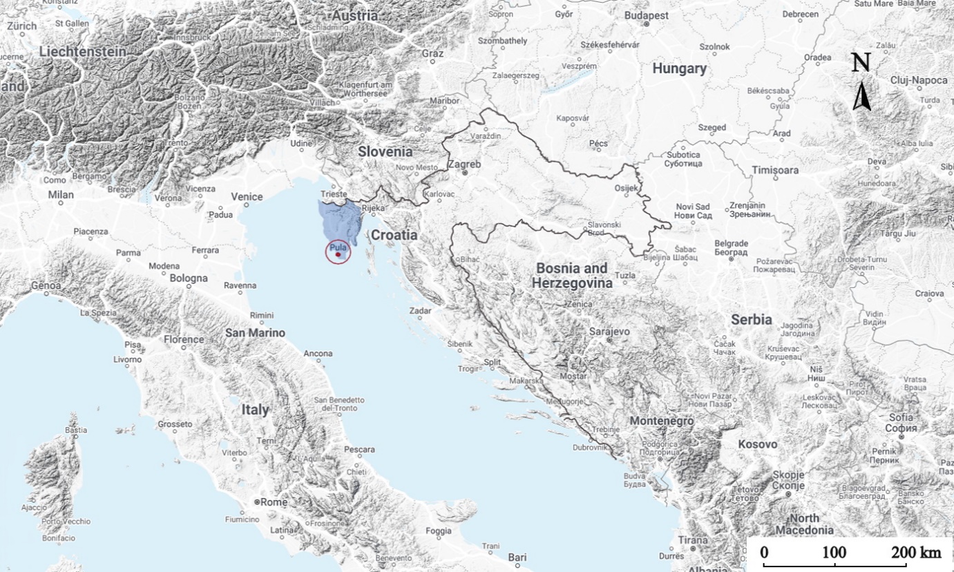
Fig. 1: The location of Istria County (blue) in Croatia and Pula (circled in red). The map was generated using the online web tool Snazzy maps (Krogh, 2019) and edited in Krita software (version 5.0.6).
Roman colonies in the territory of Istria (such as Trieste, Pula and Poreč) had a degree of administrative and economic autonomy and experienced cultural and economic growth under Caesar Augustus. Between 18-12 BCE Augustus integrated the Istrian territory into the larger region Regio X ( Venetia et Histria). Istria’s economy comprised manufacturing workshops and coastal and maritime trade, with wine, fish and oil being the most significant products. In the 1st century, the Istrian economy experienced a decline due to the politics of prioritising cereal production in the Empire. In 395, Istria became part of the Western Roman Empire (Girardi Jurkić, 1988).
The Ozad Arene site (Fig. 2) is situated in the immediate vicinity of the Roman amphitheatre (Arena) in Pula and covers the area of 430 m2. The excavation and the initial archaeological analysis were conducted between 2020 and 2021. During the excavation, the existence of the Roman period cemetery at the site, dated between the 1st and the 4th century CE, was established. The cemetery was dated based on archaeological artefacts found in burials.
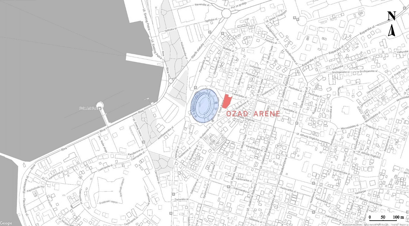
Fig. 2: The map of Pula with the location of Ozad Arene site marked in red. The position of the site directly next to the Roman amphitheatre (Arena, in blue) is clearly visible. The map was generated using the online web tool Snazzy maps (Krogh, 2019) and edited in Krita software (version 5.0.6).
Twenty-five graves containing human bone material, and additional four human bone assemblages (such as the assemblage E4-SJ049 - a bone pit containing disarticulated remains of multiple individuals found outside of the burial context) were discovered, consisting of multiple burial types, including glass cremation urns and a lead sarcophagus, with most burials at this site in stone tombs covered with roof tiles ( tegulae). Three of the graves contained cremated human remains, while the rest (22 graves and four bone assemblages) were inhumation burials (Godinović 2021).
The human skeletal material discovered at the Ozad Arene site was sent to the Laboratory for Evolutionary Anthropology and Bioarchaeology at the Institute for Anthropological Research in Zagreb, where the bioanthropological analysis was carried out. The analysis was conducted using the standard methods described by Brickley and McKinley (2004), and by Buikstra and Ubelaker (1994). Also, the inventory of all present skeletal and dental elements was made for the individuals interred in single burials. The individual’s sex and age at the time of death were established where possible (as described in White & Folkens, 2005). Adults were defined as individuals who were at least 18 years old at the time of death. All elements were examined macroscopically for any signs of pathological changes. The skeletal material was examined for signs of dental and alveolar diseases (caries and antemortem tooth loss), indicators of physiological stress (linear enamel hypoplasia, cribra orbitalia and porotic hyperostosis), vertebral pathologies (osteoarthritis and Schmorl’s nodes), non-specific periostitis, specific diseases such as scurvy and rickets, and bone fractures. The statistical analysis of dental and alveolar diseases was made by the tooth count method (the number of teeth exhibiting pathologies in relation to the total number of examined teeth). Similarly, the vertebral pathologies were statistically analysed at the vertebral level (number of the affected vertebrae in relation to the overall number of examined vertebrae). The most prominent pathological changes were measured and photographed. For multiple bone assemblages, the minimum number of individuals (MNI) was established based on the number of repeating skeletal elements (present only once in an individual), and any previously mentioned pathological changes were recorded. For cremated human remains, bone fragments were weighed, described, and analysed. The sex and age of the individual was established when possible and any pathological changes were noted.
The recorded data were compared with available bioarchaeological data for other Roman period sites from the eastern Adriatic coast. The comparison was made to consider the Pula sample in the broader context of the Roman period populations from the region.
All bioarchaeological data were recorded on a worksheet and later digitized in the graphic software Krita (version 5.0.6) and MS® Excel®. Data was statistically analysed using Microsoft® Excel® (version 2209) and IBM SPSS Statistics (version 28.0.1.1.) for Windows. Due to the small sample size, nonparametric tests were used for data analysis. For the analysis of the age at the time of death, the nonparametric One-way ANOVA test (Kruskal-Wallis test) was used with a significance level of 5% and a p value ≤ 0.05. Pathology frequencies were tested using the Chi-squared test (significance level 5%; p value ≤ 0.05). Fisher's exact test was used where Chi-squared test was not applicable due to the small sample size (level of significance of 5%). The Fisher's exact test was chosen over Chi-squared with Yates' correction, which has the tendency to skew the results towards the insignificant result (Madrigal, 2012). The obtained data was divided into groups by age (adults and subadults) and by sex (males and females; adults only).
Results
The human skeletal sample from Pula – Ozad Arene consists of the remains of a minimum of 48 individuals buried in 25 graves and four assemblages, two of which (Grave 3 and assemblage E4-SJ049) were features containing remains of multiple individuals. Three graves (graves 19, 26, and 27) contained cremated human remains. Assemblages E3-SJ047, E3-SJ048, E4-SJ049, and H2/G2 are not formal graves but are bone assemblages found outside the burial context in the targeted archaeological layers (Table 1).
Table 1: Sex and age distribution of the skeletal remains from Pula by burial/assemblage.

In individuals whose age could not be established, age is marked with X. The age is stated in years, except in the cases of infants (subadults under one year of age) where the age is expressed in months (M). Burials marked with * are not formal graves but bone assemblages outside of the burial context. Burials marked with ** contained cremated remains.
Out of 48 individuals, 50% were subadults (24/48), 14.6% (7/48) were males and 8.3% were females (4/48). The remaining 27.1% (13/48) of the sample were adults whose sex could not be established because their skeletal remains were too fragmented, only a small number of skeletal elements were present, or they were buried in mass bone assemblages. The mean age at death for adults with known sex is 33.32 years, with a standard deviation (sd) of 9.113, with the average age of death being similar for males and females – 33.8 years (sd=12.055) for males and 33.38 years (sd=8.032) for females. The highest adult mortality was in the age group of 18 to 35 years (63.6% of adults) and only one individual was older than 50 years. Half of all present subadults (12/24) died during the first year of life.
In Pula, dental caries occurs almost only in adults, the only exception being one subadult with caries lesions on two deciduous teeth. The frequency of caries per tooth is shown in Table 2. Caries is present in 21.5% (60/279) of the examined permanent adult teeth with a nonsignificant difference between the sexes (40.9% (9/22) in females and 38.8% (31/80) in males). The lesions were mainly present on molars and premolars.
Table 2: The frequency of caries and antemortem tooth loss in Pula.

n=number of teeth showing pathological changes; N=total number of teeth examined.
Antemortem tooth loss (AMTL) and abscesses are present in 5.3% (15/282) (Table 2) of all analysed adult alveoli. There is no significant difference between males (8.5% (7/82)) and females (9.8% (4/41)) in AMTL frequency. Three cases of alveolar abscesses were found in three adult individuals of unknown sex.
Linear enamel hypoplasia was found in 92.1% (93/101) of the examined teeth (only permanent incisors (I) and canines (C) were examined for the presence of LEH). Statistically, there is no difference in the frequency of LEH occurrence between males (93.3% (28/30)) and females (85.7% (6/7)), and between adults (92.5% (74/80)) and subadults (90.5% (19/21)).
Cribra orbitalia (CO) was present only in subadults and always in active form, with no signs of healing. CO was present in 27.3% (6/22) of preserved subadult frontal bones. One subadult (Grave 7; 2.5-3.5 years) had severe porosity in both orbits (Fig. 3). Porotic hyperostosis was detected in 35.7% (10/28) of the available crania. It is interesting to note that 50% of individuals with porotic hyperostosis also have porosity on the temporal bones.
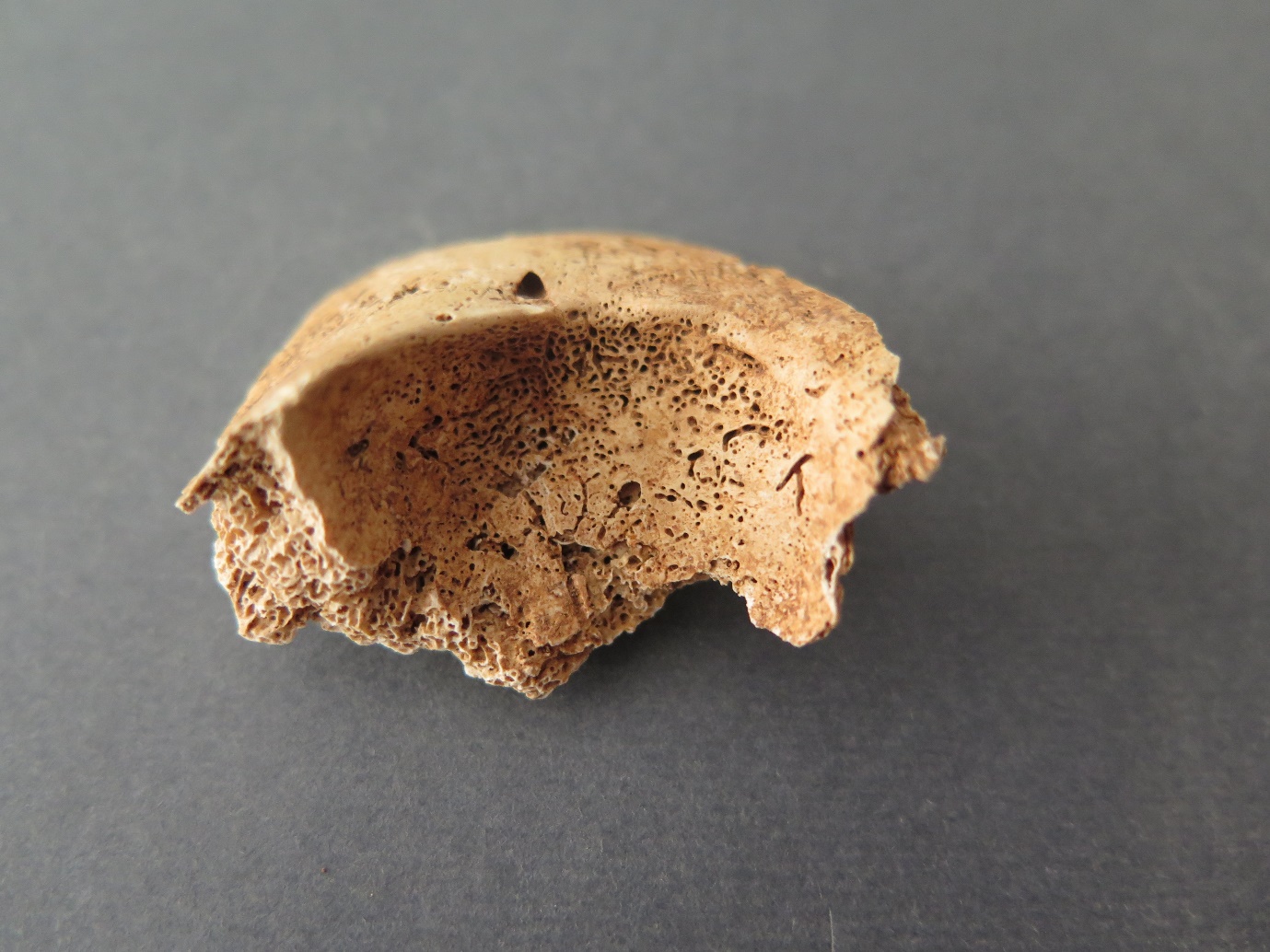
Fig. 3: Active cribra orbitalia in the right orbit of the subadult from Grave 7.
In six individuals (four adults and two subadults) the porosity was present on multiple cranial elements, which may indicate the possibility of metabolic diseases such as scurvy (each of these individuals had porosity on temporal and parietal bones). It is interesting that multiple porosity and CO were present in two subadults (graves 6 and 7).
Periostitis occurs in six out of 24 subadults (25%) and does not occur in adults, thus representing 12.5% (6/48) of the total Pula sample. In all cases, periostitis was in active form with the tibiae and the femurs as most affected elements.
In the assemblage E4-SJ049, a subadult right tibia with mild antero-posterior deviation (so-called “sabre shin tibia”) was found. The medial side of the bone also showed active signs of a new periosteal reaction. Based on the size of the bone (78.5 mm), it was estimated to belong to an individual aged between 1 and 3 months.
The frequency of Schmorl’s nodes is shown in Table 3. In the adult skeletal sample, they occur in 36.8% (50/136) of the vertebrae. There is no significant difference between the sexes (31% (13/42) in females and 39.4% (37/94) in males). Females have four times higher frequency of Schmorl’s nodes in cervical vertebrae than males (15.4% (2/13) and 3.9% (1/26) respectively), but the difference is not statistically significant. In turn, the frequency is higher in males for thoracic vertebrae – 58.3% (28/48) compared to 35% (7/20) in females. With χ2=3.077 and p=0.079 this is a borderline insignificant difference. There is no significant difference in the frequency in the lumbar vertebrae.
Table 3: The frequency of Schmorl’s nodes in Pula.

n=number of vertebrae with Schmorl’s nodes; N=number of examined vertebrae.
Vertebral osteoarthritis (OA) is present in 27.9% (38/136) of all studied vertebrae (Table 4). The frequency is slightly lower in males (24.5% (23/94)) than in females (35.7% (15/42)), but it is not significant. No cases of OA in the cervical vertebrae were found. Females have significantly higher OA rates on thoracic vertebrae than males (55% (11/20) vs. 27.1% (13/48); χ2= 4.818; p=0.028). However, for lumbar vertebrae, there is no significant difference between the sexes (50% (10/20) in males and 44.4% (4/9) in females).
Table 4: The frequency of vertebral osteoarthritis in Pula.

n=number of vertebrae with OA; N=number of examined vertebrae; significant values are underlined.
Bone fractures were recorded in at least four individuals (two of the fractures, the humerus and the rib from Grave 3 come from the same mass bone assemblage, making it impossible to tell whether they belonged to just one or two different individuals). In total, three long bone fractures, one rib fracture and one frontal bone trauma, were recorded. All fractured bones were broken antemortem and were well healed at the time of an individual’s death. One subadult had a fracture of the right ulna (Grave 18-B), one female (Grave 24) had a fracture of the left ulna (Fig. 4), one adult (Grave 3) had a fracture of the right humerus, and one adult individual had a fractured rib (Grave 3). The prevalence of long bone fractures in the adult sample is 1.2% (2/162). One case of blunt force trauma was also found – a well-healed fracture of the frontal bone (3.6% (1/28) of crania) of an adult from the assemblage E4-SJ049.
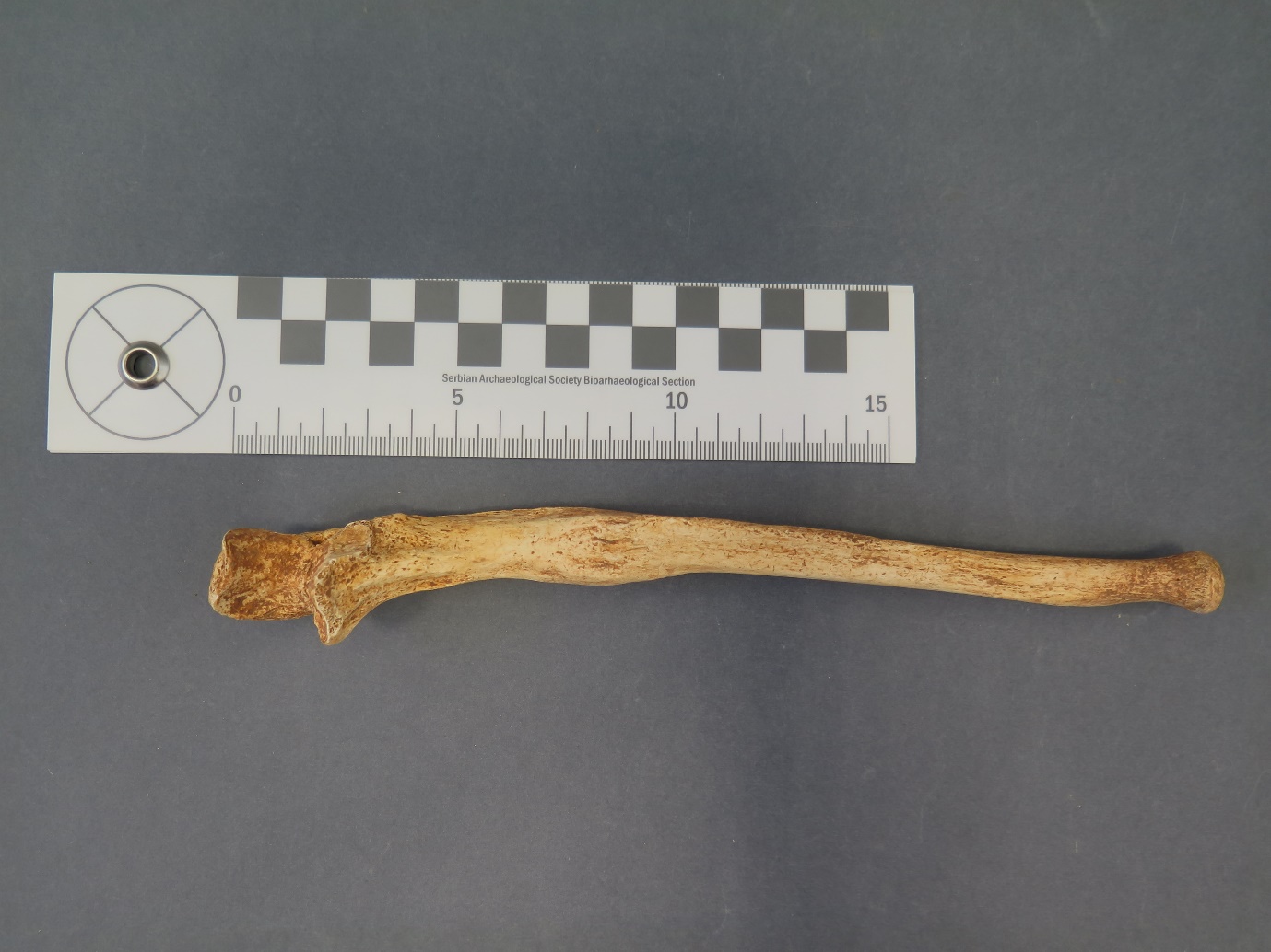
Fig. 4: Well healed fracture of the left ulna in the female from Grave 24.
Two individuals in the Pula skeletal sample had bifid ribs. In both cases, the rib anomaly is unilateral and on the left side of the individual’s body. In the case of the male from Grave 4, the bifid rib is the sixth left rib with the thickening and bifurcation of the sternal end of the rib. The sternal end is completely split into the inferior and superior arm of the rib. One male individual (Grave 15) had the bifurcation of the fourth left rib (Fig. 5). The rib is also completely split at the sternal end into an inferior and superior r arm.
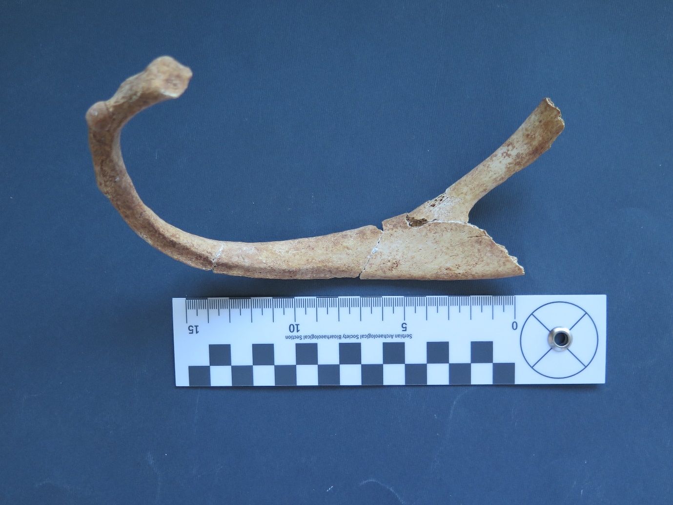
Fig. 5: Bifid rib in the male from Grave 15.
One individual (Grave 15) had multiple anomalies of the sternum in the form of sternal foramina and fused elements of the sternum (Fig. 6). This individual also has the bifid rib mentioned earlier. The first sternal foramen is located between the fourth and fifth segment of the body of the sternum, while the second foramen being at the tip of the xiphoid process. Besides the foramens, the xiphoid process is fused with the body of the sternum.
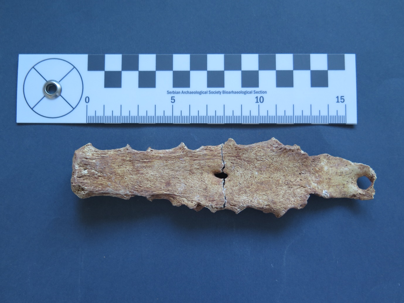
Fig. 6: Sternum of the male from Grave 15 with multiple sternal foramens and the xiphoid process fused with the body of the sternum.
Discussion
The skeletal sample from Pula comprises the minimum of 48 individuals. Such a number of samples are susceptible to random statistical variation (Bedić et al., 2013), especially when analysing individual pathologies of a smaller sample size. For this reason, the discussion and interpretation of the results of this analysis have to be taken with caution.
The demographic distribution is evenly split between the adults and subadults. Childhood, especially infancy, is considered a period of life with increased mortality (Manifold, 2014). Some estimates place subadult mortality in pre-industrial societies (including Roman period populations) at approximately 50% (Pearce, 2001). Subadult mortality is considered an important indicator of the level of development and overall health of the population (Gonzalez & Gilleskie, 2017; Esmaeilzadeh et al., 2021). Nowadays, subadult mortality is in a constant decline, but it is still more prevalent in developing countries due to unfavourable hygienic, economic, and social conditions (Esmaeilzadeh et al., 2021). In 2020, infant mortality rate was 0.4% in Croatia and 0.3% in the European Union (Rodin et al., 2021). In comparison, the subadult mortality under one year of age in Pula is much higher (50% of subadults and 25% of the total sample). This is typical of ancient populations where infant mortality is estimated between 30 and 40% (Scheidel et al., 2008; Caroll, 2011; Pilkington, 2013), mostly because of exposure to the disease and malnutrition. The overall high frequency of subadult burials in Pula is also consistent with the prevalence of subadult burials in other Roman period sites, where subadults make up 20–40% of the sample (Pearce, 2001; Novak, 2008; Novak & Šlaus, 2010a, Caroll, 2011).
Generally speaking, life expectancy in the Roman world is estimated to be around 20 to 30 years, depending on the region and the socioeconomic conditions of the population (Scheidel et al., 2008). In archaeological sites on the Croatian territory, the average age at death is between 30 and 45 years (Novak, 2008; Bedić, 2017; Hincak & Zglav Martinac, 2016; Vyroubal & Bedić, 2020; Šlaus 2021). Most adults in the Pula sample died between the ages of 20 and 35, which is slightly younger than other Croatian sites from this period, but this difference is nonsignificant and likely due to the small sample size. Overall, it seems that the skeletal sample from Pula, by its demographic characteristics, is not different from other Roman period assemblages from the region (Novak, 2008; Bedić et al., 2013).
Dental pathologies, especially caries, occur in high frequency in Pula, affecting females slightly more than males, but this difference is minimal and insignificant, and possibly because of a small sample size. The diet in the Roman period was based on grains (mostly in the form of wheat porridge/ puls, bread, millet, and barley) with mostly vegetables and fruit as an addition – this type of diet, consisting mainly of carbohydrates, is typical of a society whose economy is based on agriculture (Scheidel et al., 2008; Bedić et al., 2009). Carious lesions during the Roman period (including Croatian sites) occur with low frequency – usually less than 10% of teeth (Manzi et al., 1999; Bonfiglioli et al., 2003; Šlaus, 2006; Novak, 2008; Bedić et al., 2009; Diéguez Ramírez et al., 2017; Vyroubal & Bedić, 2020; Vergidou et al., 2021). In comparison, the frequency of caries in Pula is twice as high as expected for the period. This may also be due to the small sample size, but it may also result from poor dietary habits with low-quality and cariogenic foods, such as carbohydrates (Newbrun, 1979; Giacaman, 2018) (e.g. millet porridge).
Alveolar disease and tooth loss can result from dental plaque and caries (Bonfiglioli et al., 2003; Šlaus 2006). In the Roman Empire, dentistry was, generously stated, still in its infancy. Dental medicine consisted mainly of pain management by taking opium, saffron, and other remedies. Tooth removal was avoided and performed in case of crown destruction or infection (Fejerskov et al., 2012). These practices could easily lead to the development of a tooth abscess or other complications. The prevalence of abscesses and AMTL is low in Pula (5.3%). This makes sense given the high mortality of younger individuals at the site, as the development of severe alveolar disease requires time (Bonfiglioli et al., 2003). Individuals in Pula probably died before caries could spread and cause an infection that led to tooth loss. This is confirmed by the fact that almost all cases of caries in Pula were mild (category one or two) and affected less than half of the tooth surface. The prevalence of AMTL in Pula is consistent with other Roman period populations from the region, generally under 10% (Šlaus, 2006; Novak, 2008; Vyroubal & Bedić, 2020).
Linear enamel hypoplasia (LEH) is a good indicator of metabolic stress in childhood (Littleton & Townsend, 2005; Novak et al., 2009; Dąbrowski et al., 2021). In the Pula sample, LEH is present in almost all examined incisors and canines, representing a very high prevalence of enamel defects in this population. In other Croatian Roman period populations, LEH is present in approximately 40-60% (Šlaus, 2006; Bedić, 2017; Vyroubal & Bedić, 2020) of the studied teeth. Considering this, we can presume there were certain factors in Pula that contributed to the widespread physiological stress during childhood. This high frequency is probably not related to the small sample size, as the defects were consistently present in every individual. High rates of LEH were linked to the increased physiological stress in the population because of disease, nutritional deficiencies (Littleton & Townsend, 2005; Dąbrowski et al., 2021), lifestyle based on agriculture, increase in population (Cohen & Armelagos, 1984) and population aggregation (Ham et al., 2020). This stress could result from weaning when children transition from consuming breast milk to other foods that contain microorganisms that can cause digestive distress, reduce appetite, and cause nutritional deficiencies. Depending on the food, nutrient deficiencies can also be caused by a sudden switch to an unbalanced grain-based diet (Manifold, 2014). Individuals with LEH may have a shorter life expectancy than people without these defects, as stress exposure in early life is found to be associated with higher mortality in earlier life stages (Littleton & Townsend, 2005; Stutz et al., 2021). Lower socioeconomic status and poor living conditions that caused LEH are likely to persist into adulthood and that continuous stress could contribute to the individual’s earlier death. Furthermore, malnutrition and illnesses in childhood may weaken the individual’s immune system, making them more susceptible to other diseases later in life (Šlaus 2006).
LEH appears to be associated with an increased risk of caries development. Research on the children from the US in the 1990s found a positive correlation between LEH and caries, as weakened tooth enamel is more sensitive and more susceptible to bacteria colonisation (Hong et al., 2009). It is interesting that in the same study, LEH defects occurred in only 3.9% of subadults showing the low prevalence of LEH in the modern world, especially when compared to Roman period Pula. The high LEH prevalence in Pula points to the poor living conditions and widespread metabolic stress in childhood during the weaning phase and the transition to solid foods (first to third year of life, when the enamel of permanent teeth is formed) (Cunningham et al., 2016). LEH is probably the contributing factor leading to relatively high caries rates and is probably related to the high mortality in younger individuals in the Roman period Pula.
Cribra orbitalia is mostly associated with anaemia and iron deficiency, but various other conditions could also cause such lesions (Wapler et al., 2004; Šlaus, 2006; Brickley, 2018). In Pula, CO occurs only in subadults. Subadults are more susceptible to anaemia because of higher iron requirements due to rapid growth (Özdemir, 2015). Changes in diet, insufficient diet, parasites and diseases can also lead to the occurrence of anaemia (Facchini et al., 2004; Šlaus, 2006; Gebreweld et al., 2019). The frequency of CO in subadults in Pula is 27.3%, which is lower compared to other Roman-era sites where CO is present with 40–60% (Facchini et al., 2004; Novak & Šlaus 2010b). The absence of CO in adults from Pula could indicate severe acute illness that led to the death of the individual before any marks could form on the bone (Pine et al. 2009).
The widespread presence of porotic hyperostosis and possible cases of scurvy and rickets also shows a high level of physiological stress in the studied population (Šlaus, 2006; Rohnbogner, 2015). Scurvy is associated with vitamin C deficiency (Gandhi et al., 2023) and rickets with vitamin D deficiency (Sahay & Sahay 2012). Possible scurvy recorded in Pula points to poor living conditions – the individuals likely had a lower socioeconomic status and did not have access to balanced and varied food. Most of the graves discovered at the Ozad Arene site were burials in stone tombs covered with roof tiles and were usually reserved for the poorer citizens of the Roman society (Godinović, 2021).
The “sabre shin tibia” is an isolated find in the bone assemblage E4-SJ049 containing multiple individuals – no other similarly sized bones or bones showing similar pathological changes were unearthed. Due to the isolated nature of the find, and the mild deviation of the bone, any diagnosis of specific causes (such as syphilis, rickets or other metabolic diseases) is not viable.
Infectious diseases are one of the major causes of death in archaeological populations, particularly in childhood (Ortner & Putschar, 1981). Since in most cases specific diseases cannot be identified, these diseases are grouped as non-specific diseases that manifest themselves in a form of periostitis (Ortner & Putschar, 1981; Bedić et al., 2013). Periostitis can also occur due to number of other conditions, including trauma, infections, metabolic, congenital and genetic conditions (Pilloud & Schwitalla, 2020). Periostitis is common in Roman period sites in Croatia, especially in subadults with a frequency between 50 and 70% (Šlaus, 2006; Novak, 2008, Bedić, 2017). In Pula, only 25% of the subadults had periostitis, which is surprising considering the high prevalence of other conditions pointing to poor living conditions. As in the case of CO, it is possible that severe acute illness caused an individual’s death before periostitis changes on the bones could form. Bone fragmentation and relatively poor preservation of the cortex may also contribute to the underrepresentation of periostitis in Pula.
As a result of disc herniation (Kyere et al., 2012), Schmorl’s nodes in archaeological populations are associated with the mechanical load of the spine (Faccia & Williams, 2008; Bedić et al., 2013) and heavy labour (Hincak & Zglav Martinac, 2016). In Pula, their prevalence is twice as high as the prevalence in the composite Roman period Croatian sample (Novak, 2008). While it is possible that the Roman period inhabitants of Pula performed more physically demanding activities than their contemporaries, it is more likely that this difference results from the random statistical variation due to a small sample size.
In Croatian archaeological samples from the Roman period, males exhibit significantly higher prevalence of spinal alterations, which points to clear division of labour, where the males were engaged in more physically demanding tasks (Šlaus, 2006; Novak 2008; Vyroubal & Bedić, 2020). Therefore, it is interesting to note that there is no difference in the overall frequency of Schmorl’s nodes between the sexes in Pula. These results could be influenced by the small number of identified males and females at the site (the sex of the most adult individuals could not be established) and should be taken with caution. The absence of a difference in the frequency of Schmorl’s nodes between sexes suggests that both sexes were equally included in physically demanding work. However, it is likely that there was some sort of division in the types of tasks performed by males and females. Females exhibit Schmorl's nodes on the cervical vertebrae which are absent in males. This could be attributed to the practice of carrying heavy loads on the head, as those activities have been found to contribute to the accentuated degenerative changes in the cervical spine (Joosab et al., 1994; Dave et al., 2021). Occupational activities involving lifting heavy loads have been thought to increase the risk of spine degeneration, with some evidence linking occupational heavy labour with disc degradation and disc bulging (Macedo & Battié, 2019). Back and, to the lesser extent, neck pain has been linked to the disc degeneration due to various factors including aging and occupational activities – with disc disease in the lumbar spine causing pain because of heavy lifting, and frequent spine bending and twisting (Williams & Sambrook, 2011).
Osteoarthritic changes are a commonly found in archaeological populations and associated with the aging process. Today, OA is primarily associated with older individuals who have surpassed the age of 60 (Loeser, 2011), but OA changes can occur as early as the age of 40 (Šlaus, 2006). In the Roman period Pula, OA was present in the number of younger individuals around the age of 30. Lumbar vertebrae were most affected, suggesting that hard physical labour may be a contributing factor. When compared to other Roman period Croatian samples (around 10-15% (Šlaus, 2006; Bedić, 2017)) it is evident that the occurrence of spinal OA is more widespread in Pula. When compared, the prevalence of spinal OA in females in Pula is four times higher than in other similar samples (the results should be taken with caution as they could be impacted by the small number of identified males and females in the sample). The reason for this difference is not clear. Maybe females in the Roman period Pula were doing more strenuous labour, but it is also likely that the difference is due to the statistical variation because of the small number of females in the sample. We cannot tell with certainty whether age played a significant role (and to what extent) because of the mentioned sample size, but also due to an almost non-existent difference in average age between the sexes in Pula.
In Pula, bone fractures were not a common find, with only five recorded cases in total. All fractures were well healed, most likely occurring long before the death of the affected individual. The bones healed in more or less anatomically correct positions, thus indicating the possibility of medical treatment such as the realignment and immobilisation of the affected bone. Low number of long bone trauma is consistent with the data observed in other Roman period populations (Novak & Šlaus, 2010a; Vyroubal & Bedić, 2020). In Pula, the prevalence of skull fractures (3.6%) is notably lower than other Roman period Croatian samples (15-28%) (Novak, 2008; Novak & Šlaus, 2010a; Vyroubal & Bedić, 2020). Bone fractures can result from intentional violence or from accidents and/or occupational activities (Walker, 1989; Dittmar et al., 2021). Two cases of a distal ulna fracture in Pula could be possible indicators of violence as such fractures usually occur as a result of a defensive reflex where the person raises their arm to protect the face and head from the attacker (Šlaus, 2006, 2021). Facial and cranial fractures are also indicative of potential acts of violence, as the head is an attractive target for the attacker due to the nigh-instantaneous incapacitating effect on the victim (Walker, 1997). The potential cause of the frontal bone injury in Pula could not be established because of the fracture being discovered in the mass bone assemblage. However, this fracture could have also resulted from a fall and/or some other accident.
In one case, multiple congenital anomalies of the sternum were found. These anomalies were most likely asymptomatic and were medically insignificant. The variations in the sternal anatomy are relatively common and are asymptomatic in most cases (Choi et al., 2017; Gans et al, 2021). They can be found in 4-14% of modern populations (Choi et al., 2017; Sungur et al., 2020; Gans et al, 2021). The formation of the sternal foramen occurs due to the incomplete fusion of segments during foetal development (Choi et al., 2017).
The occurrence of two bifid ribs is an interesting find. Rib anomalies are a relatively common occurrence, occurring in approximately 2% of the human population, with bifid ribs being one of the more common anatomical anomalies (Andrea et al. 2016; Rajić Šikanjić et al., 2017). This anomaly probably occurs because of incomplete fusion of the sclerotomes during embryonic development (Rathinasabapathi & Perumallapalli, 2015). Usually, it is unilateral and occurs as an independent anatomical variation. However, in rare cases, it may occur in more complex conditions, such as the Gorlin-Goltz syndrome, Jobs syndrome, or Kindler syndrome (Rathinasabapathi & Perumallapalli, 2015; Rajić Šikanjić et al., 2017; Tsoucalas et al., 2019; Galassi et al., 2023). In Pula, both individuals that had bifid ribs were well-preserved, with most skeletal elements present. As no other major skeletal pathologies were found, bifid ribs were most likely the result of individual and pathologically insignificant anatomic variations.
Conclusion
The human skeletal sample from the Ozad Arene site in Pula comprises the remains of a minimum of 48 individuals. Based on its demographic characteristics, this skeletal sample represents a typical Roman period population from the region. The prevalence of caries in Pula is higher than expected for the period, probably due to the diet based on grain likely in the form of bread and porridge. That, in combination with poor oral hygiene, contributed to the development of the disease. The frequency of linear enamel hypoplasia is significantly higher compared to other Roman period populations indicating overall poor living conditions and widespread metabolic stress during childhood. The presence of cribra orbitalia, porotic hyperostosis, and possible cases of scurvy and rickets further support the idea of increased metabolic stress during childhood that likely persisted into the adulthood. The studied individuals from Pula probably belonged to a lower socioeconomic status, lacking access to a well-balanced and sufficiently nutritious diet. The relatively low number of cases of periostitis and CO could indicate the possibility of severe acute illnesses that could lead to a quick death of the individuals. The presence of Schmorl’s nodes and osteoarthritis changes indicate a continuous mechanical strain on the spine due to hard physical labour. Skeletal traumas are infrequent in Roman period Pula, suggesting that violence was not prevalent within the community.