Skip to the main content

Original scientific paper

https://doi.org/10.15567/mljekarstvo.2024.0307

Izazovi povezani sa mastitisom u mliječnom govedarstvu u Srbiji: analiza broja somatskih stanica i distribucije patogena

Dragana Tomanić ; University of Novi Sad, Faculty of Agriculture, Department of Veterinary Medicine, Trg Dositeja Obradovica 8, 21000 Novi Sad, Serbia
Marko Samardžija ; University of Zagreb, Faculty of Veterinary Medicine, Heinzelova 55, 10000 Zagreb, Croatia
Ivan Stančić ; University of Novi Sad, Faculty of Agriculture, Department of Veterinary Medicine, Trg Dositeja Obradovica 8, 21000 Novi Sad, Serbia
Nebojša Kladar ; University of Novi Sad, Faculty of Medicine, Department of Pharmacy, Center for Medical and Pharmaceutical Investigations and Quality Control, Hajduk Veljkova 3, 21000 Novi Sad, Serbia
Nino Maćešić ; University of Zagreb, Faculty of Veterinary Medicine, Heinzelova 55, 10000 Zagreb, Croatia
Zorana Kovačević ; University of Novi Sad, Faculty of Agriculture, Department of Veterinary Medicine, Trg Dositeja Obradovica 8, 21000 Novi Sad, Serbia *

* Corresponding author.


Full text: english pdf 671 Kb

page 239-248

downloads: 47

cite

Full text: croatian pdf 671 Kb

page 239-248

downloads: 29

cite

Download JATS file


Abstract

Mastitis predstavlja veliki problem za mliječna stada zbog svog ekonomskog utjecaja i potencijalnih zdravstvenih rizika. Broj somatskih stanica, koji odražava zdravlje vimena, igra ključnu ulogu u dijagnozi mastitisa. Cilj ovog istraživanja je određivanje distribucije somatskih stanica i korelacije s različitim uzročnicima mastitisa na mliječnim farmama u Srbiji. Analizirana su 194 pojedinačna uzorka kravljeg mlijeka. Mikrobiološka ispitivanja provedena su u aseptičnim uvjetima kako bi se izolirali i identificirali uzročnici mastitisa. Za procjenu broja somatskih stanica u uzorcima mlijeka korištena je mikroskopska referentna metoda. Kao uzročnici mastitisa, bakterije su bile prisutne u 28,87 %, gljivice u 12,38 %, dok su u 5,15 % uzoraka mlijeka bile prisutne i bakterije i gljivice. Uočena je veza između broja somatskih stanica u različitim tipovima uzoraka (negativni uzorci, uzorci sa bakterijama, uzorci s gljivicama i uzorci s bakterijama i gljivicama). Naime, uzorci s bakterijama i gljivicama imali su najviši broj somatskih stanica. Iako je broj somatskih stanica vrijedan alat za praćenje zdravlja vimena i učinkovitosti programa kontrole mastitisa, njegov odgovor na specifične patogene je složen i ne dopušta lako razlikovanje između tipova patogena. Ovo istraživanje naglašava izazove u razlikovanju tipova patogena isključivo na temelju broja somatskih stanica.

Keywords

bakterije; krave; mastitis; broj somatskih stanica; gljivice

Hrčak ID:

317723

URI

https://hrcak.srce.hr/317723

Publication date:

10.6.2024.

Article data in other languages: english

Visits: 162 *


Introduction

Mastitis significantly affects dairy herds, leading to economic losses due to high treatment costs, reduced milk production and quality, increased labor and premature culling (Magaš et al., 2013; Benić et al., 2018; Poljak et al., 2022). It also raises concerns about antimicrobial use, leading to resistant strains and antimicrobial residues in dairy products, posing public health risk (Turk et al., 2017). Therefore, monitoring mastitis in dairy cows is crucial for maintaining herd health, milk quality and public health (Maletić et al., 2017; Knežević et al., 2021).

Somatic cell count (SCC) reflects the udder health and is a quantitative method for diagnosing mastitis by enumerating different cell types in milk (leucocytes, including neutrophils, macrophages and lymphocytes) (Darbaz et al., 2023). SCC exceeding 200,000 cells/mL is typically associated with bacterial infection (Sharma et al., 2011). Both, clinical and subclinical mastitis can disrupt the SCC pattern (De Haas et al., 2004). In general, the major mastitis pathogens ( Streptococcus uberis, Escherichia coli, Staphylococcus aureus, Streptococcus dysgalactiae and Streptococcus agalactiae) elicit greater somatic cell response (Bradley and Green, 2005), than the minor pathogens ( Corynebacterium species and coagulase-negative staphylococci) (Sharma et al., 2011). Interestingly, infections involving the major pathogens are more likely to result in SCC levels over 200,000 cells/mL, whereas minor pathogens typically maintain SCC levels in the range from 50,000 to 150,000 cells/mL (Bradley and Green, 2005). Therefore, it's essential to keep these factors in mind when using SCCs as marker for making decisions regarding health status in dairy herds (Haxhiaj et al., 2022). Significantly, SCC has been linked to intramammary infection resolution, indicating its potential role in antibiotic treatment decisions (Williamson et al., 2022; De Jong et al., 2023).

Hence, this study was conducted to observe the distribution of SCCs and potential correlation to the different mastitis associated pathogens in the selected dairy farms in Serbia. Additionally, the prevalence of pathogens in the milk samples obtained from cows affected by mastitis was assessed.

Material and methods

Isolation and identification of mastitis associated pathogens

The Animal Ethics Committee of the Ministry of Agriculture, Forestry, and Water Management-Veterinary Directorate granted the approval for the experimental protocol (Approval No. 9000-689/2, dated 06 July 2020). This study was conducted from June to December 2021 on two dairy farms located in the Vojvodina Province, Serbia. A total of 194 individual cow milk samples were collected from cows with clinical and subclinical mastitis. Farm veterinarians examined the udders and milk to check for clinical mastitis, while the California Mastitis Test was used to confirm subclinical mastitis by analyzing the milk's SCC. Clinical signs of udder inflammation included swelling, pain and redness, while changes of interest in the first jets of milk were clots, color change and density. The milk samples for microbiological testing were obtained under aseptic conditions. After teat cleaning, drying and disinfection, the first jets of milk were discarded, and a 10 mL sample was collected in sterile tubes. These samples were stored in an ice container at 4 °C during transport to the Laboratory for Milk Hygiene at the Department of Veterinary Medicine, Faculty of Agriculture, University of Novi Sad. The samples were inoculated on the 2 % blood agar, and standard bacteriological diagnostic techniques, as previously described by Kovačević et al. (2021a), were employed for the isolation, identification and determination of mastitis pathogens. Yeast strains were cultured on Sabouraud dextrose agar plates, which were then incubated at 30 °C for 48 hours. Isolates were subsequently identified, using the “API 20 C AUX” (bio Meraux, France).

Determination of the somatic cell count

The SCC in the milk samples was assessed following the microscopic reference method as per the Institute for Standardization of Serbia (SRPS EN ISO 13366-1:2010) (ISO, 2010). To determine the SCC, 0.01 mL of mixed milk from each sample was spread across a 1 cm2 area on a glass slide. These slides were then air-dried, stained with the Newman-Lampert stain, and examined under a microscope. The SCC result below 200,000 cells/mL was considered low, indicating a healthy mammary gland (Piccinini et al., 2005). In contrast, SCC exceeding 200,000 cells/mL was classified as high, signifying the presence of an intramammary infection.

Statistical analysis

The obtained data were summarized by application of Microsoft Office Excel and statistically processed by Tibco Statistica (v. 13.5). Data were analysed by descriptive statistical methods, while differences between evaluated groups in terms of number of somatic cells were assessed by application of ANOVA.

Results and discussion

Prevalence of mastitis-associated pathogens

Based on laboratory results, 104 (53.60 %) milk samples were negative for both yeast and bacteria (group 1), while the rest of samples (46.40 %) were positive on pathogens presence. Of those, 56 (28.87 %) were positive for bacteria (group 2), 24 (12.38 %) for yeast (group 3), and 10 (5.15 %) were positive for yeast and bacteria (group 4).

Table 1. Prevalence of mastitis-associated pathogens in the milk samples

image1.jpeg

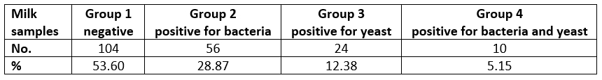
Of the isolated bacteriological causes of mastitis, Streptococcus spp. was isolated in 20 cases (30.30 %), E. coli and S. marcescens were present in 12 samples (18.18 %) each, followed by 8 cases (12.12 %) of S. aureus, while Proteus mirabilis was isolated in 6 cases (9.10 %). Klebsiella spp., Staphylococcus spp., S. uberis and β-haemolytic Streptococcus spp. were isolated in 2 samples, each (3.03 %). With regard to yeast, we isolated Candida spp. in 28 samples (82.35 %) of total samples positive for yeast, and C. albicans in 6 cases (17.64 %).

image2.jpeg

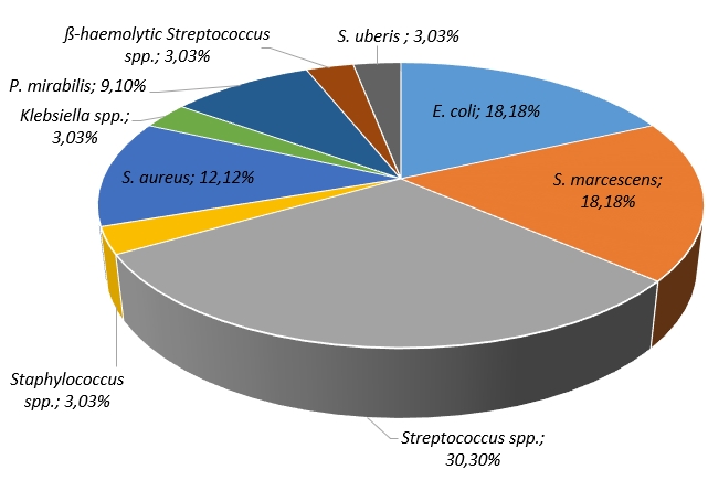
Figure 1. Frequency of the mastitis-associated bacteria isolates in the tested samples

Bovine mastitis involves range of microorganisms with changing prevalence linked to factors like sample timing and region-specific variations in infection patterns (Malinowski et al., 2006). Since this disease has gained great attention, mastitis causative agents are well described in the studies conducted in Serbia (Milanov et al., 2014; Kovačević et al., 2021a, 2021b; Kovačević et al., 2022) and worldwide (Janosi and Baltay, 2004; Malinowski et al., 2006; Tenhagen et al., 2006; Bi et al., 2016; Abed et al., 2021). While some researchers explored the relationship between the SCC and mastitis-associated pathogens (Moretti et al., 1998; De Haas et al., 2004; Huang and Kusaba, 2022), to our knowledge no similar studies have been performed yet in Serbia.

Our findings indicate that Streptococcus spp. were prevalent in 30.30 % of evaluated samples, which is consistent with study in Italy (33.84 %) (Ceniti et al., 2017). These bacteria are globally recognized as major mastitis pathogens (Kaczorek et al., 2017) with the prevalence of 50 % in Australia, followed by Europe (38 %) (Kabelitz et al., 2021). Smistad et al. (2023) suggests that strategies to control infectious pathogens, like the five-point and 10-point plans, reduced contagious mastitis but had limited impact on environmental pathogens like Streptococcus spp. and coliform bacteria. Our findings show a 12.12 % prevalence of S. aureus, despite being considered as one of the most common causes of mastitis (Liu et al., 2020). In addition, lower prevalence of S. aureus was also reported in Croatia, being isolated in 4.48 % of udder quarter samples (Cvetnić et al., 2021). According to McDougall et al. (2022) prevalence of S. aureus can vary with the age being more prevalent and of greater duration in older animals due to its contagious nature.

Coliform-associated mastitis ( E. coli and S. marcescens) in our study had a relatively high prevalence (18.18 %) being comparable to study in Norway (14.50 %) (Smistad et al., 2023), but lower than 21.9 % prevalence in North West Cameroon, where it was significantly associated with factors such as lactation stage, cow breed, history of mastitis and contaminated environment (Abegewi et al., 2022). Furthermore, E. coli is recognized as the most common gram-negative coliform bacteria in intensive milk production systems on dairy farms (Morales-Ubaldo et al., 2023). Apart from E. coli, S. uberis and K. oxytoca were also found, supporting the increasing presence of environmental pathogens (Cervinkova et al., 2013).

The prevalence of S. uberis in our study (3.03 %) aligns with other reports ranging from 2 % to 9 % (Bi et al., 2016; Smistad et al., 2023). This pathogen's ability to produce biofilms, capsules, invade mammary gland cells and resist phagocytosis makes controlling S. uberis mastitis challenging, even in case of a low farm-level presence (Wente et al., 2019). Furthermore, Klebsiella spp., particularly K. oxytoca, plays a significant role in bovine mastitis (Song et al., 2023), primarily originating from environmental sources. Improved hygiene practices can reduce transmission (Song et al., 2023). In our study, K. oxytoca was found in 3.03 % of samples, but it's less significant in milk samples when compared to S. aureus and E. coli, as noted by other authors (Song et al., 2023).

Yeast, although infrequently implicated, have been linked to mastitis in dairy cattle. While data on yeast prevalence in Serbian dairy farms is limited, our study's results (12.38 %) are relatively comparable with those from (Milanov et al., 2014), who reported yeast isolation in 6.02 % of milk samples. Candida is a significant pathogen in mycotic mastitis among dairy cows, particularly non-albicans species, as supported by the literature (Zhou et al., 2013). Our study found all yeast to be Candida spp., including C. albicans. In contrast, Milanov et al. (2014) pointed out that among all the yeast species recovered from cow's milk , C. albicans rarely takes dominant role. However, it's important to note that extensive production systems, environmental temperatures and disease duration are significant risk factors contributing to the prevalence of mycotic mastitis (Zhou et al., 2013).

Somatic cell count in relation to type of pathogens

The application of ANOVA has demonstrated a negative statistically significant differences in SCC of milk samples (Figure 2), while positive on bacteria, positive on yeast and positive on bacteria and yeast (F(3, 98)=10.895, p=0.00). Moreover, the recorded difference was a result of high SCC in milk samples being positive both for bacteria and yeast.

SCC levels and bacteriological examinations serve as different methods for evaluating the mammary gland's condition (Schwarz et al., 2010; Tommasoni et al., 2023). Besides, using the test-day SCC records helps identify deviations from the usual SCC pattern, indicating potential mastitis-causing pathogens (De Haas et al., 2004). Numerical increase in lactations associated to isolated pathogens, compared with unaffected cows have been reported worldwide (De Haas et al., 2004; Skrzypek et al., 2004; Malinowski et al., 2006; Lopes Júnior et al., 2012; Sumon et al., 2020). Yet, comparing milk SCC in relation to specific bacterial species in the literature is challenging due to variations in methodologies, including the analysis of different milk types by different authors. Typically, the major pathogens are associated to the most significant increase in SCC, while infection by minor pathogens leads to a notably lower SCC rise and rarely to the clinical manifestation of the mastitis (Supré et al., 2011). Our study results indicated that there was a statistically significant difference between the SCC observed among all tested samples (negative, positive for bacteria, positive for yeast and positive both for bacteria and yeast) (Figure 2). Notably, the highest SCC was recorded in milk samples that tested positive for both bacteria and yeast.

image3.jpeg

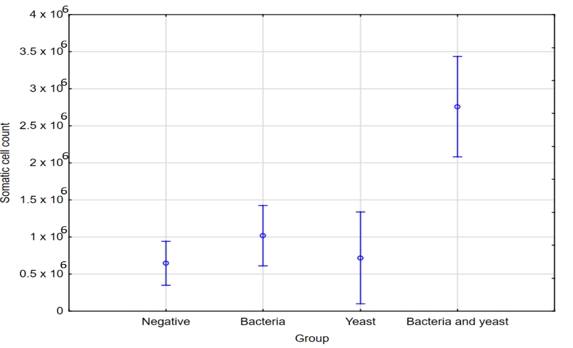
Figure 2. ANOVA - SCC in relation to type of pathogens

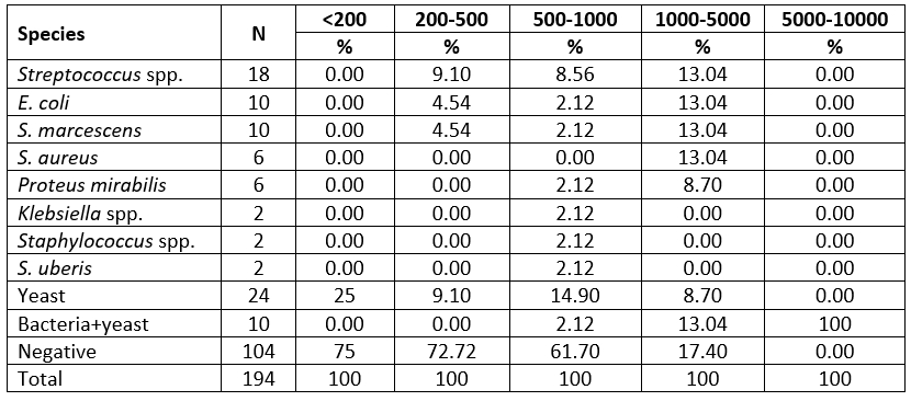
Milk samples with SCC levels below 200,000 cells/mL were mostly cultured negative (75 %). In contrast to these results, some of the mastitis cases without detectable bacterial growth exhibited an increase in SCC. The majority of samples showing no bacterial growth indicated SCC levels in the range from 200,000 to 500,000 cells/mL. Besides, samples where SCC levels exceeded 1,000,000 cells/mL were primarily associated with infections caused by E. coli, S. marcescens and S. aureus. Furthermore, notably elevated SCC levels were linked to infections attributed to both yeast and bacteria, whereas yeast-only infections resulted in a comparatively smaller increase in SCC, typically measuring less than 1 million cells per milliliter.

Passing over the limit of 200,000 cells/mL indicates the transition from health to disease (Skrzypek et al., 2004). Our study revealed that milk samples with SCC below 200,000 cells/mL were mainly culture negative, consistent with other research (Malinowski et al., 2006). We have also observed higher SCC in cases of mastitis with no bacterial growth, aligning with previous findings suggesting that high SCCs may not always indicate the presence of mastitis pathogens (Souza et al., 2016; Sumon et al., 2020). According to McDougall et al. (2001), the presence of high SCC in milk samples, even in the absence of microorganisms, does not necessarily indicate the udder's health. Nonetheless, even though we couldn't detect any pathogens in those quarters, they could still be infected. Elevated SCC in cases without detected bacteria could result from undetectable bacterial levels during sampling or effective immune elimination (Campos et al., 2022). Negative bacteriological findings may also be influenced by sporadic pathogen shedding, antimicrobials, growth-inhibiting substances, or intracellular survival (Schwarz et al., 2010; Kandeel et al., 2018). It must be pointed out, that SCC can also be affected by non-infectious factors, such as animal’s age, stage of lactation, the time of year, milking frequency and nutrition (Bradley and Green, 2005). Our study confirmed mastitis pathogens presence in cows with lower SCC, emphasizing the need for bacteriological culture even when SCC suggests lower likelihood of subclinical mastitis (Katsande et al., 2013; Huang and Kusaba, 2022). It is important to note that SCC and bacteriology may not always produce matching results, as infected udders may not consistently release pathogens, leading to negative test outcomes (De Haas, 2005).

In the group of cows with SCC between 200,000 and 500,000 per milliliter, a substantial number did not test positive for any of pathogens. This pattern is consistent with other authors (Janosi and Baltay, 2004; Souza et al., 2016). Samples with SCC exceeding 1,000,000/mL were primarily associated with E. coli, S. marcescens, and S. aureus infections. Janosi and Baltay (2004) reported that nearly 50 % of cows infected by coliform bacteria had milk SCC exceeding 400,000/mL. In Serbian study, milk samples had SCC of over 2,000,000 cells per milliliter due to S. aureus infection (Radinović et al., 2008). This SCC elevation was also observed in other countries with S. aureus infections (Souza et al., 2016; Karzis et al., 2017) De Haas et al. (2004) found that different pathogens affect lactation SCC differently. For instance, S. aureus mastitis leads to a long-lasting SCC increase, while E. coli mastitis results in a short-term elevation. Infections early in lactation, particularly if caused by persistent pathogens such as S. aureus, have a significant impact on lactation SCC (De Haas et al., 2004).

Our study found that infections attributed to both bacteria and yeast were associated with significantly elevated SCC, which is consistent with other studies (Malinowski et al., 2006; Lopes Júnior et al., 2012; Sumon et al., 2017). This increased response may result of interaction between these pathogens as the immune system simultaneously combats both, compared to single-pathogen infections. SCC increases depend on bacteria pathogenicity and affected udder tissue (Pyörälä, 2003). Variation in susceptibility to different bacteria is linked to individual immune responses and distinct infection mechanisms (Campos et al., 2022). Based on findings by Safak et al. (2022), it was determined that maintaining strong cellular immunity can be beneficial in preventing E. coli-induced mastitis, while strong humoral immunity is advantageous in reducing the occurrence of S. aureus and S. agalactiae-induced mastitis. Additionally, SCC response to significant pathogens varies among individual cows, making it impractical to identify pathogen types based solely on SCC (Sharma et al., 2011). As Hariharan et al. (2004) stated, there is weak correlation between SCC and bacteriological results.

Table 2. Microorganisms isolated from milk samples in relation to SCC (x 103/mL)

image4.jpeg

Somatic cell count in relation to specific isolated mastitis associated pathogens.

The application of ANOVA did not show statistically significant differences in SCC in the milk samples when the specific isolated pathogens, as well as samples being negative were taken into account (Figure 3, F(11, 90)=.87, p=0.57).

image5.jpeg

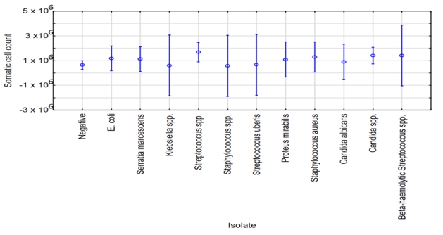
Figure 3. ANOVA - SCC in relation to specific isolated pathogens

Conclusion

In conclusion, mastitis is significant challenge in the dairy industry. Monitoring udder health using SCC is crucial and analyzing SCC records in the mastitis control programs is effective. However, distinguishing specific pathogens based on SCC is challenging. Current study offers insights into the complex management of the mastitis in the dairy herds.

References


Abed, A.H., Menshawy, A.M., Zeinhom, M.M., Hossain, D., Khalifa, E., Wareth, G., Awad, M.F. 2021 Subclinical mastitis in selected bovine dairy herds in North Upper Egypt: Assessment of prevalence, causative bacterial pathogens, antimicrobial resistance and virulence-associated genes. Microorganisms. 9(6):1175


Abegewi, U.A., Esemu, S.N., Ndip, R.N., Ndip, L.M. 2022 Prevalence and risk factors of coliform-associated mastitis and antibiotic resistance of coliforms from lactating dairy cows in North West Cameroon. PLoS One. 17(7):0268247https://doi.org/10.1371/journal.pone.0268247


Benić, M., Maćešić, N., Cvetnić, L., Habrun, B., Cvetnić, Ž., Turk, R., Đuričić, D., Lojkić, M., Dobranić, V., Valpotić, H., Grizelj, J., Gračner, D., Grbavac, J., Samardžija, M. 2018 Bovine mastitis: a persistent and evolving problem requiring novel approaches for its control-a review. Veterinarski arhiv. 88(4):535–557. https://doi.org/10.24099/vet.arhiv.0116


Bi, Y., Wang, Y.J., Qin, Y., Guix Vallverdú, R., Maldonado García, J., Sun, W., Li, S., Cao, Z. 2016 Prevalence of bovine mastitis pathogens in bulk tank milk in China. PLoS One. 11(5):0155621https://doi.org /10.1371/journal.pone.0155621


Bradley, A., Green, M. 2005 Use and interpretation of somatic cell count data in dairy cows.In Practice. 27(6):p. 310–315. https://doi.org/10.1136/inpract.27.6.310


Campos, B., Pickering, A.C., Rocha, L.S., Aguilar, A.P., Fabres-Klein, M.H., de Oliveira Mendes, T.A., Fitzgerald J.R., de Oliveira Barros Ribon, A. 2022 Diversity and pathogenesis of Staphylococcus aureus from bovine mastitis: Current understanding and future perspectives. BMC Veterinary Research. 18(1):1–16. https://doi.org/10.1186/s12917-022-03197-5


Ceniti, C., Britti, D., Santoro, A.M.L., Musarella, R., Ciambrone, L., Casalinuovo, F., Costanzo, N. 2017 Phenotypic antimicrobial resistance profile of isolates causing clinical mastitis in dairy animals. Italian Journal of Food Safety. 6(2):6612https://doi.org/10.4081/ijfs.2017.6612


Cervinkova, D., Vlkova, H., Borodacova, I., Makovcova, J., Babak, V., Lorencova, A., Vrtkova, I., Marosevic, D., Jaglic, Z. 2013 Prevalence of mastitis pathogens in milk from clinically healthy cows. Veterinární Medicína. 58(11):567–575. https://doi.org/10.17221/7138-VETMED


Cvetnić, L., Samardžija, M., Duvnjak, S., Habrun, B., Cvetnić, M., Jaki Tkalec, V., Đuričić, D.,Benić, M. 2021 Multi Locus Sequence Typing and spa typing of Staphylococcus aureus isolated from the milk of cows with subclinical mastitis in Croatia. Microorganisms. 9(4):725


Darbaz, I., Ulusoy, B. H., Darbaz, T., Hecer, C., Aslan, S. 2023 The importance of somatic cell count in dairy technology. Mljekarstvo. 73(2):75–84. https://doi.org/10.15567/mljekarstvo.2023.0201


de Haas, Y., Barkema, H.W., Schukken, Y.H., de Jong, G., Veerkamp, R.F. 2005 Somatic cell count patterns to improve udder health by genetics and management.Mastitis in Dairy Production. p. 439–444. https://doi.org/10.3920/9789086865505_065


de Haas, Y., Veerkamp, R., Barkema, H., Gröhn, Y., Schukken, Y. 2004 Associations between pathogen-specific cases of clinical mastitis and somatic cell count patterns. Journal of Dairy Science. 87(1):95–105. https://doi.org/10.3168/jds.S0022-0302(04)73146-X


de Jong, E., McCubbin, K.D., Speksnijder, D., Dufour, S., Middleton, J.R., Ruegg, P.L., Lam, T.J., Kelton, D.F., McDougall, S., Godden, S.M. Lago, A. 2023 Invited review: Selective treatment of clinical mastitis in dairy cattle. Journal of Dairy Science. 106(6):3761–3778. https://doi.org/10.3168/jds.2022-22826


Hariharan, H., Donachie, W., Macaldowie, C., Keefe, G. 2004 Bacteriology and somatic cell counts in milk samples from ewes on a Scottish farm. Canadian journal of veterinary research. 68(3):188


Haxhiaj, K., Wishart, D.S., Ametaj, B.N. 2022 Mastitis: What it is, current diagnostics, and the potential of metabolomics to identify new predictive biomarkers. Dairy. 3(4):722–746. https://doi.org/10.3390/dairy3040050


Huang, C.H., Kusaba, N. 2022 Association between differential somatic cell count and California Mastitis Test results in Holstein cattle. JDS Communications. 3(6):441–445. https://doi.org/10.3168/jdsc.2022-0249


ISO. 13366-1;2008 2010 Milk - Enumeration of Somatic Cells - Part 1: Microscopic Method (Reference Method). Institute for Standardization of Serbia:. (Beograd, Serbia.).


Janosi, S., Baltay, Z. 2004 Correlations among the somatic cell count of individual bulk milk, result of the California Mastitis Test and bacteriological status of the udder in dairy cows. Acta Veterinaria Hungarica. 52(2):173–183. https://doi.org/10.1556/AVet.52.2004.2.6


Kabelitz, T., Aubry, E., van Vorst, K., Amon, T., Fulde, M. 2021 The role of Streptococcus spp. in bovine mastitis. Microorganisms. 9(7):1497


Kaczorek, E., Małaczewska, J., Wójcik, R., Rękawek, W., Siwicki, A. 2017 Phenotypic and genotypic antimicrobial susceptibility pattern of Streptococcus spp. isolated from cases of clinical mastitis in dairy cattle in Poland. Journal of Dairy Science. 100(8):6442–6453. https://doi.org/10.3168/jds.2017-12660


Kandeel, S.A., Morin, D.E., Calloway, C.D. and Constable, P.D. 2018 Association of California mastitis test scores with intramammary infection status in lactating dairy cows admitted to a veterinary teaching hospital. Journal of Veterinary Internal Medicine. 32(1):497–505. https://doi.org/10.1111/jvim.14876


Karzis, J., Donkin, E.F., Petzer, I.-M., Webb, E.C., Etter, E.M. 2017 Validity of somatic cell count as indicator of pathogen-specific intramammary infections. Journal of the South African Veterinary Association. 88(1):1–10. https://doi.org/10.4102/jsava.v88i0.1465


Katsande, S., Matope, G., Ndengu, M., Pfukenyi, D.M. 2013 Prevalence of mastitis in dairy cows from smallholder farms in Zimbabwe. Onderstepoort Journal of Veterinary Research. 80(1):1–7. https://doi.org/10.4102/ojvr.v80i1.523


Knežević, K., Dobranić, V., Đuričić, D., Samardžija, M., Benić, M., Getz, I., Efendić, M., Cvetnić, L., Šavorić, J., Butković, I., Cvetnić, M., Mazić, M., Maćešić, N. 2021 Primjena broja somatskih stanica za dijagnostiku mastitisa i utjecaj na kakvoću mlijeka. Veterinarska stanica. 52(6):751–764. https://doi.org/10.46419/vs.52.6.11


Kovačević, Z., Kladar, N., Čabarkapa, I., Radinović, M., Maletić, M., Erdeljan, M., Božin, B. 2021 New perspective of Origanum vulgare L. and Satureja montana L. essential oils as bovine mastitis treatment alternatives. Antibiotics. 10(12):1460https://doi.org/10.3390/antibiotics10121460


Kovačević, Z., Radinović, M., Čabarkapa, I., Kladar, N., Božin, B. 2021 Natural agents against bovine mastitis pathogens. Antibiotics. 10(2):205https://doi.org/10.3390/antibiotics10020205


Kovačević, Z., Samardžija, M., Horvat, O., Tomanić, D., Radinović, M., Bijelić, K., Vukomanović, A.G., Kladar, N. 2022 Is there a relationship between antimicrobial use and antibiotic resistance of the most common mastitis pathogens in dairy cows? Antibiotics. 12(1):3https://doi.org/10.3390/antibiotics12010003


Liu, K., Tao, L., Li, J., Fang, L., Cui, L., Li, J., Meng, X., Zhu, G., Bi, C., Wang, H. 2020 Characterization of Staphylococcus aureus isolates from cases of clinical bovine mastitis on large-scale Chinese dairy farms. Frontiers in Veterinary Science. 7:580129https://doi.org/10.3389/fvets.2020.580129


Lopes Júnior, J.E.F., Lange, C.C., Brito, M.A.V.P., Santos, F.R., Silva, M.A.S., Moraes, L.C.D., Souza, G.N.: 2012 Relationship between total bacteria counts and somatic cell counts from mammary quarters infected by mastitis pathogens. Ciência Rural. 42:691–696. https://doi.org/10.1590/S0103-84782012000400019


Magaš, V., Vakanjac, S., Pavlović, V., Velebit, B., Mirilović, M., Maletić, M., Đurić, M., Nedić, S. 2013 Efficiency evaluation of a bivalent vaccine in the prophylaxis of mastitis in cows. Acta Veterinaria-Beograd. 6356:525–536. https://doi.org/10.2298/AVB1306525M


Maletić, M., Magaš, V., Maletić, J. 2017 The role of veterinarian in the monitoring programs of mastitis control. IOP Conference Series: Earth and Environmental Science. 85:012035https://doi.org/10.1088/1755-1315/85/1/012035


Malinowski, E., Lassa, H., Klossowska, A., Markiewicz, H., Kaczmarowski, M., Smulski, S. 2006 : Relationship between mastitis agents and somatic cell count in foremilk samples. Bulletin-Veterinary Institute In Pulawy. 50(3):349–352


McDougall, S., Murdough, P., Pankey, W., Delaney, C., Barlow, J., Scruton, D. 2001 Relationships among somatic cell count, California mastitis test, impedance and bacteriological status of milk in goats and sheep in early lactation. Small Ruminant Research. 40(3):245–254. https://doi.org/10.1016/s0921-4488(01)00185-7


McDougall, S., Clausen, L.M., Hussein, H.M. and Compton, C.W. 2022 Therapy of subclinical mastitis during lactation. Antibiotics. 11(2):209https://doi.org/10.3390/antibiotics11020209


Milanov, D., Prunic, B., Velhner, M., Bojkovski, J. 2014 Diagnosis of yeast mastitis in dairy cows. Lucrari Stiintifice Medicina Veterinara. 47(1):56–64


Morales-Ubaldo, A.L., Rivero-Perez, N., Valladares-Carranza, B., Velázquez-Ordoñez, V., Delgadillo-Ruiz, L., Zaragoza-Bastida, A. 2023 Bovine mastitis, a worldwide impact disease: prevalence, antimicrobial resistance, and viable alternative approaches. Veterinary and Animal Science. 21:100306https://doi.org/10.1016/j.vas.2023.100306


Moretti, A., Pasquali, P., Mencaroni, G., Boncio, L., Fioretti, D.P. 1998 Relationship between cell counts in bovine milk and the presence of mastitis pathogens (yeasts and bacteria). Journal of Veterinary Medicine, Series B. 45110:129–132. https://doi.org/10.1111/j.1439-0450.1998.tb00775.x


Piccinini, R., Binda, E., Belotti, M., Casirani, G., Zecconi, A. 2005 Comparison of blood and milk non-specific immune parameters in heifers after calving in relation to udder health. Veterinary Research. 3656:747–757. https://doi.org/10.1051/vetres:2005030


Poljak, F., Šperanda, M., Špehar, M., Đidara, M., Gantner, V. 2022 Co-variability of daily milk production traits, haematological and biochemical parameters in the blood serum and milk of Holstein cows. Mljekarstvo. 72(3):131–139. https://doi.org 10.15567/mljekarstvo.2022.0302


Pyörälä, S. 2003 Indicators of inflammation in the diagnosis of mastitis. Veterinary Research. 34(5):565–578. https://doi.org/10.1051/vetres:2003026


Radinović, M., Boboš S., Ukropina, M.M., Nešić, M. 2008 Uticaj patogena prvog reda na higijensku ispravnost mleka. Contemporary Agriculture. 5734:39–45


Safak, T., Rısvanli, A., Asci-Toraman, Z. 2022 Th1/Th2 cytokine polarization in milk according to different pathogens causing subclinical mastitis in cows. Mljekarstvo. 72(2):105–113. https://doi.org/10.15567/mljekarstvo.2022.0204


Schwarz, D., Diesterbeck, U. S., Failing, K., König, S., Bruegemann, K., Zschöck, M., Wolter, W., Czerny, C.P. 2010 Somatic cell counts and bacteriological status in quarter foremilk samples of cows in Hesse, Germany - A longitudinal study. Journal of Dairy Science. 93(12):5716–5728. https://doi.org/10.3168/jds.2010-3223


Sharma, N., Singh, N., Bhadwal, M. 2011 Relationship of somatic cell count and mastitis: An overview. Asian-Australasian journal of animal sciences. 24(3):429–438. https://doi.org/10.5713/ajas.2011.10233


Skrzypek, R., Wojtowski, J., Fahr, R.D. 2004 Factors affecting somatic cell count in cow bulk tank milk - a case study from Poland. Journal of Veterinary Medicine Series A. 51(3):127–131. https://doi.org/10.1111/j.1439-0442.2004.00611.x


Smistad, M., Bakka, H.C., Sølverød, L., Jørgensen, H.J., Wolff, C. 2023 Prevalence of udder pathogens in milk samples from Norwegian dairy cows recorded in a national database in 2019 and 2020. Acta Veterinaria Scandinavica. 65(1):19https://doi.org/10.1186/s13028-023-00681-2


Song, J., Xiang, W., Wang, Q., Yin, J., Tian, T., Yang, Q., Zhang, M., Ge, G., Li, J., Diao, N., Liu, F., Shi, K., Cai, R., Du, R., Gong, Q. 2023 Prevalence and risk factors of Klebsiella spp. in milk samples from dairy cows with mastitis - A global systematic review. Frontiers in Veterinary Science. 10:1143257https://doi.org/10.3389/fvets.2023.1143257


Souza, F. N., Cunha, A.F., Rosa, D.L., Brito, M. A. V., Guimarães, A. S., Mendonça, L.C., Souza, G.N., Lage, A.P., Blagitz, M.G., Della Libera, A.M., Heinemann, M.B. 2016 Somatic cell count and mastitis pathogen detection in composite and single or duplicate quarter milk samples. Pesquisa Veterinária Brasileira. 36:811–818. https://doi.org/10.1590/S0100-736X2016000900004


Sumon, S., Ehsan, M., Islam, M. 2017 Subclinical mastitis in dairy cows: somatic cell counts and associated bacteria in Mymensingh,. Bangladesh. Journal of the Bangladesh Agricultural University. 15(2):266–271. https://doi.org/10.3329/jbau.v15i2.35073


Sumon, S.M.R., Parvin, M.S., Ehsan, M.A., Islam, M.T. 2020 Relationship between somatic cell counts and subclinical mastitis in lactating dairy cows. Veterinary World. 13(8)https://doi.org/10.14202/vetworld.2020.1709-1713


Supré, K., Haesebrouck, F., Zadoks, R., Vaneechoutte, M., Piepers, S., De Vliegher, S. 2011 Some coagulase-negative Staphylococcus species affect udder health more than others. Journal of Dairy Science. 94(5):2329–2340. https://doi.org/10.3168/jds.2010-3741


Tenhagen, B.-A., Köster, G., Wallmann, J., Heuwieser, W. 2006 Prevalence of mastitis pathogens and their resistance against antimicrobial agents in dairy cows in Brandenburg, Germany. Journal of Dairy Science. 89(7):2542–2551. https://doi.org/10.3168/jds.S0022-0302(06)72330-X


Tommasoni, C., Fiore, E., Lisuzzo, A., Gianesella, M. 2023 Mastitis in dairy cattle: On-farm diagnostics and future perspectives. Animals. 13(15):2538https://doi.org/10.3390/ani13152538


Turk, R., Koledić, M., Maćešić, N., Benić, M., Dobranić, V., Đuričić, D., Cvetnić, L., Samardžija, M. 2017 The role of oxidative stress and inflammatory response in the pathogenesis of mastitis in dairy cows. Mljekarstvo. 67(2):91–101. https://doi.org/10.15567/mljekarstvo.2017.0201


Wente, N., Klocke, D., Paduch, J.-H., Zhang, Y., Tho Seeth, M., Zoche-Golob, V., Reinecke, F., Mohr, E., Krömker, V. 2019 Associations between Streptococcus uberis strains from the animal environment and clinical bovine mastitis cases. Journal of Dairy Science. 102(10):9360–9369. https://doi.org/10.3168/jds.2019-16669


Williamson, J., Callaway, T., Rollin, E., Ryman, V. 2022 Association of milk somatic cell count with bacteriological cure of intramammary infection - a review. Agriculture. 12(9):1437https://doi.org/10.1016/j.rvsc.2022.09.015


Zhou, Y., Ren, Y., Fan, C., Shao, H., Zhang, Z., Mao, W., Wei, C., Ni, H., Zhu, Z., Hou, X., Piao, F., Cui, Y. 2013 Survey of mycotic mastitis in dairy cows from Heilongjiang Province, China. Tropical Animal Health and Production. 45(8):1709–1714. https://doi.org/10.1007/s11250-013-0419-y

Acknowledgements

This research was supported by the Science Fund of the Republic of Serbia, PROMIS, #GRANT No 6066966, InfoBomat and the Ministry of Science, Technological Development and Innovation Republic of Serbia [Grant Number 451-03-65/2024-03/ 200117].


This display is generated from NISO JATS XML with jats-html.xsl. The XSLT engine is libxslt.