Skoči na glavni sadržaj

Pregledni rad

https://doi.org/10.5599/admet.2162

Label-free and label-based electrochemical detection of disease biomarker proteins

Tias Febriana Hanifa Lestari
Irkham Irkham
Uji Pratomo
Shabarni Gaffar
Salma Nur Zakiyyah
Isnaini Rahmawati
Seda Nur Topkaya
Yeni Wahyuni Hartati


Puni tekst: engleski pdf 788 Kb

str. 463-486

preuzimanja: 0

citiraj

Preuzmi JATS datoteku


Sažetak

Abstract
Introduction
Biosensors, analytical devices integrating biological sensing elements with physicochemical transducers, have gained prominence as rapid and convenient tools for monitoring human health status using biochemical analytes. Due to its cost-effectiveness, simplicity, portability, and user-friendliness, electrochemical detection has emerged as a widely adopted method in biosensor applications. Crucially, biosensors enable early disease diagnosis by detecting protein biomarkers associated with various conditions. These biomarkers offer an objective indication of medical conditions that can be accurately observed from outside the patient.
Method
This review comprehensively documents both label-free and labelled detection methods in electrochemical biosensor techniques. Label-free detection mechanisms elicit response signals upon analyte molecule binding to the sensor surface, while labelled detection employs molecular labels such as enzymes, nanoparticles, and fluorescent tags.
Conclusion
The selection between label-free and labelled detection methods depends on various factors, including the biomolecular compound used, analyte type and biological binding site, biosensor design, sample volume, operational costs, analysis time, and desired detection limit. Focusing on the past six years, this review highlights the application of label-free and labelled electrochemical biosensors for detecting protein biomarkers of diseases.

Ključne riječi

Biosensor; biomarker; electrochemical detection; protein detection

Hrčak ID:

320009

URI

https://hrcak.srce.hr/320009

Datum izdavanja:

5.8.2024.

Posjeta: 0 *


Introduction

A biosensor is a chemical sensor that uses the recognition properties of biological compounds in a sensitive layer [1]. Biosensors are analytical devices that combine sensing elements of biological compounds (enzymes, antibodies, proteins, nucleic acids, tissues or receptors, and cells) and are closely related to physiochemical transducers. Biosensors consist of three main interconnected components, i.e., (1) bioreceptors or biological recognition systems, (2) transducers, and (3) electronic devices. A biological recognition system provides a sensor with high selectivity to the analyte being measured. The basic principle of biosensors is the recognition of biological compounds and sensing so that when a biological compound is recognized by a recognition compound, a signal change occurs by the transducer [2-7]. The key to a biosensor device is the transducer used. Transducers take advantage of the physical changes that accompany the reaction. The working principle of a biosensor based on the transducer used is divided into calorimetric biosensors (based on heat released or absorbed by the reaction), electrochemical biosensors (based on changes in voltage, current, or conductance), piezoelectric biosensors (based on the change of mass of products or reactants), optical biosensors (based on light output or differences in light absorbance of a product or reactant) [8]. Electrochemical transducers are widely used in point-of-care testing because they are simple, portable, cost-effective, and easy to use [8].

Detection of disease biomarkers, such as proteins or metabolites in human body fluids, is one of the diagnostic applications [9]. Biomarker detection is increasingly in demand due to its high demand in various fields, such as biotechnology, health care and life sciences [10]. Currently, there is a pressing need for monitoring human health status by analyzing biochemical markers such as glucose, galactose, cholesterol, uric acid, and urea. In recent years, biosensors have been widely used as a fast and convenient alternative to conventional analytical methods, which play a role in monitoring human health status with biochemical analytes [11]. The utilization of biosensors for disease detection, particularly in the case of cancer, is extensive due to their exceptional performance and real-time detection capabilities. Furthermore, biosensors possess a notably low minimum detection limit, enabling the measurement of biomarkers at extremely low levels in physiological samples for early-stage disease diagnosis. Additionally, biosensors can concurrently detect multiple biomarkers [12]. Notably, biosensors also offer the advantage of simultaneous detection of multiple biomarkers, presenting a versatile and efficient approach to disease diagnosis.

Several review articles have discussed electrochemical biosensors for various disease biomarkers that have been reported. However, no one has discussed the details of the electrochemical label-free or labeled detection mechanism. This review covers protein biomarkers of diseases, label-free and labeled biosensors detection techniques, and the utilization of electrochemical biosensors for detecting disease-related protein biomarkers over the past six years to help other researchers in developing biosensors. Furthermore, the future perspective was also discussed.

Electrochemical biosensors

Electrochemical measurements are based on electrochemical processes or changes in electrical signals that occur due to electrochemical reactions on the electrode surface. The reaction occurs due to the influence of a given current or potential [13,14]. Electrochemical detection is widely used in biosensor applications because of its low cost, simple construction, portability and ease of use. Electrochemical detection can be used to achieve low detection limits, either with or without sample preparation [15,16]. Nevertheless, electrochemical-biosensor platforms are still limited by the multiple steps involved in the testing process, including sample introduction, repetitive washing, and additional signaling-agent introduction. Further, a large sample volume is required, and the analysis is time-intensive [17]. Electrochemical measurements are grouped into four categories, i.e., voltammetry, potentiometry, impedance and conductometry. The grouping is based on changes in electrochemical properties detected during the biological attachment process [18].

The voltammetry method is the application of time-dependent potential to electrochemical cells. The function of the potential is to measure the resulting current. The results of voltammetry measurements are displayed in the form of a voltammogram. The principle of voltammetry measurement is based on measuring anode/cathode currents resulting from the oxidation/reduction reaction of an electroactive species at a selected potential window [19]. To obtain an electrochemical signal, it is common to use electroactive indicators such as ferricyanide ([Fe(CN)6]3-/4-) and hexaammineruthenium(III) chloride (Ru(NH3)6]3-/4-). Based on the potential scanned, the types of voltammetry are divided into cyclic voltammetry (CV), differential pulse voltammetry (DPV), square wave voltammetry (SWV), and linear sweep voltammetry (LSV) [20]. In the voltammetry method, the change in current resulting from electrochemical reduction or oxidation is monitored directly with time, while a constant potential is maintained at the working electrode in relation to the reference electrode [15]. The potentiometry method is an electroanalytic technique that detects ionic activity in samples [21]. Potential measurements are made when no current flows (I = 0) [13]. In the potentiometry method, two reference electrodes are used to measure the potential as it passes through the membrane and selectively reacts with the desired charged ion [15].

The impedance method generally describes an electrochemical analysis technique that, in its application, uses an alternating current or voltage (AC) in the system under investigation. This is followed by measuring the response in the form of AC current or voltage as a function of frequency [22]. During the experiment, the response of the system to the AC signal is recorded. This response is usually represented in a complex plane as impedance (Z), resistance value (Rs), electron transfer resistance (Rct), double-layer capacitance (Cdl) and angular frequency. The frequency can cover a wide spectrum, from very low frequencies (typically mHz) to high frequencies [23]. Electrochemical impedance measurement is carried out using a small excitation signal so that the cell response is pseudolinear [13].

The conductometry method is used to measure changes in the electrical conductivity of a solution or medium due to changes in the composition of the solution or medium during a chemical reaction. In its use, enzymes whose products are charged are used to produce changes in ionic strength, thereby increasing conductivity [15]. The advantages of using the conductometry method are the use of alternating voltages with low amplitudes that allow the Faraday process to be avoided at the electrodes, the use of a simple reference electrode, the low production costs and a high degree of integration with cheap film standards [24]. However, there is a drawback of the conductometric method: the ionic species produced must be able to change the total ionic strength significantly to obtain reliable measurements [13]. There are several methods that have been developed in the analysis of protein biomarkers of a disease, including gel electrophoresis, mass spectrometry and ELISA (enzyme-linked immunosorbent assay), but in their use, they require a lot of time, are expensive, and even require highly skilled human resources and other preparations [25]. For example, the cancer biomarker EpCAM (epithelial cell adhesion molecule) is generally detected using ELISA, PCR and cytometry, but researchers developed an electrochemical biosensor using rGO@TiO2 nanocomposites [26]. This artificial biosensor showed promising results for the detection of cancer biomarkers in serum samples, as shown by excellent electronic properties, selectivity and serves as a suitable sensing layer. So, it is believed that the biosensor created has the potential to be used in monitoring other cancer biomarkers.

Protein biomarkers

Proteins are biological macromolecules in the body that play an important role in the metabolic catalysis process, transporting molecules across cells and cell apoptosis. Overexpression of a protein can be associated with certain types of pathogens [27]. Detecting biomarkers involves measuring the immune response and hormonal changes associated with a developing disease [28]. Biomarker proteins are only present at the molecular level during the early stages of a disease [27]. Protein biomarkers have been widely used in the field of applied research related to genomics and proteomics techniques [29-31]. Proteins are key compounds in different biological cells, tissues and organs [32]. Protein is a very informative type of biomarker, so many protein biomarkers are used in the application of clinical diagnosis and treatment of a disease. Biomarker proteins are expressed differently depending on the type of disease and provide various information about disorders that occur in the body. Different expressions of each biomarker protein can occur due to different protein processing in the body [33]. The research of Hartati et al. [34] reported using a gold bioconjugate modified electrochemical biosensor to detect epithelial sodium channel protein (ENaC), a protein biomarker of hypertension.

Molecular biomarkers are a broad scope for all biomarkers, both existing and to be discovered, and which can be measured or detected based on molecular characteristics (gene arrangement, proteomic analysis and complex multiplex analysis) and modified versions of the analytes [35]. According to the World Health Organization [36], biological markers are substances, processes, or structures in the body and can be measured to predict a disease. Apart from that, it can also be said to be a collection of certain molecules that can help with the diagnosis or prognosis of an abnormality in the body [37]. Biomarkers are used in clinical practice to provide different treatments or health care for each individual depending on the type of disease [38], so they are objective indications of medical conditions that can be observed accurately from outside the patient [39]. Generally, research or clinical practice using biomarkers can clinically predict a disorder or disease [40], demonstrate knowledge of clinical pharmacology, and provide a design basis for safe, rapid, and definitive clinical trials [41].

Determining biomarkers must be considered to obtain feasibility and ease of clinical use, so preclinical and clinical validation studies are needed first [39]. Biomarkers are classified into prognostic and diagnostic biomarkers. Prognostic biomarkers are related to disease recurrence information, while diagnostic biomarkers are related to the detection of a disease [12,42,43]. These biomarkers are used to predict the future clinical progression, severity, or risk of recurrence of a disease in a patient. Prognostic biomarkers play a crucial role in personalized medicine, allowing healthcare providers to tailor treatment plans to individual patients based on their predicted disease outcomes. Biomarkers have various functions, including the detection of disease, detection of abnormal conditions in the body (i.e., elevated blood glucose levels), monitoring of health status, and monitoring of clinical response to interventions (e.g., blood cholesterol) [41].

Based on their characteristics, biomarkers are grouped into imaging biomarkers and molecular biomarkers. Imaging biomarkers are specific characteristics or features identified through medical imaging techniques that provide valuable information about the presence, progression, or severity of disease, as well as treatment response. Imaging biomarkers include positron emission tomography, magnetic resonance imaging, and computerized axial tomography. They enhance diagnostic accuracy, inform clinical decisions, and contribute to better patient outcomes. Meanwhile, the molecular biomarkers include proteins, DNA, RNA, small metabolites, and lipids [44]. Biomarkers are found in several biological fluids, such as serum and plasma, whole blood, sweat, nasal secretions, urine, sputum, lacrimal, bronchial, amniotic fluid, pleura, seminal fluid, and cerebrospinal fluid [33]. Protein biomarkers are of great interest because they can be detected in various analytical instruments to identify and measure proteins in complex biological samples [45]. The best use of biomarkers is demonstrated by tests that are accurate, easy to perform, and relatively non-invasive [46].

The following are several examples of protein biomarkers widely used for disease detection. Biomarker proteins in pancreatic cancer include KRAS (Kirsten Rat Sarcoma Viral Oncogene), MBD3L2 (Methyl-CpG Binding Domain Protein 3 Like 2), DPMI mRNAs (Dolichol phosphate mannose synthase), and ACRV1 (Acrosomal Vesicle Protein 1) [47]. Biomarkers for cervical cancer, namely Ki-67, BD ProEx C, and Cytoactiv HPV-L1 (Human Papillomavirus) [48]. In breast cancer, the most widely used biomarkers include HER2 (Human epidermal growth factor receptor 2), BRCA1 and BRCA2 (Breast cancer type 1 and 2 susceptibility proteins), CEA (Carcinoembryonic antigen), MUC1 (Mucin 1), VEGF (Vascular endothelial growth factor), CA15-3 (Cancer antigen 15-3), microRNA (miRNAs) [49]. The biomarkers for liver cancer are AFP (Alpha fetoprotein) and CEA, and the biomarker for prostate cancer is PSA (Prostate-specific antigen) [50]. The emergence of the novel SARS-CoV-2 disease in late 2019 has also led to the identification of numerous protein biomarkers associated with the disease, such as receptor-binding domain (RBD) protein, spike protein, and nucleocapsid protein (N protein).

Biosensors detection mechanism of protein biomarker

The biosensor protein detection mechanism can be carried out in two steps, i.e., directly (label-free) and indirectly (labeled), depending on the output signal caused by the binding of the analyte to the labeled compound. Label-free detection is simpler, where antibodies are immobilized on the electrode surface, and changes in their electronic properties are immediately detected due to the formation of immunocomplexes [51]. In using the label-free detection technique, the signal response changes when the analyte molecule binds to the transducer surface. There are weaknesses in label-free detection, such as the occurrence of binding of non-analyte components to the sample matrix on the sensor surface, which affects false positive results. In label-free detection, it must be ensured that only the analyte bound to the appropriate biologically identifiable compound is immobilized on the electrode surface to obtain a significant change in signal response [52].

In labeled detection, further labeling of antibodies using enzymes or other molecules is required. In addition to enzymes, other labels are used, such as nanoparticles, fluorescent or electrochemiluminescent probes, and radionuclides [52]. Labeling is time-consuming, complex, has many steps and does not allow real-time detection and the use of labels can interfere with analyte binding, which can lead to distorted results [51]. The main advantage of the label detection method is that it has a higher potential for detection at lower concentrations. In addition, labeled detection can minimize false positive results because in labeled biosensors the final result is determined by the labeled compound whose binding is independent of the matrix [52].

Detection of protein biomarkers in a label-free biosensor

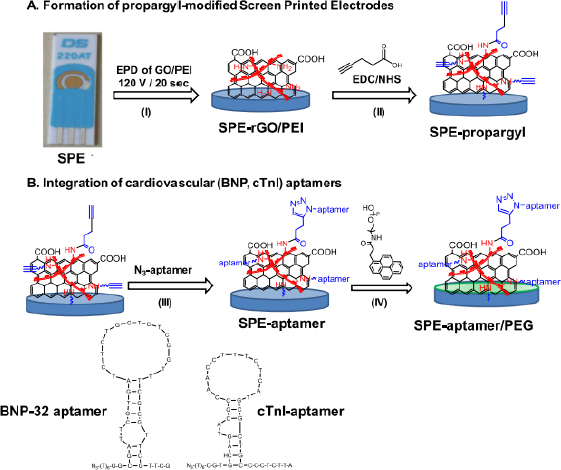
The following are several examples of label-free electrochemical biosensor research that has been conducted (shown inTable 1). Grabowska et al. [53] developed an aptamer-based electrochemical biosensor for early detection of cardiovascular disorders using two biomarkers, namely brain natriuretic peptide (BNP-32) and cardiac troponin I (cTnI). This study used commercial gold-based screen-printing electrodes (SPE) modified by electrophoretic deposition (EPD) using reduced polyethylenimine (PEI)/reduced graphene oxide (rGO) nanocomposite films, resulting in a robust and sensitive electrochemical platform for BNP-32 and cTnI sensing without the need for any labels (seeFigure 1). The presence of the amine group on PEI facilitates the binding of the BNP-32 and cTnI aptamers via a propargylacetic acid linker followed by Cu(I)-based click chemical attachment to the azide and ending with the aptamer. Apart from that, modifications were also made by adding pyrene anchors carrying polyethylene glycol (PEG) units to ensure that the sensor is low in biofouling and has high specificity. Electrochemical measurements were carried out using differential pulse voltammetry with a [Fe(CN)6]4− redox probe. This sensor has a detection limit of 0.9 pg/mL.

Table 1. application of label-free biosensors for detection of protein biomarkers of a disease in the last six years.
Working electrodeBioreceptorsProtein biomarkersType of diseaseDetection limitRef.
Gold electrode (GE)D-fructose 6-phosphate (F6P)Phosphoglucose isomerase from rabbit muscle (RmPGI)Cancer in human plasma6.6×10-15 M[55]
GESynthetic Peptides PCT BP3Procalcitonin (PCT)Sepsis12.5 ng/mL[56]
Screen-printed gold electrode (SPGE)Molecularly imprinted polymers (MIPs)Hemeprotein myoglobinCardiovascular disease (CVD)2.1×10⁻³ and 14×10-3 ng/mL[57]
Printed circuit board (PCB)BSA antibodyBovine serum albumin (BSA)Inflammation2.89 ng/mL[58]
Gold disk electrode (GDE)Anti-tau antibodyTau-441Neurodegeneration-[59]
GEEGFR antibodyEGFR antigenBreast cancer6.9×10-3 ng/mL[60]
GETNF- α antibodyTumor necrosis factor alpha (TNF- α) proteinInflammation10-12 M[61]
GES100 beta antibody and CRP antibodyC-reactive proteins (CRP) and S100 beta proteinsCVD10 ng/mL[62]
ZnOα-cTnT and α-cTnI antibodyCardiac troponin T (cTnT) and cTnIMyocardial infarction (MI)10-3 ng/mL[63]
ITOAnti-EpCAMEpCAMTumour6.5×10-3 ng/mL[26]
ITOAnti-TNF-αTNF-αCancer1.39×10-3 ng/mL[64]
GDETdT -mediated G-quadrupplex complex of 3’-OH terminalHeminDiseases of thrombin31×10-13 M[65]
GEAnti-IgGImmunoglobulin G (IgG)Inflammation6×10-18 M[66]
GEAnti-CRPCRPCVD and Inflammatory diseases (an acute-phase protein)2.25×10⁻⁶ ng/mL[67]
GEN-(5-phosphate-D-arabinoyl)-2-aminoethanamine (5PAED)Autocrine motility factor-phosphoglucose isomerase (AMF-PGI)Cancer4.3×10-14 M[68]
Gold-based SPEBNP-32 aptamer and cTnI aptamerBrain natriuretic peptide (BNP-32) and cTnICVD0.9×10-3 ng/mL[53]
Titanium foilCobalt-functionalized TiO2 nanotubes (Co-TNTs)SARS-CoV-2 S-RBD proteinSARS-CoV-27×10-10 M[69]
GEPeptide ligan (H-C-acp-acp-FALGEA-NH2)Glioblastoma (GBM)-derived exosomesGlioblastoma - the most fatal tumors in the brain7.83×103 particle/μL[70]
Screen-printed carbon electrode (SPCE)Anti-ENaC antibodyENaC proteinSalt-sensitive hypertension0.198 ng/mL[71]
GEAntibody-tau-441Tau-441Dementia4.6×10-16 M[72]
Glassy carbon electrode (GCE)pyrrole-3-carboxylic acid monomerBRCA1 geneBreast cancer3×10-15 M[73]
SPGESelf-assembled monolayer (SAM) of cysteamine (CA)α-amilaseStress-related changes in the body< 3.0×10² ng/mL[74]
Glass capillaryMolecularly imprinted polymers (MIPs)Trypsin enzymeDigestive disease< 4.1×10 ng/mL[75]
SPEβHBA and NEFA antibodiesΒ-hydroxybutyrate (βHBA) and non-esterified fatty acid (NEFA)Dairy cow metabolic diseases0.00011 M and 0.000111 M[76]
GCEIgE-aptamerImmunoglobulin E (IgE)Allergic reactions and parasitic diseases4.2×10-5 ng/mL[77]
Graphene oxide (GO)/gold nanoparticles (GNPs) hydrogelThiolated cellular prion protein (PrPC) peptide probeAmyloid-beta oligomers (AβO)Alzheimer’s disease10-13 M[78]
Au nanostructured gold disc electrodeAnti-rhuEPO AntibodyRecombinant human erythropoietin (rhuEPO)Erythropoiesis (formation of erythrocytes in the bone marrow)10-12 M[79]
GCEmiRNA-21 aptamermiRNA-21Breast cancer2.3×10-15 M[80]
Glassy electrodeAnti-VEGF antibodyVEGFAngiogenesis, vasculogenesis, and endothelial cell growth81.46×10-3 ng/mL[81]
SPCEAnti-ENaC antibodyENaC proteinSalt-senstive hypertension0.037 ng/ml[82]
GEDGV peptideDENV-2-NS1 proteinDengue1.49×10-3 ng/mL[54]
Gold chip electrode2008s aptamerPlasmodium falciparum lactate dehydrogenase (PfLDH)Malaria8.4×10-13 M[83]
GEAnti-NUMA1 antibody and anti-CFHR1 antibodyNuclear mitotic apparatus protein 1 (NUMA1) and complement factor H-related 1 (CFHR1)Bladder cancer1.29 ng/mL and 0.97 ng/mL[84]
GCEAnti-CA15-3CA15-3Breast cancer0.32 mU/mL[85]
Gold interdigitated micro-electrode arrays (IDμE)Anti-HER4 affimerHER4Tumour< 10-12 M[86]
PlatinumAnti-PARK7/DJ-1 antibodyParkinson’s disease protein 7/protein deglycase DJ-1 (PARK7/DJ1)Parkinson’s disease7.5 ng/mL[87]
GCESpike SARS-CoV-2 antibodySpike protein SARS-CoV-2 antigenSARS-CoV-210-11 ng/mL[88]
SPCEAnti-EnaC antibodyENaC proteinHypertension8.4×10-2 ng/mL[34]
GEAnti-VEGF antibody (VEGFab) and anti-PSA antibody (PSAab)VEGF and PSAProstate cancer (PCa)50 pg/mL and 1 ng/mL[89]
GDESelf-assembled monolayer from 11-ferrocenyl-undecanethiol (11FcC) and polyethylene glycol (PEG) containing the thiol (PEG thiol)Human prostatic acid phosphatase (hPAP)PCa1.119×10-11 M[90]
GENGAL peptideNeutrophil gelatinase-associated lipocalin (NGAL)Acute kidney injury and the diabetic3.93 ng/mL (SWV) and 1.74 ng/mL (EIS)[91]
Multiwall carbon nanotube (MWCNT) electrodesAnti-OV6-AbOV6 markerCancer-[92]
Au micro-gap electrodeBioprobe DNA 3 way-junction (3WJ)cTnICVD10-12 M[93]
GEHER2-specific hybrid aptamer-polyclonal antibody and antibody-based sandwichHER2Breast cancer1 ng/mL[94]
GEDual-functional hairpin dNA probe which consists miR-16 complementary sequence and AFP aptamer sequencemiRNA-16 and AFPHCC1.4×10-10 M[95]
Carbon electrodeAnti-lysozyme aptamerLysozymeBreast Cancer, alzheimer’s, malaria90 ng/mL[96]
3D nanoprinted gold micropillar array electrodeSARS-CoV-2 spike RBD proteinAnti-spike antibodies CR3022SARS-CoV-20.4 BAU/mL[97]
ITOAnti-Aβ42Aβ42Alzheimer3.7×10-4 ng/mL[98]
316 L stainless-steel plate electrodeGelsolin-actinLysophosphatidic acid (LPA)Ovarian cancer7×10-7 M[99]
ITO micro-electrode arrayAnti-plasma phosph-orylated-tau threonine 181 (p-tau181) antibodiesPlasma phosphorrylated-tau threonine 181 (p-tau181)Alzheimer and mild cognitive impairment (MCI)9.2×10⁻⁷ ng/mL[100]
MGCE modified Mg0.5Cu0.5Fe2O4-AuDNA-aptamerCA125Ovarian cancer4.4 U/mL[101]
FTO electrode modified graphene oxide (GO) decorated with gold nano-flower nanostructures (GO@Au-NS)Thiolated DNA capture probe against miRNA-223 (Cap-223)miRNA-223Colorectal cancer1.2×10-20 M[102]
GE modified gold nanoparticles-black phosphorus (AuNPs@BP@PDA)Synthetic peptide receptor (C-terminus incorporated to gold binding peptide (GBP)CRPCrohn’s disease0.7 ng/mL[103]
α-Fe2O3/carbon cloth yarnAnti-IL-6 antibodiesInterleukin-6 (IL-6)Cancer2.6×10-4 ng/mL[104]
Magnetic glassy carbon electrode (MGCE)Peptide nucleic acid (PNA)TP53 geneTumour, cancer2.6×10-13 M[105]
Gold-interdigitated microelectrodes (IdμEs) modified VS2MMP-9 antibodyMMP-9 antigenOcular inflammatory1.344×10-9 ng/mL[106]
GEHRP-conjugated antibody of telomeraseTelomerase antigenCancer0.078 IU/mL[107]
ITOAnti-CYFRA 21-1Cytokeratin subunit 19 (CYFRA 21-1)Lung cancer4.7×10-6 ng/mL[108]
ITOAnti-SP17 antibodiesSperm protein-17 (SP17)Cancer47.57×10-3 ng/mL[109]
ITO modified AuNPs/Ti3C2-mxenescTnI-specific aptamer (SH-AptcTnI)cTnIAcute myocardial infarction1.4×10-7 ng/mL[110]
GO/ amino substituted polypyrrole polymer modified disposable electrodeAnti-CALR antibodiesCalreticulin (CALR)Cancer10.4×10-6 ng/mL[111]
SPCE modified AuNPs/GO-COOHCRP aptamer probesCRPCVD and inflammation0.001 ng/mL[112]
GCE modified PtNi nanocubes assembliesHE4 antibody (HE4-Ab)Human epididymis protein 4 (HE4)Epithelial ovarian cancer (EOC)0.11×10⁻³ ng/mL[113]
GECis P-tau monoclonal antibody (mAb)Cis phosphorylated tau (cis P-tau)Alzheimer2×10-14 M[114]
SPCEAptamer ENaCENaC proteinSalt-sensitive hypertension0.012 ng/mL[115]
SPCEAnti-ENaC antibodyENaC proteinSalt-sensitive hypertension0.113 ng/mL[116]
SPCE modified PdAntibodies specific to HER2 (anti-HER2)HER2Breast cancer1 ng/mL[117]
GCE modified carbon nanofiberAptamerCytochrome c (Cyt c)Cancer7.4×10-10 M[118]
GEAptamer-based specific recognition with CRISPR-Cas12aSARS-CoV-2 nucleocapsid antigenSARS-CoV 20.077 ng/mL[119]
GDEAnti-MCM5Mini chromosome maintenance protein 5, MCM5Cervical cancer2.9×10-12 M[120]
SPCEAnti-ENaC antibodyENaC proteinSalt-sensitive hypertension0.0372 ng/mL[116]
SPCEBicyclic peptidesHuman urokinase-type plasminogen activator (h-uPA)Cancer9 ng/mL[121]
SPCEAnti-ENaC antibodyENaC proteinSalt-sensitive hypertension0.110 ng/mL[122]
Figure 1. Schematic (A) surface modification of the gold SPE sensor by electrophoretic deposition of GO/PEI solution forms a thin layer of rGO/PEI; (B) integration of aptamers and passivation with synthetic pyrene-PEG (shown with green layer). Reproduction from [53] with copyright permission.
ADMET-12-2162-g001.jpg

Jalil et al. [26] developed a label-free electrochemical biosensor for early detection of a tumor biomarker EpCAM. This is the first research study regarding the creation of a transducer platform based on rGO@TiO2 nanocomposites for determining cancer biomarkers. In this study, antibodies (anti-EpCAM) were immobilized directly on the surface of the rGO@TiO2/indium tin oxide (ITO) electrode and were ready to capture the EpCAM antigen. The development of the biosensor was carried out using reduced graphene oxide (rGO) modified with titanium dioxide (TiO2) nanoparticles to form rGO@TiO2 nanocomposites, which were synthesized through a hydrothermal process. The rGO@TiO2 nanocomposite was deposited on an ITO-coated glass substrate by electrophoretic deposition method so that the modification became rGO@TiO2/ITO electrode. Spectroscopy techniques, microscopic identification and electrochemical measurements were used to determine the success of the deposition stage. After the electrodes were modified, they were used for covalent immobilization of the EpCAM monoclonal antibody (anti-EpCAM/rGO@TiO2/ITO electrode). After modification was completed, EpCAM was immobilized at the electrodes. Bovine serum albumin was used as a blocking agent to avoid non-specific binding of EpCAM. Electrochemical measurements were carried out using DPV and EIS with the electroactive indicator ferricyanide ([Fe(CN)6]3-/4-). The detection range is 0.01-60 ng/mL, with a detection limit of 0.0065 ng/mL.

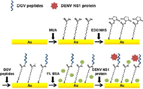
In addition, Kim et al. [54] also developed a label-free electrochemical biosensor for the detection of nonstructural dengue virus protein (DENV) 1 (NS1), which is a specific and sensitive biomarker for the diagnosis of dengue fever (shown inFigure 2). In this research, a series of synthetic peptides substituted with amino acids was designed. This synthetic peptide acts as a recognition compound that will recognize the DENV-NS1 target. Five synthetic peptide derivatives (DGV BP1, BP2, BP3, BP4 and BP5) were used, rationally designed and chemically synthesized. Modification of the biosensor was carried out with an Au substrate prepared by evaporation of gold on a clean silicon wafer, then coated with titanium. The gold substrate was placed in a piranha solution to remove residual substances, which was washed with distilled water. Then, the Au substrate was dried under nitrogen flow and immersed in an ethanol solution of 1-mercaptodecanoic acid (MUA) overnight. The activated gold substrate was dried under a nitrogen stream. MUA-activated Au substrates were immersed in ethyl(dimethylaminopropyl)carbodiimide (EDC) and N-hydroxysuccinimide (NHS) solutions in methanol. The active Au substrate was rinsed using methanol and immersed in PBS. After functionalization of the Au substrate, electrode assembly was carried out, and the synthetic peptide was dropped on the surface of the Au substrate. Synthetic peptides are covalently immobilized onto the gold sensor surface. The biomarker protein DENV-NS1 was dripped onto the surface of the modified electrode. In practice, the performance of the biosensors is monitored using SWV and EIS. Electrochemical analysis was carried out using PBS solution containing ferro/ferricyanide. The detection limit for NS1 was 1.49 g/mL.

Figure 2. label-free biosensor schematic for detection of DENV NS1 protein. Reproduction from [54] with copyright permission.
ADMET-12-2162-g002.jpg

Detection of protein biomarkers by labelled biosensor

The following are several examples of the development of labeled electrochemical biosensors for the detection of protein biomarkers of diseases. Kasturi et al. [123] developed an electrochemical biosensor using a thiol-labeled probe DNA to detect microRNA-122 (miRNA-122), which is a biomarker of liver diseases, including hepatocellular carcinoma (HCC). This study developed an easy, effective, and sensitive RNA electrochemical biosensor for the detection of Au-loaded reduced graphene oxide (rGO) miRNA-122 synthesized by a simple hydrothermal reflux method. The thiol-labeled DNA probe was anchored at the rGO/Au nanocomposite binding site and recognized the target miRNA-122. This biosensor is shown inFigure 3. The rGo/Au nanocomposite serves to improve the performance of the biosensor due to the significant electron conductivity of the electrochemical surface area. Modification of the biosensor was carried out with a glass wafer. Gold (Au) was sprayed on the glass wafer by a sputtering system.

Figure 3. schematic of a thiol-labeled biosensor for the detection of miRNA-122 protein. Redraw from [123].
ADMET-12-2162-g003.jpg

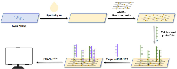
The Au-sputtered glass wafers were then cleaned with deionized water and dried under a nitrogen stream. Furthermore, the surface of the Au-modified glass wafer was dripped with rGO/Au nanocomposite. After the electrode surface was modified by the rGO/Au nanocomposite, the thiol-labeled DNA probe solution in tris-EDTA was dispersed on the electrode surface and incubated for 12 h under moist conditions. Then, BSA 1 % solution was used to block the electrode surface. Next, the target miRNA in the Tris-EDTA solution dripped on the surface of the electrode, where the probe DNA was immobilized. The thiol was used as a linker to label the DNA probe solution dispersed on the surface of the rGO nanocomposite modified Au electrode to identify miRNA-122 targets. Electrochemical measurements were carried out using CV and DPV using electrolyte solution [Fe(CN)6]-3/-4 to determine the success of immobilizing DNA probes and target miRNA hybridization. This biosensor showed a linear response for various target concentrations of miRNA-122 in the range of 10 μM to 10 pM with a detection limit of 1.73 pM.

Muñoz-San Martín et al. [124] developed an electrochemical peptide biosensor based on the on-off method used for the detection of pancreatic cancer using a biomarker in the form of trypsin, which belongs to the protease family. Double-labeled short synthetic peptides were used in this biosensor modification. Double labeling was performed using fluorescein isothiocyanate (FITC) and biotin. The biosensor development strategy was carried out using electrodes in the form of an SPCE modified using magnetic microbeads (MBs). The MBs surface was double-labeled with FITC immobilization, and a biotin-labeled peptide probe immobilized through the biotinylated end of the neutravidin-MBs surface. MBs are used to support the performance of biosensors with lower non-specific adsorption capabilities and higher affinity for biotin. Further cleavage is carried out using a target enzyme (trypsin) that cleaves the peptide at the C-terminal end of arginine so that the FITC-bound peptide moiety is released from the MBs. Enzymatic labeling was performed using anti-FITC conjugated with horseradish peroxidase (HRP) to (HRP-anti-FITC). The modified MBs were magnetically captured on the surface of the SPCE, thus modifying the biosensor to become HRP-anti-FITC/Peptide/Neutravidin-MBs/SPCE. In the development of this biosensor, an on-off approach was used due to the lower amount of HRP-anti-FITC, which will attach to the missing peptide fragment that carries the FITC moiety from the surface of MBs. Electrochemical measurements were carried out using the amperometry method with a redox medium in the form of hydroquinone (HQ) and the enzymatic substrate H2O2. The sample used for trypsin detection is human cell lysate. The high sensitivity of the biosensor can determine trypsin in clinical samples and quantify the trypsin content in cell lysates with the ability to differentiate between pancreatic and non-pancreatic cancer cells. The results of the biosensor showed a detection limit of 0.16 g/mL.

Recently, researchers have increasingly focused on simultaneous biomarker detection, enabling the analysis of multiple analytes in a single assay. This approach offers several advantageous features, addressing the limitations associated with single-analyte detection methods and catering to the need for comprehensive and efficient analysis. Simultaneous biomarker detection is particularly valuable when faced with limited sample volumes and contributes to more accurate diagnoses. Anabalagan et al. [125] conducted a study that exemplifies this trend. They simultaneously developed an innovative approach for detecting two cancer biomarkers, CEA and AFP, through the design of two distinct redox-labelled detection probes. Specifically, silver NPs functionalized with CEAAb2 and 1-amino anthraquinone were employed for CEA detection, while polyaniline NPs were functionalized with ferrocenecarboxaldehyde (Fc-CHO) for detecting AFP. The detection process involved applying different voltage pulses in a sequence, including 0 V for 10 s, -0.75 V (potential AQ) for 10 s, 0 V for 10 s, and +0.5 V (Potential Fc) for 10 s, hence exhibited excellent sensitivity, specificity, and minimal cross-reactivity between the two targeted biomarkers, with detection limits of 30 pg/mL for AFP and 80 pg/mL for CEA. Moreover, the proposed sensor was used to determine APF and CEA in human blood serum. In addition to several examples already described,Table 2 shows other examples of the use of labeled biosensors for protein biomarker detection.

Table 2. application of labeled biosensor for detection of protein biomarkers of disease in the last six years.
Working electrodeBio-receptorsProtein biomarkersType of diseaseDetection methodDetection limitRef.
GEDNA ProbemiRNA-375, miRNA-141, and PSAPCaMethyl blue labeledmiRNA-141: 8×10⁻¹⁰ M, miRNA-375: 8×10⁻¹⁰ nM, PSA: 10-12 M[126]
GEHER2 antibodyHER2Breast cancerNanoprobe catalytic labeled10-5 ng/mL[25]
GEProbe DNAmiRNA-122Liver diseases including HCCThiol-labeled1.73×10-12 M[123]
GEDNA ProbeDNA H1-MB and H2-MB sampleLiver diseaseMethylen blue labeled4.1×10-5 ng/mL[127]
GEAnti-TNF-α antibodyProtein TNF-αInflammationBiotin labeled11.21×10-3 ng/mL[128]
SPEHeparin (Hep) from Hep-Au@Fe3O4Eosinophil cationic proteinAsthmaHeparin labeled3×10-10 M[129]
SPCEHRP-anti-FITCTrypsinCancerFluorescein isothiocyanate (FITC) and biotin labeled160 ng/mL[124]
GCEAβO-specific aptamerAβOsAlzheimer's diseaseThiol labeled1.22×10-3 ng/mL[130]
GCECatalytic hairpin assembly (CHA)miRNA-1246 and miRNA-4521HemophiliaQDs@ZIF-8 labeledmiRNA-1246: 1.9×10⁻¹⁶ M miRNA-4521: 2.8×10⁻¹⁶ M[131]
GEssDNA aptamerTransforming growth factor b1 (TGF-b1)HPV-16 and parovovirus B19 (PB-19)N-succinimidyl S-acetylthioacetat Labeled2×10-10 M[132]
Gold nanostructured electrodesAnti-OTOL1 Dan anti-PRESOtolin-1 and prestin proteinsHearing disordersMethylen blue labeled-[133]
GEProbe sequencemiRNA-155Breast cancerPolyethyleneimine-silver nanoparticles (PEI-Ag NPs)20 zmol[134]
GETetrahedral DNA nanostructure (TDNs)-aptamerHER2Breast cancerHorseradish peroxidase-labeled0.08 ng/mL[135]
GEApt15 and Apt29 aptamerThrombinHemostasisFerrocene labeled7.6×10-13 M[136]
GEAnti-CA 15-3 monoclonal antibodyCA15-3Breast cancerMagnetic beads labeled15×10-6 U/mL[137]
GEBiotin-DNA-biotinmiRNAsCancerEnzyme labeled10-17 M[138]
SPCEAnti-AFPAFPLiver cancerMethylene blue labeled8.5×10-5 ng/mL[139]
ITOAnti CA125 antibodiesCA125Ovarian cancerSilver@polypyrrole (Ag@PPy) labeled10-7 ng/mL[140]
SPCES9.6 antibodies (one anti-DNA/RNA antibody)multiple miRNA biomarkers (miRNA-21, miRNA-155 and miRNA-10b)Cancertitanium phosphate nanospheres with different heavy metal ions (zinc, cadmium, lead),1.3×10-16 M, 1.9×10⁻¹⁶ M, and 2.3×10⁻¹⁶ M[141]
SPGEAntibodies against HER-1 and HER-2HER-1 and HER-2Breast cancerHorseradish peroxidase-labeled1.06 ng/mL and 0.95 ng/mL[142]
SPCECEA antibody and AFP antibodyCEA and AFPCancerSilver nanoparticles and anthraquinone for CEA; and ferrocene for AFP8×10-2 ng/mL for CEA and 3×10⁻² ng/mL for AFP[125]
GCEAnti-miRNA-141 complementary sequence (ACP--141) and nti-miRNA-21 DNA probemiRNA-141.and miRNA-21Lung cancerMethylene blue and ferrocene8.9×10-16 M for miRNA-141 and 1.24×10⁻¹⁵ M for miRNA-21[143]
GECA199 antibodyCarbohydrate antigen-199Pancreatic cancerGlucose oxidase-amino magnetic nanoparticles (AMNP) and gold- horseradish peroxidase0.2 U/mL[144]
Graphene/SPCEN protein SARS-CoV-2IgG-SARS-CoV-2 nucleocapsidSARS-CoV2Secondary antibody labeled with horseradish peroxidase1:4947 v/v[145]
GCEg-C3N4/Fe3O4/ /aptamerCA15-3Breast cancerMethylene blue-labeled0.2 U/mL[3]
SPCEHuman eukaryotic myelin basic protein (MBP)anti-MBPMultiple sclerosis autoimmune diseaseSecondary antibody labelled with horseradish peroxidase (HRP-anti-hIgG)0.016 ng/mL[146]

Future perspectives

Early detection of a disease can help control the infection of a disease more effectively so that it can treat patients on time [147]. The development of biosensor technology in the future will develop rapidly as the use of biosensors increases as a device for monitoring a person's health status. Biosensors are increasingly in demand due to their wide use in healthcare and medicinal applications, paving the way for better development [148]. A broad strategy for developing biosensors for protein biomarker detection can be carried out by establishing an economical, straightforward, reusable biosensor construction that has the potential for large-scale manufacture and rapid operation of biosensors. In the future, this strategy can be applied in the development of biosensors in general and can be used for the detection of various protein biomarkers of disease [75].

Nowadays, label-free biosensor detection techniques have made advances in the use of newer signal detection schemes. The use of nanotechnology-based transducers allows label-free biosensors to have high sensitivity, little analyte damage, and use little sample. Label-free biosensor techniques have excellent potential to meet the demand for higher-quality biosensors and have been widely developed over the last few years [149]. So far, the technical use of label detection in biosensors has increased detection potential at lower concentrations. However, the use of labeled compounds usually has high operational costs and longer testing times. In addition, real-time analysis is not possible, and the use of labels can disrupt the binding of analytes, causing distorted results [52]. The rapid development of biosensors in the future depends on the innovation of researchers to accept the opportunities and challenges in the development of electrochemical biosensors. The fields of electrochemistry, proteomics and biotechnology that continue to develop will have an impact on the development of reliable electrochemical biosensors in the diagnosis of protein biomarkers of a disease on the spot [47]. Electrochemical biosensors will grow yearly as a reliable analytical tool [150].

Conclusions

Protein biomarkers are useful in the clinical detection of disease and monitoring health status that can indicate abnormal conditions in the body. Electrochemical biosensors have been widely used for early diagnosis applications of disease in recent years. Using electrochemical biosensors with simple construction, low cost, easy to use, portability, and low detection limits makes biosensors an alternative method for early detection of a disease. Label-free or labeled detection techniques on biosensors can be used according to research needs and requirements, such as the biomolecular compound used, the type of analyte and its biological binding site, biosensor construction, sample volume, operational costs, analysis time, and the desired detection limit and effectiveness of the use of biosensors.

Acknowledgements

We would like to give an acknowledgment for this review article to the Indonesian Ministry of Education and Culture for Penelitian Tesis Magister No. 3018/UN6.3.1/PT.00/2023 and Academic Leadership Grant of Universitas Padjadjaran, Indonesia No. 1549/UN6.3.1/PT.00/2023.

Notes

[1] Conflicts of interest Conflict of interest: The authors declare no conflict of interest.

References

[1] 

Ziegler C.; Gopel W.. Biosensor Development, Institute of Physical and Theoretical Chemistry, University of Tiibingen, D-72076 Tiibingen, Germany, 2022, p.585. https://dx.doi.org/10.1016/B978-0-12-822548-6.00112-6. https://doi.org/10.1016/B978-0-12-822548-6.00112-6

[2] 

Karunakaran C.; Rajkumar R.; Bhargava K.. Introduction to Biosensors, Elsevier Inc., Delhi, 2015, p.3. http://dx.doi.org/10.1016/B978-0-12-803100-1.00001-3. https://doi.org/10.1016/B978-0-12-803100-1.00001-3

[3] 

Pourmadadi M.; Yazdian F.; Ghorbanian S.; Shamsabadipour A.; Khandel E.; Rashedi H.; Rahdar A.; Diez-Pascual A.. Construction of Aptamer-Based Nanobiosensor for Breast. Biosensors 12 (2022) 921. https://doi.org/10.3390/bios12110921. https://doi.org/10.3390/bios12110921

[4] 

Röhlen D.L.; Pilas J.; Dahmen M.; Keusgen M.; Selmer T.; Schöning M.J.. Toward a hybrid biosensor system for analysis of organic and volatile fatty acids in fermentation processes. Frontiers in Chemistry 6 (2018) 284. https://dx.doi.org/10.3389/fchem.2018.00284. https://doi.org/10.3389/fchem.2018.00284

[5] 

Kulkarni M.B.; Ayachit N.H.; Aminabhavi T.M.. Biosensors and Microfluidic Biosensors: From Fabrication to Application. Biosensors 12 (2022) 543. https://dx.doi.org/10.3390/bios12070543. https://doi.org/10.3390/bios12070543

[6] 

Kulkarni M.B.; Ayachit N.H.; Aminabhavi T.M.. Recent Advancements in Nanobiosensors: Current Trends, Challenges, Applications, and Future Scope. Biosensors 12 (2022) 892. https://dx.doi.org/10.3390/bios12100892. https://doi.org/10.3390/bios12100892

[7] 

Kulkarni M.B.; Ayachit N.H.; Aminabhavi T.M.. A Short Review on Miniaturized Biosensors for the Detection of Nucleic Acid Biomarkers. Biosensors 13 (2023) 412. https://dx.doi.org/10.3390/bios13030412. https://doi.org/10.3390/bios13030412

[8] 

Thakur M.S.; Ragavan K. V.. Biosensors in food processing. Journal of Food Science and Technology 50 (2013) 625-641. https://dx.doi.org/10.1007/s13197-012-0783-z. https://doi.org/10.1007/s13197-012-0783-z

[9] 

Gruhl F.J.; Rapp B.E.; Länge K.. Biosensors for Diagnostic Applications, Springer-Verlag, Berlin Heidelberg, 2011. https://dx.doi.org/10.1007/10_2011_130. https://doi.org/10.1007/10_2011_130

[10] 

Kumar S.; Bukkitgar S.D.; Singh S.; Pratibha, Singh V.; Reddy K.R.; Shetti N.P.; Venkata Reddy C.; Sadhu V.; Naveen S.. Electrochemical Sensors and Biosensors Based on Graphene Functionalized with Metal Oxide Nanostructures for Healthcare Applications. ChemistrySelect 4 (2019) 5322-5337. https://dx.doi.org/10.1002/slct.201803871. https://doi.org/10.1002/slct.201803871

[11] 

Arya S.K.; Datta M.; Singh S.P.; Malhotra B.D.. Biosensor for total cholesterol estimation using N-(2-aminoethyl)-3- aminopropyltrimethoxysilane self-assembled monolayer. Analytical and Bioanalytical Chemistry 389 (2007) 2235-2242. https://dx.doi.org/10.1007/s00216-007-1655-7. https://doi.org/10.1007/s00216-007-1655-7

[12] 

Jayanthi V.S.P.K.S.A.; Das A.B.; Saxena U.. Recent advances in biosensor development for the detection of cancer biomarkers. Biosensors and Bioelectronics 91 (2017) 15-23. https://dx.doi.org/10.1016/j.bios.2016.12.014. https://doi.org/10.1016/j.bios.2016.12.014

[13] 

Arya S.K.; Singh S.P.; Malhotra B.D.. Handbook of Biosensors and Biochips, John Wiley & Sons, New Delhi, 2008, p.1-2. https://dx.doi.org/10.1002/9780470061565.hbb032. https://doi.org/10.1002/9780470061565.hbb032

[14] 

Keçili R.; Denizli A.. Molecular Imprinting for Nanosensors and Other Sensing Applications, Inc., Delhi, 2021, p.19-43. https://doi.org/10.1016/B978-0-12-822117-4.00002-2. https://doi.org/10.1016/B978-0-12-822117-4.00002-2

[15] 

Ronkainen N.J.; Halsall H.B.; Heineman W.R.. Electrochemical biosensors. Chemical Society Reviews 39 (2010) 1747-1763. https://dx.doi.org/10.1039/b714449k. https://doi.org/10.1039/b714449k

[16] 

Baranwal J.; Barse B.; Gatto G.; Broncova G.; Kumar A.. Electrochemical Sensors and Their Applications: A Review. Chemosensors 10 (2022) 363. https://dx.doi.org/10.3390/chemosensors10090363. https://doi.org/10.3390/chemosensors10090363

[17] 

Deenin W.; Yakoh A.; Pimpitak U.; Pasomsub E.; Rengpipat S.; Crespo G.A.; Chaiyo S.. Electrochemical lateral-flow device for rapid COVID-19 antigen-diagnostic testing. Bioelectrochemistry 152 (2023) 108438. https://dx.doi.org/10.1016/j.bioelechem.2023.108438. https://doi.org/10.1016/j.bioelechem.2023.108438

[18] 

Perumal V.; Hashim U.. Advances in biosensors: Principle, architecture and applications. Journal of Applied Biomedicine 12 (2014) 1-15. https://doi.org/10.1016/j.jab.2013.02.001. https://doi.org/10.1016/j.jab.2013.02.001

[19] 

Harvey D.. 11.4: Voltammetric and Amperometric Methods - Chemistry LibreTexts. The LibreTexts Libraries (2020) 285-289. https://chem.libretexts.org/Bookshelves/Analytical_Chemistry/Book%3A_Analytical_Chemistry_2.1_(Harvey)/11%3A_Electrochemical_Methods/11.04%3A_Voltammetric_and_Amperometric_Methods.

[20] 

Ghanam A.; Mohammadi H.; Amine A.; Haddour N.; Buret F.. Chemical Sensors: Electrochemical Sensors; Voltammetry/Amperometry. Encyclopedia of Sensors and Biosensors (2021) 03327589. https://doi.org/10.1016/B978-0-12-822548-6.00032-7. https://doi.org/10.1016/B978-0-12-822548-6.00032-7

[21] 

Bakker E.; Pretsch E.. Potentiometric sensors for trace-level analysis. TrAC - Trends in Analytical Chemistry 24 (2005) 199-207. https://dx.doi.org/10.1016/j.trac.2005.01.003. https://doi.org/10.1016/j.trac.2005.01.003

[22] 

El-Azazy M.. Electrochemical Impedance Spectroscopy (EIS) in Food, Water, and Drug Analyses: Recent Advances and Applications. Electrochemical Impedance Spectroscopy (2020) c.8. https://dx.doi.org/10.5772/intechopen.92333. https://doi.org/10.5772/intechopen.92333

[23] 

Pohanka M.; Skládal P.. Electrochemical biosensors - Principles and applications. Journal of Applied Biomedicine 6 (2008) 57-64. https://dx.doi.org/10.32725/jab.2008.008. https://doi.org/10.32725/jab.2008.008

[24] 

Soldatkin O.O.; Peshkova V.M.; Dzyadevych S. V.; Soldatkin A.P.; Jaffrezic-Renault N.; El’skaya A. V.. Novel sucrose three-enzyme conductometric biosensor. Materials Science and Engineering C 28 (2008) 959-964. https//dx.doi.org/10.1016/j.msec.2007.10.034. https://doi.org/10.1016/j.msec.2007.10.034

[25] 

Fu Y.; Wang N.; Yang A.; wai Law H.K.; Li L.; Yan F.. Highly Sensitive Detection of Protein Biomarkers with Organic Electrochemical Transistors. Advanced Materials 29 (2017) 1703787. https://dx.doi.org/10.1002/adma.201703787. https://doi.org/10.1002/adma.201703787

[26] 

Jalil O.; Pandey C.M.; Kumar D.. Electrochemical biosensor for the epithelial cancer biomarker EpCAM based on reduced graphene oxide modified with nanostructured titanium dioxide. Microchimica Acta 187 (2020) 275. https://dx.doi.org/10.1007/s00604-020-04233-7. https://doi.org/10.1007/s00604-020-04233-7

[27] 

Chong K.C.; Hu F.; Liu B.. AIEgen bioconjugates for specific detection of disease-related protein biomarkers. Materials Chemistry Frontiers 3 (2019) 12-24. https://dx.doi.org/10.1039/c8qm00383a. https://doi.org/10.1039/c8qm00383a

[28] 

Pepe M.S.; Etzioni R.; Feng Z.; Potter J.D.; Lou Thompson M.; Thornquist M.; Winget M.; Yasui Y.. Phases of biomarker development for early detection of cancer. Journal of the National Cancer Institute 93 (2001) 1054-1061. https://dx.doi.org/10.1093/jnci/93.14.1054. https://doi.org/10.1093/jnci/93.14.1054

[29] 

Perco P.; Pleban C.; Kainz A.; Lukas A.; Mayer G.; Mayer B.; Oberbauer R.. Protein biomarkers associated with acute renal failure and chronic kidney disease: Review. European Journal of Clinical Investigation 36 (2006) 753-763. https://dx.doi.org/10.1111/j.1365-2362.2006.01729.x. https://doi.org/10.1111/j.1365-2362.2006.01729.x

[30] 

Reilly L.; Seddighi S.; Singleton A.B.; Cookson M.R.; Ward M.E.; Qi Y.A.. Variant biomarker discovery using mass spectrometry-based proteogenomics. Frontiers in Aging 4 (2023) 1191993. https://dx.doi.org/10.3389/fragi.2023.1191993. https://doi.org/10.3389/fragi.2023.1191993

[31] 

Chase Huizar C.; Raphael I.; Forsthuber T.G.. Genomic, proteomic, and systems biology approaches in biomarker discovery for multiple sclerosis. Cellular Immunology 358 (2020) 104219. https://dx.doi.org/10.1016/j.cellimm.2020.104219. https://doi.org/10.1016/j.cellimm.2020.104219

[32] 

Hasan M.R.; Ahommed M.S.; Daizy M.; Bacchu M.S.; Ali M.R.; Al-Mamun M.R.; Saad Aly M.A.; Khan M.Z.H.; Hossain S.I.. Recent development in electrochemical biosensors for cancer biomarkers detection. Biosensors and Bioelectronics: X 8 (2021) 100075. https://doi.org/10.1016/j.biosx.2021.100075. https://doi.org/10.1016/j.biosx.2021.100075

[33] 

Karaulov A. V.; Garib V.; Garib F.; Valenta R.. Protein Biomarkers in Asthma. International Archives of Allergy and Immunology 175 (2018) 189-208. https://dx.doi.org/10.1159/000486856. https://doi.org/10.1159/000486856

[34] 

Hartati Y.W.; Gaffar S.; Alfiani D.; Pratomo U.; Sofiatin Y.; Subroto T.. A voltammetric immunosensor based on gold nanoparticle - Anti-ENaC bioconjugate for the detection of epithelial sodium channel (ENaC) protein as a biomarker of hypertension. Sensing and Bio-Sensing Research 29 (2020) 100343. https://doi.org/10.1016/j.sbsr.2020.100343. https://doi.org/10.1016/j.sbsr.2020.100343

[35] 

Laterza O.F.; Hendrickson R.C..; Wagner J... Molecular Biomarkers. Biomarkers. 41 (2007) 573-585. https://doi.org/10.1177/009286150704100504. https://doi.org/10.1177/009286150704100504

[36] 

Biomarkers in risk assesment: validity and validation. World Health Organization (2001). https://apps.who.int/iris/handle/10665/42363 (accessed October 25, 2021).

[37] 

Shi K.; Lin W.; Zhao X.M.. Identifying Molecular Biomarkers for Diseases with Machine Learning Based on Integrative Omics. IEEE/ACM Transactions on Computational Biology and Bioinformatics 18 (2021) 2514-2525. https://dx.doi.org/10.1109/TCBB.2020.2986387. https://doi.org/10.1109/TCBB.2020.2986387

[38] 

Bodaghi A.; Fattahi N.; Ramazani A.. Biomarkers: Promising and valuable tools towards diagnosis, prognosis and treatment of Covid-19 and other diseases. Heliyon 9 (2023) e13323. https://dx.doi.org/10.1016/j.heliyon.2023.e13323. https://doi.org/10.1016/j.heliyon.2023.e13323

[39] 

Frank R.; Hargreaves R.. Clinical biomarkers in drug discovery and development. Nature Reviews Drug Discovery 2 (2003) 566-580. https://dx.doi.org/10.1038/nrd1130. https://doi.org/10.1038/nrd1130

[40] 

Strimbu K.; Tavel J.A.. What are biomarkers? Current Opinion in HIV and AIDS. National Institutes of Health 5 (2010) 463-466. https://dx.doi.org/10.1097/COH.0b013e32833ed177. https://doi.org/10.1097/COH.0b013e32833ed177

[41] 

Atkinson A.J.; Colburn W.A.; DeGruttola V.G.; DeMets D.L.; Downing G.J.; Hoth D.F.; Oates J.A.; Peck C.C.; Schooley R.T.; Spilker B.A.; Woodcock J.; Zeger S.L.. Biomarkers and surrogate endpoints: Preferred definitions and conceptual framework. Clinical Pharmacology and Therapeutics 69 (2001) 89-95. https://dx.doi.org/10.1067/mcp.2001.113989. https://doi.org/10.1067/mcp.2001.113989

[42] 

Liu L.; Pang H.; He Q.; Pan B.; Sun X.; Shan J.; Wu L.; Wu K.; Yao X.; Guo Y.. A novel strategy to identify candidate diagnostic and prognostic biomarkers for gastric cancer. Cancer Cell International 21 (2021) 335. https://dx.doi.org/10.1186/s12935-021-02007-6. https://doi.org/10.1186/s12935-021-02007-6

[43] 

Wallington-Beddoe C.T.; Mynott R.L.. Prognostic and predictive biomarker developments in multiple myeloma. Journal of Hematology and Oncology 14 (2021) 151. https://dx.doi.org/10.1186/s13045-021-01162-7. https://doi.org/10.1186/s13045-021-01162-7

[44] 

Fathi E.; Mesbah-namin S.A.. Biomarkers in Medicine: An Overview. British Journal of Medicine and Medical Research 4 (2013) 1701-1718. https://dx.doi.org/10.9734/bjmmr/2014/6917. https://doi.org/10.9734/bjmmr/2014/6917

[45] 

Michalski A.; Cox J.; Mann M.. More than 100,000 detectable peptide species elute in single shotgun proteomics runs but the majority is inaccessible to data-dependent LC-MS/MS. Journal of Proteome Research 10 (2011) 1785-1793. https://dx.doi.org/10.1021/pr101060v. https://doi.org/10.1021/pr101060v

[46] 

Hewitt S.M.; Dear J.; Star R.A.. Discovery of protein biomarkers for renal diseases. Journal of the American Society of Nephrology 15 (2004) 1677-1689. https://dx.doi.org/10.1097/01.ASN.0000129114.92265.32. https://doi.org/10.1097/01.ASN.0000129114.92265.32

[47] 

Campuzano S.; Yánez-Sedeño P.; Pingarrón J.M.. Electrochemical bioaffinity sensors for salivary biomarkers detection. TrAC - Trends in Analytical Chemistry 86 (2017) 14-24. https://dx.doi.org/10.1016/j.trac.2016.10.002. https://doi.org/10.1016/j.trac.2016.10.002

[48] 

Brown C.A.; Bogers J.; Sahebali S.; Depuydt C.E.; De Prins F.; Malinowski D.P.. Role of protein biomarkers in the detection of high-grade disease in cervical cancer screening programs. Journal of Oncology 2012 (2012) 289315. https://dx.doi.org/10.1155/2012/289315. https://doi.org/10.1155/2012/289315

[49] 

Hasanzadeh M.; Shadjou N.; de la Guardia M.. Early stage screening of breast cancer using electrochemical biomarker detection. TrAC - Trends in Analytical Chemistry 91 (2017) 67-76. https://dx.doi.org/10.1016/j.trac.2017.04.006. https://doi.org/10.1016/j.trac.2017.04.006

[50] 

Bohunicky B.; Mousa S.A.. Biosensors: The new wave in cancer diagnosis. Nanotechnology, Science and Applications 4 (2011) 1-10. https://dx.doi.org/10.2147/NSA.S13465. https://doi.org/10.2147/NSA.S13465

[51] 

Rahman M.A.; Shiddiky M.J.A.; Park J.S.; Shim Y.B.. An impedimetric immunosensor for the label-free detection of bisphenol A. Biosensors and Bioelectronics 22 (2007) 2464-2470. https://dx.doi.org/10.1016/j.bios.2006.09.010. https://doi.org/10.1016/j.bios.2006.09.010

[52] 

Rapp B.E.; Gruhl F.J.; Länge K.. Biosensors with label-free detection designed for diagnostic applications. Analytical and Bioanalytical Chemistry 398 (2010) 2403-2412. https://dx.doi.org/10.1007/s00216-010-3906-2. https://doi.org/10.1007/s00216-010-3906-2

[53] 

Grabowska I.; Sharma N.; Vasilescu A.; Iancu M.; Badea G.; Boukherroub R.; Ogale S.; Szunerits S.. Electrochemical Aptamer-Based Biosensors for the Detection of Cardiac Biomarkers. ACS Omega 3 (2018) 12010-12018. https://dx.doi.org/10.1021/acsomega.8b01558. https://doi.org/10.1021/acsomega.8b01558

[54] 

Kim J.H.; Cho C.H.; Ryu M.Y.; Kim J.G.; Lee S.J.; Park T.J.; Park J.P.. Development of peptide biosensor for the detection of dengue fever biomarker, nonstructural 1. PLoS ONE 14 (2019) e0222144. https://dx.doi.org/10.1371/journal.pone.0222144. https://doi.org/10.1371/journal.pone.0222144

[55] 

Devillers M.; Ahmad L.; Korri-Youssoufi H.; Salmon L.. Carbohydrate-based electrochemical biosensor for detection of a cancer biomarker in human plasma. Biosensors and Bioelectronics 96 (2017) 178-185. https://dx.doi.org/10.1016/j.bios.2017.04.031. https://doi.org/10.1016/j.bios.2017.04.031

[56] 

Lim J.M.; Ryu M.Y.; Kim J.H.; Cho C.H.; Park T.J.; Park J.P.. An electrochemical biosensor for detection of the sepsis-related biomarker procalcitonin. RSC Advances 7 (2017) 36562-36565. https://dx.doi.org/10.1039/c7ra06553a. https://doi.org/10.1039/c7ra06553a

[57] 

Ribeiro J.A.; Pereira C.M.; Silva A.F.; Sales M.G.F.. Electrochemical detection of cardiac biomarker myoglobin using polyphenol as imprinted polymer receptor. Analytica Chimica Acta 981 (2017) 41-52. https://dx.doi.org/10.1016/j.aca.2017.05.017. https://doi.org/10.1016/j.aca.2017.05.017

[58] 

Janssen J.; Lambeta M.; White P.; Byagowi A.. Carbon nanotube-based electrochemical biosensor for label-free protein detection. Biosensors 9 (2019) 144. https://dx.doi.org/10.3390/bios9040144. https://doi.org/10.3390/bios9040144

[59] 

Carlin N.; Martic-Milne S.. Anti-Tau Antibodies Based Electrochemical Sensor for Detection of Tau Protein Biomarkers. Journal of The Electrochemical Society 165 (2018) G3018-G3025. https://dx.doi.org/10.1149/2.0041812jes. https://doi.org/10.1149/2.0041812jes

[60] 

Bakshi S.; Mehta S.; Kumeria T.; Shiddiky M.J.A.; Popat A.; Choudhury S.; Bose S.; Nayak R.. Rapid fabrication of homogeneously distributed hyper-branched gold nanostructured electrode based electrochemical immunosensor for detection of protein biomarkers. Sensors and Actuators, B: Chemical 326 (2021) 128803. https://doi.org/10.1016/j.snb.2020.128803. https://doi.org/10.1016/j.snb.2020.128803

[61] 

Kim B.Y.; Lee H.B.; Lee N.E.. A durable, stretchable, and disposable electrochemical biosensor on three-dimensional micro-patterned stretchable substrate. Sensors and Actuators, B: Chemical 283 (2019) 312-320. https://dx.doi.org/10.1016/j.snb.2018.12.045. https://doi.org/10.1016/j.snb.2018.12.045

[62] 

Kuo Y.C.; Lee C.K.; Lin C.T.. Improving sensitivity of a miniaturized label-free electrochemical biosensor using zigzag electrodes. Biosensors and Bioelectronics 103 (2018) 130-137. https://dx.doi.org/10.1016/j.bios.2017.11.065. https://doi.org/10.1016/j.bios.2017.11.065

[63] 

Radha Shanmugam N.; Muthukumar S.; Chaudhry S.; Anguiano J.; Prasad S.. Ultrasensitive nanostructure sensor arrays on flexible substrates for multiplexed and simultaneous electrochemical detection of a panel of cardiac biomarkers. Biosensors and Bioelectronics 89 (2017) 764-772. https://dx.doi.org/10.1016/j.bios.2016.10.046. https://doi.org/10.1016/j.bios.2016.10.046

[64] 

Sri S.; Lakshmi G.B.V.S.; Gulati P.; Chauhan D.; Thakkar A.; Solanki P.R.. Simple and facile carbon dots based electrochemical biosensor for TNF-α targeting in cancer patient’s sample. Analytica Chimica Acta 1182 (2021) 338909. https://dx.doi.org/10.1016/j.aca.2021.338909. https://doi.org/10.1016/j.aca.2021.338909

[65] 

Lei S.; Liu Z.; Xu L.; Zou L.; Li G.; Ye B.. A “signal-on” electrochemical biosensor based on DNAzyme-driven bipedal DNA walkers and TdT-mediated cascade signal amplification strategy. Analytica Chimica Acta (2019) 237291. https://doi.org/10.1016/j.aca.2019.12.008. https://doi.org/10.1016/j.aca.2019.12.008

[66] 

Macchia E.; Romele P.; Manoli K.; Ghittorelli M.; Magliulo M.; Kovacks-Vajna Z.; Torricelli F.; Torsi L.. Ultra-sensitive protein detection with organic electrochemical transistors printed on plastic substrate. Flexible and Printed Electronics 3 (2018) 034002. https://doi.org/10.1088/2058-8585/aad0cb. https://doi.org/10.1088/2058-8585/aad0cb

[67] 

Vilian A.T.E.; Kim W.; Park B.; Oh S.Y.; Kim T.Y.; Huh Y.S.; Hwangbo C.K.; Han Y.K.. Efficient electron-mediated electrochemical biosensor of gold wire for the rapid detection of C-reactive protein: A predictive strategy for heart failure. Biosensors and Bioelectronics 142 (2019) 111549. https://doi.org/10.1016/j.bios.2019.111549. https://doi.org/10.1016/j.bios.2019.111549

[68] 

Ahmad L.; Salmon L.; Korri-Youssoufi H.. Electrochemical detection of the human cancer biomarker ‘autocrine motility factor-phosphoglucose isomerase’ based on a biosensor formed with a monosaccharidic inhibitor. Sensors and Actuators, B: Chemical 299 (2019) 126933. https://dx.doi.org/10.1016/j.snb.2019.126933. https://doi.org/10.1016/j.snb.2019.126933

[69] 

Vadlamani B.S.; Uppal T.; Verma S.C.; Misra M.. Functionalized TiO2 nanotube-based electrochemical biosensor for rapid detection of SARS-CoV-2. MedRxiv (2020) 5871. https://dx.doi.org/10.1101/2020.09.07.20190173. https://doi.org/10.1101/2020.09.07.20190173

[70] 

Sun Z.; Wang L.; Wu S.; Pan Y.; Dong Y.; Zhu S.; Yang J.; Yin Y.; Li G.. An Electrochemical Biosensor Designed by Using Zr-Based Metal-Organic Frameworks for the Detection of Glioblastoma-Derived Exosomes with Practical Application. Analytical Chemistry 92 (2020) 3819-3826. https://dx.doi.org/10.1021/acs.analchem.9b05241. https://doi.org/10.1021/acs.analchem.9b05241

[71] 

Hartati Y.W.; Yusup S.F.; Fitrilawati, Wyantuti S.; Sofiatin Y.; Gaffar S.. A voltammetric epithelial sodium channels immunosensor using screen-printed carbon electrode modified with reduced graphene oxide. Current Chemistry Letters 9 (2020) 151-160. https://dx.doi.org/10.1016/j.sbsr.2020.100343. https://doi.org/10.1016/j.sbsr.2020.100343

[72] 

Li X.; Jiang M.; Cheng J.; Ye M.; Zhang W.; Jaffrezic-Renault N.; Guo Z.. Signal multi-amplified electrochemical biosensor for voltammetric determination of tau-441 protein in biological samples using carbon nanomaterials and gold nanoparticles to hint dementia. Microchimica Acta 187 (2020) 302. https://dx.doi.org/10.1007/s00604-020-04273-z. https://doi.org/10.1007/s00604-020-04273-z

[73] 

Shahrokhian S.; Salimian R.. Ultrasensitive detection of cancer biomarkers using conducting polymer/electrochemically reduced graphene oxide-based biosensor: Application toward BRCA1 sensing. Sensors and Actuators, B: Chemical 266 (2018) 160-169. https://dx.doi.org/10.1016/j.snb.2018.03.120. https://doi.org/10.1016/j.snb.2018.03.120

[74] 

Rebelo T.S.C.R.; Miranda I.M.; Brandão A.T.S.C.; Sousa L.I.G.; Ribeiro J.A.; Silva A.F.; Pereira C.M.. A Disposable Saliva Electrochemical MIP-Based Biosensor for Detection of the Stress Biomarker α-Amylase in Point-of-Care Applications. Electrochem 2 (2021) 427-438. https://dx.doi.org/10.3390/electrochem2030028. https://doi.org/10.3390/electrochem2030028

[75] 

Arabi M.; Ostovan A.; Zhang Z.; Wang Y.; Mei R.; Fu L.; Wang X.; Ma J.; Chen L.. Label-free SERS detection of Raman-Inactive protein biomarkers by Raman reporter indicator: Toward ultrasensitivity and universality. Biosensors and Bioelectronics 174 (2021) 112825. https://dx.doi.org/10.1016/j.bios.2020.112825. https://doi.org/10.1016/j.bios.2020.112825

[76] 

Tuteja S.K.; Duffield T.; Neethirajan S.. Graphene-based multiplexed disposable electrochemical biosensor for rapid on-farm monitoring of NEFA and βhBA dairy biomarkers. Journal of Materials Chemistry B 5 (2017) 6930-6940. https://dx.doi.org/10.1039/c7tb01382e. https://doi.org/10.1039/c7tb01382e

[77] 

Wang Y.; Cui M.; Jiao M.; Luo X.. Antifouling and ultrasensitive biosensing interface based on self-assembled peptide and aptamer on macroporous gold for electrochemical detection of immunoglobulin E in serum. Analytical and Bioanalytical Chemistry 410 (2018) 5871-5878. https://dx.doi.org/10.1007/s00216-018-1201-9. https://doi.org/10.1007/s00216-018-1201-9

[78] 

Sun L.P.; Zhong Y.; Gui J.; Wang X.W.; Zhuang X.R.; Weng J.. Ahydrogel biosensor for high selective and sensitive detection of amyloid-beta oligomers. International Journal of Nanomedicine 13 (2018) 843-856. https://dx.doi.org/10.2147/IJN.S152163. https://doi.org/10.2147/IJN.S152163

[79] 

Hassanain W.A.; Sivanesan A.; Izake E.L.; Ayoko G.A.. An electrochemical biosensor for the rapid detection of erythropoietin in blood. Talanta 189 (2018) 636-640. https://dx.doi.org/10.1016/j.talanta.2018.07.045. https://doi.org/10.1016/j.talanta.2018.07.045

[80] 

Wang G.; Han R.; Li Q.; Han Y.; Luo X.. Electrochemical Biosensors Capable of Detecting Biomarkers in Human Serum with Unique Long-Term Antifouling Abilities Based on Designed Multifunctional Peptides. Analytical Chemistry 92 (2020) 7186-7193. https://dx.doi.org/10.1021/acs.analchem.0c00738. https://doi.org/10.1021/acs.analchem.0c00738

[81] 

Xu M.; Yadavalli V.K.. Flexible Biosensors for the Impedimetric Detection of Protein Targets Using Silk-Conductive Polymer Biocomposites. ACS Sensors 4 (2019) 1040-1047. https://dx.doi.org/10.1021/acssensors.9b00230. https://doi.org/10.1021/acssensors.9b00230

[82] 

Hartati Y.W.; Satriana N.; Gaffar S.; Mulyana J.; Wyantuti S.; Sofiatin Y.. Electrochemical Label-Free Immunosensor for The Detection of Epithelial Sodium Channels Using Gold Modified Screen-Printed Carbon Electrode. ICONISTECH (2019) 1-12. https://dx.doi.org/10.4108/eai.11-7-2019.2298070. https://doi.org/10.4108/eai.11-7-2019.2298070

[83] 

Figueroa-Miranda G.; Feng L.; Shiu S.C.C.; Dirkzwager R.M.; Cheung Y.W.; Tanner J.A.; Schöning M.J.; Offenhäusser A.; Mayer D.. Aptamer-based electrochemical biosensor for highly sensitive and selective malaria detection with adjustable dynamic response range and reusability. Sensors and Actuators, B: Chemical 255 (2018) 235-243. https://dx.doi.org/10.1016/j.snb.2017.07.117. https://doi.org/10.1016/j.snb.2017.07.117

[84] 

Arya S.K.; Estrela P.. Electrochemical ELISA-based platform for bladder cancer protein biomarker detection in urine. Biosensors and Bioelectronics 117 (2018) 620-627. https://dx.doi.org/10.1016/j.bios.2018.07.003. https://doi.org/10.1016/j.bios.2018.07.003

[85] 

Han R.; Wang G.; Xu Z.; Zhang L.; Li Q.; Han Y.; Luo X.. Designed antifouling peptides planted in conducting polymers through controlled partial doping for electrochemical detection of biomarkers in human serum. Biosensors and Bioelectronics 164 (2020) 112317. https://doi.org/10.1016/j.bios.2020.112317. https://doi.org/10.1016/j.bios.2020.112317

[86] 

Zhurauski P.; Arya S.K.; Jolly P.; Tiede C.; Tomlinson D.C.; Ko Ferrigno P.; Estrela P.. Sensitive and selective Affimer-functionalised interdigitated electrode-based capacitive biosensor for Her4 protein tumour biomarker detection. Biosensors and Bioelectronics 108 (2018) 10303. https://dx.doi.org/10.1016/j.bios.2018.02.041. https://doi.org/10.1016/j.bios.2018.02.041

[87] 

de Oliveira G.C.M.; de S. Carvalho J.H.; Brazaca L.C.; Vieira N.C.S.; Janegitz B.C.. Flexible platinum electrodes as electrochemical sensor and immunosensor for Parkinson’s disease biomarkers. Biosensors and Bioelectronics 152 (2020) 112016. https://dx.doi.org/10.1016/j.bios.2020.112016. https://doi.org/10.1016/j.bios.2020.112016

[88] 

Liv L.. Electrochemical immunosensor platform based on gold-clusters, cysteamine and glutaraldehyde modified electrode for diagnosing COVID-19. Microchemical Journal 168 (2021) 106445. https://doi.org/10.1016/j.microc.2021.106445. https://doi.org/10.1016/j.microc.2021.106445

[89] 

Pan L.H.; Kuo S.H.; Lin T.Y.; Lin C.W.; Fang P.Y.; Yang H.W.. An electrochemical biosensor to simultaneously detect VEGF and PSA for early prostate cancer diagnosis based on graphene oxide/ssDNA/PLLA nanoparticles. Biosensors and Bioelectronics 89 (2017) 598-605. https://dx.doi.org/10.1016/j.bios.2016.01.077. https://doi.org/10.1016/j.bios.2016.01.077

[90] 

Fernandes F.C.B.; Bueno P.R.. Optimized electrochemical biosensor for human prostatic acid phosphatase. Sensors and Actuators, B: Chemical 253 (2017) 1106-1112. https://dx.doi.org/10.1016/j.snb.2017.06.035. https://doi.org/10.1016/j.snb.2017.06.035

[91] 

Cho C.H.; Kim J.H.; Song D.K.; Park T.J.; Park J.P.. An affinity peptide-incorporated electrochemical biosensor for the detection of neutrophil gelatinase-associated lipocalin. Biosensors and Bioelectronics 142 (2019) 111482. https://dx.doi.org/10.1016/j.bios.2019.111482. https://doi.org/10.1016/j.bios.2019.111482

[92] 

Damiati S.; Peacock M.; Leonhardt S.; Damiati L.; Baghdadi M.A.; Becker H.; Kodzius R.; Schuster B.. Embedded disposable functionalized electrochemical biosensor with a 3D-printed flow cell for detection of hepatic oval cells (HOCs). Genes 9 (2018) 89. https://dx.doi.org/10.3390/genes9020089. https://doi.org/10.3390/genes9020089

[93] 

Lee T.; Lee Y.; Park S.Y.; Hong K.; Kim Y.; Park C.; Chung Y.H.; Lee M.H.; Min J.. Fabrication of electrochemical biosensor composed of multi-functional DNA structure/Au nanospike on micro-gap/PCB system for detecting troponin I in human serum. Colloids and Surfaces B: Biointerfaces 175 (2019) 343-350. https://dx.doi.org/10.1016/j.colsurfb.2018.11.078. https://doi.org/10.1016/j.colsurfb.2018.11.078

[94] 

Jarczewska M.; Trojan A.; Gągała M.; Malinowska E.. Studies on the Affinity-based Biosensors for Electrochemical Detection of HER2 Cancer Biomarker. Electroanalysis 31 (2019) 1125-1134. https://dx.doi.org/10.1002/elan.201900041. https://doi.org/10.1002/elan.201900041

[95] 

Gao T.; Zhi J.; Mu C.; Gu S.; Xiao J.; Yang J.; Wang Z.; Xiang Y.. One-step detection for two serological biomarker species to improve the diagnostic accuracy of hepatocellular carcinoma. Talanta 178 (2018) 89-93. https://dx.doi.org/10.1016/j.talanta.2017.09.011. https://doi.org/10.1016/j.talanta.2017.09.011

[96] 

Khan N.I.; Maddaus A.G.; Song E.. A low-cost inkjet-printed aptamer-based electrochemical biosensor for the selective detection of lysozyme. Biosensors 8 (2018) 7. https://dx.doi.org/10.3390/bios8010007. https://doi.org/10.3390/bios8010007

[97] 

Shoute L.C.T.; Abdelrasoul G.N.; Ma Y.; Duarte P.A.; Edwards C.; Zhuo R.; Zeng J.; Feng Y.; Charlton C.L.; Kanji J.N.; Babiuk S.; Chen J.. Label-free impedimetric immunosensor for point-of-care detection of COVID-19 antibodies. Microsystems and Nanoengineering 9 (2023) 3. https://dx.doi.org/10.1038/s41378-022-00460-5. https://doi.org/10.1038/s41378-022-00460-5

[98] 

Altay D.N.; Yagar H.; Ozcan H.M.. A new ITO-based Aβ42 biosensor for early detection of Alzheimer’s disease. Bioelectrochemistry 153 (2023) 37421689. https://dx.doi.org/10.1016/j.bioelechem.2023.108501. https://doi.org/10.1016/j.bioelechem.2023.108501

[99] 

Ahmadi S.; Lotay N.; Thompson M.. Affinity-based electrochemical biosensor with antifouling properties for detection of lysophosphatidic acid, a promising early-stage ovarian cancer biomarker. Bioelectrochemistry 153 (2023) 108466. https://dx.doi.org/10846610.1016/j.bioelechem.2023.108466. https://doi.org/10.1016/j.bioelechem.2023.108466

[100] 

Tieu M.V.; Choi S.H.; Le H.T.N.; Cho S.. Electrochemical impedance-based biosensor for label-free determination of plasma P-tau181 levels for clinically accurate diagnosis of mild cognitive impairment and Alzheimer’s disease. Analytica Chimica Acta 1273 (2023) 341535. https://doi.org/10.1016/j.aca.2023.341535. https://doi.org/10.1016/j.aca.2023.341535

[101] 

Yue Y.; Chen X.; Wang J.; Ma M.; He A.; Liu R.. Label-free electrochemical biosensor with magnetically induced self-assembly for the detection of cancer antigen 125. Arabian Journal of Chemistry 16 (2023) 105070. https://doi.org/10.1016/j.arabjc.2023.105070. https://doi.org/10.1016/j.arabjc.2023.105070

[102] 

Akbari A.; Hashemzadeh H.; Eshkiki Z.S.; Masoodi M.; Tabaeian S.P.; Naderi-Manesh H.; Zare A.A.; Agah S.. Detection of plasma miR-223 by a novel label-free graphene oxide/gold nanocomposite immunosensor in colorectal cancer patients: An electrochemical biosensor approach. Biosensors and Bioelectronics: X 14 (2023) 100331. https://dx.doi.org/10.1016/j.biosx.2023.100331. https://doi.org/10.1016/j.biosx.2023.100331

[103] 

Yang H.J.; Kim M.W.; Raju C.V.; Cho C.H.; Park T.J.; Park J.P.. Highly sensitive and label-free electrochemical detection of C-reactive protein on a peptide receptor−gold nanoparticle−black phosphorous nanocomposite modified electrode. Biosensors and Bioelectronics 234 (2023) 115382. https://dx.doi.org/10.1016/j.bios.2023.115382. https://doi.org/10.1016/j.bios.2023.115382

[104] 

Prabhu K.; Lakshminarayanan M.; Mohankumar G.; Ponpandian N.; Viswanathan C.. Vertically pillared α-Fe2O3 nanorods on carbon yarn as a textile-based stable immunosensor electrode for selective electrochemical sensing of interleukin-6 cancer biomarker. Sensors and Actuators A: Physical 357 (2023) 114419. https://doi.org/10.1016/j.sna.2023.114419. https://doi.org/10.1016/j.sna.2023.114419

[105] 

Liu R.; Zhang Y.; Liu M.; Ni Y.; Yue Y.; Wu S.; Li S.. Electrochemical sensor based on Fe3O4/α-Fe2O3@Au magnetic nanocomposites for sensitive determination of the TP53 gene. Bioelectrochemistry 152 (2023) 108429. https://dx.doi.org/10.1016/j.bioelechem.2023.108429. https://doi.org/10.1016/j.bioelechem.2023.108429

[106] 

Ghosh T.N.; Rotake D.; Kumar S.; Kaur I.; Singh S.G.. Tear-based MMP-9 detection: A rapid antigen test for ocular inflammatory disorders using vanadium disulfide nanowires assisted chemi-resistive biosensor. Analytica Chimica Acta 1263 (2023) 341281. https://dx.doi.org/10.1016/j.aca.2023.341281. https://doi.org/10.1016/j.aca.2023.341281

[107] 

Kohansal F.; Mobed A.; Aletaha N.; Ghaseminasab K.; Dolati S.; Hasanzadeh M.. Biosensing of telomerase antigen using sandwich type immunosensor based on poly(β-Cyclodextrin) decorated by Au@Pt nanoparticles: An innovative immune-platform toward early-stage identification of cancer. Microchemical Journal 190 (2023) 108649. https://doi.org/10.1016/j.microc.2023.108649. https://doi.org/10.1016/j.microc.2023.108649

[108] 

Aydın E.B.; Aydın M.; Sezgintürk M.K.. A novel electrochemical impedance immunosensor for the quantification of CYFRA 21-1 in human serum. Microchimica Acta 190 (2023) 235. https://dx.doi.org/10.1007/s00604-023-05813-z. https://doi.org/10.1007/s00604-023-05813-z

[109] 

Yadav A.K.; Verma D.; Kumar A.; Bhatt A.N.; Solanki P.R.. Biocompatible epoxysilane substituted polymer-based nano biosensing platform for label-free detection of cancer biomarker SP17 in patient serum samples. International Journal of Biological Macromolecules 239 (2023) 124325. https://doi.org/10.1016/j.ijbiomac.2023.124325. https://doi.org/10.1016/j.ijbiomac.2023.124325

[110] 

Mi X.; Li H.; Tu Y.. An Aptamer Biosensing Strategy for Label-Free Assay of Dual Acute Myocardial Infarction Biomarkers Built upon AuNPs/Ti3C2-MXenes. Chemosensors 11 (2023) 157. https://dx.doi.org/10.3390/chemosensors11030157. https://doi.org/10.3390/chemosensors11030157

[111] 

Aydin E.B.; Aydin M.; Sezgintürk M.K.. A Simple and Low-Cost Electrochemical Immunosensor for Ultrasensitive Determination of Calreticulin Biomarker in Human Serum. Macro Molecular Bioscience (2022) 2200390. https://doi.org/10.1002/mabi.202200390. https://doi.org/10.1002/mabi.202200390

[112] 

Gao H.; Bai Y.; He B.; Tan C.S.. A Simple Label-Free Aptamer-Based Electrochemical Biosensor for the Sensitive Detection of C-Reactive Proteins. Biosensors 12 (2022) 1180. https://dx.doi.org/10.3390/bios12121180. https://doi.org/10.3390/bios12121180

[113] 

Chen D.N.; Jiang L.Y.; Zhang J.X.; Tang C.; Wang A.J.; Feng J.J.. Electrochemical label-free immunoassay of HE4 using 3D PtNi nanocubes assemblies as biosensing interfaces. Microchimica Acta 189 (2022) 455. https://dx.doi.org/10.1007/s00604-022-05553-6. https://doi.org/10.1007/s00604-022-05553-6

[114] 

Shiravandi A.; Yari F.; Tofigh N.; Kazemi Ashtiani M.; Shahpasand K.; Ghanian M.H.; Shekari F.; Faridbod F.. Earlier Detection of Alzheimer’s Disease Based on a Novel Biomarker cis P-tau by a Label-Free Electrochemical Immunosensor. Biosensors 12 (2022) 879. https://dx.doi.org/10.3390/bios12100879. https://doi.org/10.3390/bios12100879

[115] 

Hartati Y.W.; Komala D.R.; Hendrati D.; Gaffar S.; Hardianto A.; Sofiatin Y.; Bahti H.H.. An aptasensor using ceria electrodeposited-screen-printed carbon electrode for detection of epithelial sodium channel protein as a hypertension biomarker. Royal Society Open Science 8 (2021) 202040. https://dx.doi.org/10.1098/rsos.202040. https://doi.org/10.1098/rsos.202040

[116] 

Zakiyyah S.N.; Eddy D.R.; Firdaus M.L.; Subroto T.; Hartati Y.W.. Screen-printed carbon electrode/natural silica-ceria nanocomposite for electrochemical aptasensor application. Journal of Electrochemical Science and Engineering 12 (2022) 1225-1242. https://dx.doi.org/10.5599/jese.1455. https://doi.org/10.5599/jese.1455

[117] 

Joshi A.; Vishnu A.G.K.; Dhruv D.; Kurpad V.; Pandya H.J.. Morphology-Tuned Electrochemical Immunosensing of a Breast Cancer Biomarker Using Hierarchical Palladium Nanostructured Interfaces. ACS Omega 7 (2022) 34177-34189. https://dx.doi.org/10.1021/acsomega.2c03532. https://doi.org/10.1021/acsomega.2c03532

[118] 

Sadrabadi E.A.; Benvidi A.; Yazdanparast S.; Amiri-zirtol L.. Fabrication of a label-free electrochemical aptasensor to detect cytochrome c in the early stage of cell apoptosis. Microchimica Acta 189 (2022) 279. https:/dx.doi.org/10.1007/s00604-022-05373-8. https://doi.org/10.1007/s00604-022-05373-8

[119] 

Liu N.; Liu R.; Zhang J.. CRISPR-Cas12a-mediated label-free electrochemical aptamer-based sensor for SARS-CoV-2 antigen detection. Bioelectrochemistry 146 (2022) 108105. https://dx.doi.org/10810510.1016/j.bioelechem.2022.108105. https://doi.org/10.1016/j.bioelechem.2022.108105

[120] 

Barhoum A.; Forster R. J.. Label-free electrochemical immunosensor for picomolar detection of the cervical cancer biomarker MCM5. Analytica Chimica Acta 1225 (2022) 340226. https://dx.doi.org/34022610.1016/j.aca.2022.340226. https://doi.org/10.1016/j.aca.2022.340226

[121] 

Moro G.; Ferrari L.; Angelini A.; Polo F.. An Impedimetric Biosensing Strategy Based on BicyclicPeptides as Bioreceptors for Monitoring h-uPA Cancer Biomarkers. Chemosensors 11 (2023) 234. https://dx.doi.org/10.3390/chemosensors11040234. https://doi.org/10.3390/chemosensors11040234

[122] 

Fazrin E.I.; Sari A.K.; Setiyono R.; Gaffar S.; Sofiatin Y.; Bahti H.H.; Hartati Y.W.. The Selectivity and Stability of Epithelial Sodium Channel (ENaC) Aptamer as an Electrochemical Aptasensor. Anal. Bioanal. Electrochem. 14 (2022) 715-729. https://www.researchgate.net/publication/365977169_The_Selectivity_and_Stability_of_Epithelial_Sodium_Channel_ENaC_Aptamer_as_an_Electrochemical_Aptasensor.

[123] 

Kasturi S.; Eom Y.; Torati S.R.; Kim C.G.. Highly sensitive electrochemical biosensor based on naturally reduced rGO/Au nanocomposite for the detection of miRNA-122 biomarker. Journal of Industrial and Engineering Chemistry 93 (2021) 186-195. https://dx.doi.org/10.1016/j.jiec.2020.09.022. https://doi.org/10.1016/j.jiec.2020.09.022

[124] 

Muñoz-San Martín C.; Pedrero M.; Gamella M.; Montero-Calle A.; Barderas R.; Campuzano S.; Pingarrón J.M.. A novel peptide-based electrochemical biosensor for the determination of a metastasis-linked protease in pancreatic cancer cells. Analytical and Bioanalytical Chemistry 412 (2020) 6177-6188. https://dx.doi.org/10.1007/s00216-020-02418-w. https://doi.org/10.1007/s00216-020-02418-w

[125] 

Chellachamy Anbalagan A.; Sawant S.N.. Redox-labelled detection probe enabled immunoassay for simultaneous detection of multiple cancer biomarkers. Microchimica Acta 190 (2023) 86. https://dx.doi.org/10.1007/s00604-023-05663-9. https://doi.org/10.1007/s00604-023-05663-9

[126] 

Xu T.; Song Y.; Gao W.; Wu T.; Xu L.P.; Zhang X.; Wang S.. Superwettable Electrochemical Biosensor toward Detection of Cancer Biomarkers. ACS Sensors 3 (2018) 72-78. https://dx.doi.org/10.1021/acssensors.7b00868. https://doi.org/10.1021/acssensors.7b00868

[127] 

Han B.; Dong L.; Li L.; Sha L.; Cao Y.; Zhao J.. Mild reduction-promoted sandwich aptasensing for simple and versatile detection of protein biomarkers. Sensors and Actuators, B: Chemical 325 (2020) 128762. https://doi.org/10.1016/j.snb.2020.128762. https://doi.org/10.1016/j.snb.2020.128762

[128] 

Arya S.K.; Kongsuphol P.; Park M.K.. Off surface matrix based on-chip electrochemical biosensor platform for protein biomarker detection in undiluted serum. Biosensors and Bioelectronics 92 (2017) 542-548. https://dx.doi.org/10.1016/j.bios.2016.10.063. https://doi.org/10.1016/j.bios.2016.10.063

[129] 

Lee C.Y.; Wu L.P.; Chou T.T.; Hsieh Y.Z.. Functional magnetic nanoparticles-assisted electrochemical biosensor for eosinophil cationic protein in cell culture. Sensors and Actuators, B: Chemical 257 (2018) 672-677. https://dx.doi.org/10.1016/j.snb.2017.11.033. https://doi.org/10.1016/j.snb.2017.11.033

[130] 

You M.; Yang S.; An Y.; Zhang F.; He P.. A novel electrochemical biosensor with molecularly imprinted polymers and aptamer-based sandwich assay for determining amyloid-β oligomer. Journal of Electroanalytical Chemistry 862 (2020) 114017. https://dx.doi.org/10.1016/j.jelechem.2020.114017. https://doi.org/10.1016/j.jelechem.2020.114017

[131] 

Rezaei H.; Motovali-bashi M.; Radfar S.. An enzyme-free electrochemical biosensor for simultaneous detection of two hemophilia A biomarkers: Combining target recycling with quantum dots-encapsulated metal-organic frameworks for signal amplification. Analytica Chimica Acta 1092 (2019) 66-74. https://dx.doi.org/10.1016/j.aca.2019.09.037. https://doi.org/10.1016/j.aca.2019.09.037

[132] 

Dai Y.; Somoza R.A.; Wang L.; Welter J.F.; Li Y.; Caplan A.I.; Liu C.C.. Exploring the Trans-Cleavage Activity of CRISPR-Cas12a (cpf1) for the Development of a Universal Electrochemical Biosensor. Angewandte Chemie 131 (2019) 17560-17566. https://dx.doi.org/10.1002/ange.201910772. https://doi.org/10.1002/ange.201910772

[133] 

Mahshid S.S.; Dabdoub A.. Development of a novel electrochemical immuno-biosensor for circulating biomarkers of the inner ear. Biosensors and Bioelectronics 165 (2020) 112369. https://dx.doi.org/10.1016/j.bios.2020.112369. https://doi.org/10.1016/j.bios.2020.112369

[134] 

Hakimian F.; Ghourchian H.. Ultrasensitive electrochemical biosensor for detection of microRNA-155 as a breast cancer risk factor. Analytica Chimica Acta 1136 (2020) 1-8. https://dx.doi.org/10.1016/j.aca.2020.08.039. https://doi.org/10.1016/j.aca.2020.08.039

[135] 

Ou D.; Sun D.; Lin X.; Liang Z.; Zhong Y.; Chen Z.. A dual-aptamer-based biosensor for specific detection of breast cancer biomarker HER2 via flower-like nanozymes and DNA nanostructures. Journal of Materials Chemistry B 7 (2019) 3661-3669. https://dx.doi.org/10.1039/c9tb00472f. https://doi.org/10.1039/c9tb00472f

[136] 

Zhu J.; Gan H.; Wu J.; Ju H.. Molecular Machine Powered Surface Programmatic Chain Reaction for Highly Sensitive Electrochemical Detection of Protein. Analytical Chemistry 90 (2018) 5503-5508. https://dx.doi.org/10.1021/acs.analchem.8b01217. https://doi.org/10.1021/acs.analchem.8b01217

[137] 

Akbari Nakhjavani S.; Khalilzadeh B.; Samadi Pakchin P.; Saber R.; Ghahremani M.H.; Omidi Y.. A highly sensitive and reliable detection of CA15-3 in patient plasma with electrochemical biosensor labeled with magnetic beads. Biosensors and Bioelectronics 122 (2018) 8-15. https://dx.doi.org/10.1016/j.bios.2018.08.047. https://doi.org/10.1016/j.bios.2018.08.047

[138] 

Liu L.; Deng D.; Wu D.; Hou W.; Wang L.; Li N.; Sun Z.. Duplex-specific nuclease-based electrochemical biosensor for the detection of microRNAs by conversion of homogeneous assay into surface-tethered electrochemical analysis. Analytica Chimica Acta 1149 (2021) 338199. https://dx.doi.org/33819910.1016/j.aca.2021.338199. https://doi.org/10.1016/j.aca.2021.338199

[139] 

Chanarsa S.; Jakmunee J.; Ounnunkad K.. A sandwich-like configuration with a signal amplification strategy using a methylene blue/aptamer complex on a heterojunction 2D MoSe2/2D W/Sable2 electrode: Toward a portable and sensitive electrochemical alpha-fetoprotein immunoassay. Frontiers in Cellular and Infection Microbiology 12 (2022) 1-16. https://dx.doi.org/10.3389/fcimb.2022.916357. https://doi.org/10.3389/fcimb.2022.916357

[140] 

Krathumkhet N.; Imae T.; ming Wang F.; Yuan C.C.; Manidae Lumban Gaol J.; Paradee N.. Electrochemical immunosensing by carbon ink/carbon dot/ZnO-labeled-Ag@polypyrrole composite biomarker for CA-125 ovarian cancer detection. Bioelectrochemistry 152 (2023) 108430. https://dx.doi.org/10.1016/j.bioelechem.2023.108430. https://doi.org/10.1016/j.bioelechem.2023.108430

[141] 

Ghanbari R.; Attaripour Isfahani A.; Pirmoradian S.; Rezaei H.; Radfar S.; Kheirollahi M.. A rapid and simple method for simultaneous determination of three breast cancer related microRNAs based on magnetic nanoparticles modified with S9.6 antibody. Analytical Biochemistry 665 (2023) 115052. https://doi.org/10.1016/j.ab.2023.115052. https://doi.org/10.1016/j.ab.2023.115052

[142] 

Wignarajah S.; Chianella I.; Tothill I.E.. Development of Electrochemical Immunosensors for HER-1 and HER-2 Analysis in Serum for Breast Cancer Patients. Biosensors 13 (2023) 355. https://dx.doi.org/10.3390/bios13030355. https://doi.org/10.3390/bios13030355

[143] 

Khodadoust A.; Nasirizadeh N.; Seyfati S.M.; Taheri R.A.; Ghanei M.; Bagheri H.. High-performance strategy for the construction of electrochemical biosensor for simultaneous detection of miRNA-141 and miRNA-21 as lung cancer biomarkers. Talanta 252 (2023) 123863. https://dx.doi.org/10.1016/j.talanta.2022.123863. https://doi.org/10.1016/j.talanta.2022.123863

[144] 

Wang H.; Yuan W.; Zhang L.; Xie B.; Cheng Q.. Dual-Labeled Octahedral Gold and Magnetic Microsphere Electrochemical Immunosensor for Ultrasensitive Determination of Carbohydrate Antigen-199 (CA199) by Differential Pulse Voltammetry (DPV). Analytical Letters (2023) 841-854. https://doi.org/10.1080/00032719.2023.2227907. https://doi.org/10.1080/00032719.2023.2227907

[145] 

Freire L.D.S.; Ruzo C.M.; Salgado B.; Mar A.; Gandarilla D.; Romaguera-barcelay Y.; Tavares A.P.M.; Goreti M.; Sales F.; Cordeiro I.; Dias J.; Lalwani B.; Matos R.; Filho H.F.; Astolfi-filho S.. An Electrochemical Immunosensor Based on Carboxylated Graphene / SPCE for IgG-SARS-CoV-2. Biosensors 12 (2022) 1161. https://doi.org/10.3390/bios12121161. https://doi.org/10.3390/bios12121161

[146] 

Guerrero S.; Sánchez-Tirado E.; Agüí L.; González-Cortés A.; Yáñez-Sedeño P.; Pingarrón J.M.. Monitoring autoimmune diseases by bioelectrochemical detection of autoantibodies. Application to the determination of anti-myelin basic protein autoantibodies in serum of multiple sclerosis patients. Talanta 243 (2022) 123304. https://dx.doi.org/10.1016/j.talanta.2022.123304. https://doi.org/10.1016/j.talanta.2022.123304

[147] 

Antiochia R.. Developments in biosensors for CoV detection and future trends. Biosensors and Bioelectronics 173 (2021) 112777. https://doi.org/10.1016/j.bios.2020.112777. https://doi.org/10.1016/j.bios.2020.112777

[148] 

Haleem A.; Javaid M.; Singh R.P.; Suman R.; Rab S.. Biosensors applications in medical field: A brief review. Sensors International 2 (2021) 100100. https://doi.org/10.1016/j.sintl.2021.100100. https://doi.org/10.1016/j.sintl.2021.100100

[149] 

Sang S.; Wang Y.; Feng Q.; Wei Y.; Ji J.; Zhang W.. Progress of new label-free techniques for biosensors: A review. Critical Reviews in Biotechnology 36 (2016) 465-481. https://dx.doi.org/10.3109/07388551.2014.991270. https://doi.org/10.3109/07388551.2014.991270

[150] 

Pereira da Silva Neves M.M.; González-García M.B.; Hernández-Santos D.; Fanjul-Bolado P.. Future trends in the market for electrochemical biosensing. Current Opinion in Electrochemistry 10 (2018) 107-111. https://dx.doi.org/10.1016/j.coelec.2018.05.002. https://doi.org/10.1016/j.coelec.2018.05.002


This display is generated from NISO JATS XML with jats-html.xsl. The XSLT engine is libxslt.