Skip to the main content

Review article

https://doi.org/10.5599/admet.2442

Flavonoids from Clerodendrum genus and their biological activities

Meiske Naomi Mamuaja
Tati Herlina
Rymond Jusuf Rumampuk
Iman Permana Maksum
Yaya Rukayadi


Full text: english pdf 1.416 Kb

page 843-879

downloads: 20

cite

Download JATS file


Abstract

Abstract
Background and purpose
Many studies have been performed to identify new sources, their optimal isolation, and the biological activities of flavonoids due to nutraceutical, pharmaceutical, and cosmeceutical properties.
Experimental approach
This review describes the method for flavonoid isolation and characteristic from the Clerodendrum genus and their biological activities with the indication of the most active ones. To perform a comprehensive review, a thorough literature review using Google Scholar, Scopus, and Science Direct was performed with keyword alone or in combination with other words.
Key results
The isolation and identification of flavonoids from the Clerodendrum genus have revealed a variety of compounds using various methods. Various studies conducted in vivo, in vitro and in silico also reported bioactivities of these flavonoids.
Conclusion
Several factors determine the flavonoid content in the Clerodendrum genus, among others, the different parts of the plant, extraction techniques, and solvent combination used. Isolated flavonoids also show significant biological activities, such as antioxidant, anti-inflammatory, antimicrobials, antidiabetic, anticancer, anti-tyrosinase, and neuroprotective agents.

Keywords

Traditional medicine; secondary metabolites; isolation; bioactivities

Hrčak ID:

324855

URI

https://hrcak.srce.hr/324855

Publication date:

10.1.2025.

Visits: 56 *


Introduction

Clerodendrum genus has been used as ornaments in celebration and traditional medicine in India, China, Korea, Japan, Thailand, Indonesia, and Africa [1,2]. Linnaeus was the first to identify Clerodendrum genus and later named it Clerodendrum informatum in 1753 in India. At first, the genus was put into Family Verbenaceae and later after a phylogenetic analysis through molecular data, it was included in Family Lamiaceae in 1990 [3,4]. The word Clerodendrum itself comes from Greek ‘kleros’ (destiny) and ‘dendron’ (tree), which might relate to two definitions of the use of species of the genus at that time; some of the species are believed to have curing properties, and others have toxic properties [5].

Clerodendrum genus has at least 500 species and grows in tropical and warm regions of Africa, eastern and southern Asia, and also in America and the northern part of Australia [6]. They are evergreen shrubs, lianas, small and woody vines [7]. The members of the Clerodendrum genus have been reported for traditional medicines, for example, Clerodendrum trichotomum for rheumatic and headaches [8], Clerodendrum serratum for asthma and malaria [9] and inflammation [10], Clerodendrum inerme for hepatitis and antidote [11], Clerodendrum colebrookianum for hypertension [12] and Clerodendrum philippinum for anti-diabetic [13]. Based on these therapeutic properties, Clerodendrum members have been studied for more than decades, resulting in extraction, isolation, purification, and identification of some compounds, including flavonoids and their glycosides, terpenoids, phenylethanoid glycosides, steroids and their derivates, cyclo-hexyl ethanoic and cyanogenic glycosides [14]. There are more than 300 compounds have been isolated and identification from the Clerodendrum genus, where pharmacological studies indicate that the crude extract and several monomers have various biological activities such as antioxidants, antidiabetic, anti-inflammatory, anticancer, antimicrobe, antihypertension, anti-obesity, antidiarrhea, liver protection, improving memories and neuroprotective [15].

Flavonoids are a group of secondary metabolites abundant in plants and represent the third largest group of natural products after alkaloids and terpenoids [16]. Flavonoids have a polyphenolic structure and are produced for plant growth and defines mechanisms [17] by acting as attractants for pollinators, sunscreen to protect against solar radiation, antimicrobial and antiherbivore [18]. The basic structure of flavonoids is C6-C3-C6, where two aromatic rings, A and B, are connected by a unit of three carbon atoms [19]. Flavonoids are classified based on the connection model of ring A and ring B, the position of ring B connection, the oxidation level of ring C substructure and the degree of polymerization, where the main structure is as inFigure 1 [20]. Variations in the structure of these flavonoids produce several biological activities that are important for medicine [21].
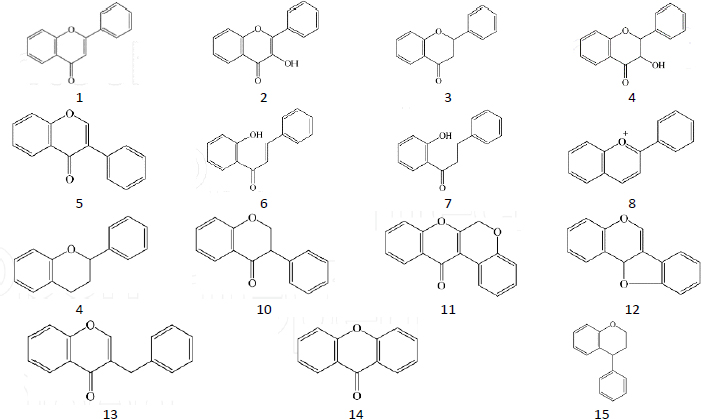
Figure 1. Main structure of flavonoid: flavone (1), flavonol (2), flavonone (3), flavanonol (4), isoflavone (5), chalcone (6), dihydrochalcone (7), anthocyanidin (8), aurone (9), isoflavanone (10), rotenoid (11), pterocarpan (12), homoisoflavanone (13), xanthone (14) and neoflavonoid (15).
ADMET-12-2442-g001.jpg

Research shows that flavonoids have nutraceutical, pharmaceutical, medicinal and cosmetic functions due to their ability to act as antioxidants, anti-inflammatory, antimutant and anticarcinogenic [22]. At the cellular level, they combine to modulate key cellular enzyme functions [23]. The flavonoid content in Clerodendrum genus has been reported in several studies with varying bioactivities [24,25]. Therefore, this review examines the members of the Clerodendrum genus and their flavonoid presence, highlighting their isolated methods and their biological potential in the area of the most often studied activities, such as antioxidant, antidiabetic, anti-inflammatory, antitumor, and others.

Isolation and characterization of flavonoids from Clerodendrum genus

Based on flavonoids potential for human health, several techniques and conditions for the isolation of this compounds have been implemented [26]. The common procedure for the isolation process involves plant preparation, extraction and fractionation, and purification [27]. The type of solvent and its polarity, solvent combination, and the ratio of liquid-solid have a significant impact on the process [28]. Various solvents, methods and parts of the plants used for isolation from the Clerodendrum genus are given inTables 1-6, based on the Clerodendrum species.

Table 1. Flavonoid Isolated from Clerodendrum paniculatum Linn
MaterialSolvent and Extraction MethodIsolated flavonoidActivityRef.
RootsMaceration with ethanol; fractination, and ethyl acetate fraction was subject to column chromatography with DCM:MeOH = 7.5:2.5Quercetin-[30]
FlowerMaceration with alcohol, no available data for CC solventQuercetinAntioxidant
Hepatoprotective
[31]
LeavesMaceration with n-hexane, fractionation with ethyl acetate and ethanolApigenin scutellarinAntibacterial[32]
Table 2. Flavonoid Isolated from Clerodendrum inerme
MaterialSolvent and extraction methodIsolated flavonoidActivityRef.
LeavesExtraction with 95 % ethanol, partition with hexane, chloroform and butanol. CC with silica gel for chloroform using MeOH/CH2Cl from 0 %, 1 %, 2 %, 10 % to get 9 fractions; a crystal of fraction 7 is the subject for NMR compound identificationHispidulinPreventing hyperdopaminergic activity[40]
Leaves and stemsMaceration with ethanol, fractionation, CC for ethyl-acetate with chloroform = 25:75 weight ratioApigenin, Salvigenin, acacetin-[41]
Leaves and stemsVLC n-hexane: ethyl-acetate, CC with n-hexane: ethyl-acetate5-hydroxy-6,7, 40-trimethoxyflavoneAntioxidant, anti-inflammatory[42]
Table 3. Flavonoid isolated from Clerodendrum phlomidis leaves
MaterialSolvent and Extraction MethodIsolated flavonoidActivityRef.
LeavesFractionation with chloroform, hexane and ethyl acetate; chloroform extract was subject to column chromatography with hexane-ethyl acetate as eluent; found 12 fraction, fraction 5 was subject to re-column with combination of chloroform: ethyl acetate = 9:1. The final fraction was subject to HPLC and NMR checked.PectolinaringeninNatural mosquito larvicidal agent[52]
LeavesDefatted by PE; extracted with methanol; partition in butanol. The butanol extract was subject for CC with chloroform: methanol; the flavonoid fraction was re-column using the same solvent; and found the compound in combination of 85:15 % and 80:20 %, chloroform: methanolPectolinaringenin unnamed flavonoid 1 and 2-[53]
Table 4. Flavonoid Isolated from Clerodendrum petasites
MaterialSolvent and extraction methodIsolated flavonoidActivityRef.
AerialMaceration with 96 % ethanol, separated in ethyl-acetate : methanol : water (43:22:35), subsequent fraction by centrifugal partition chromatography (CPC) using chloroform : methanol : n-propanol : water (45:60:10:40). Fraction than subject to TLC using ethyl-acetate : formic acid : acetic acid : water (100:11:11:27) Active fraction was then subject to NMRHispidulinTreatment for asthma[65]
RootMaceration with 80 % ethanol, partitioned with water, ethyl acetate and dichloromethane then determined by HPLCHispidulin
Hesperetin
Hesperidin
Inhibits SARS-CoV-2 spike protein[67]
Leaves stem, rootExtracted with methanol and subjected to HPLC with the combination of acetonitrile and acetic acid for the mobile phaseQuercetin
Hispidulin
Anti- inflammatory[68]
Aerial partsMaceration with 80 % ethanol, partitioned with water, butanol, ethyl acetate and petroleum ether; the ethyl acetate and butanol fractions are subject to column chromatography and examined by NMRHispidulin
Apigenin
Luteolin
Nepetin
Skin treatment[69]
Table 5. Flavonoid Isolated from Clerodendrum volubile
MaterialSolvent and extraction methodIsolated flavonoidActivityRef.
LeavesMethanol extract was partition with n-hexane, DCM, ethyl-acetate and butanol.
DCM fraction was subject to CC with n-hexane : DCM (92.5:7.5)
PectolinaringeninAntioxidant, anti-inflammatory[74]
Table 6. Flavonoid isolated from Clerodendrum glandulosum leaves
MaterialSolvent and Extraction MethodIsolated flavonoidActivityRef.
LeavesMaceration with methanol 95 %; the extract was then subject for HPLC analysisScutellarin, luteolin, apigeninAntioxidant
Antidiabetic
[83]
LeavesMaceration with methanol 80 fractionation to hexane, followed by chloroform, the residual brown aqueous phase was considered as the polyphenol-rich fraction that subject for HPLC analysisApigeninAntioxidant
Antidiabetic
[84]

Clerodendrum paniculatum Linn

Traditionally, Clerodendrum paniculatum Linn (pagoda flower) has been used for medicine in many Asian and African countries, especially for the treatment of fever, asthma, hypertension, rheumatic, microorganism infection, leprosy and tumours [29]. Leena and Aleykutty [30] isolated quercetin (3,3’,4’,5,7- pentahydroxy flavone) from the roots of this plant, while Koppilakal et al. [31] isolated it from the flower (Table 1) and reported antioxidant activity and hepatoprotective effects of the flower in white male rats induced with CCl4. Using LC-MS/MS, Pertiwi et al. [32]reported the presence of several flavonoids in the ethyl acetate extract. These flavonoids include apigenin, apigenin 7-O-glucuronide, 6-O-methylscutellarin, apigetrin (7-(β-D-glucopyranosyloxy)-4′,5-dihydroxyflavone), 4-Coumaric acid, 7-Hydroxycoumarine and scutellarin. They also reported the strong antibacterial activity of the plant leaf extract.

Research on Clerodendrum paniculatum Linn has elicited a wide range of biological activities. Hafiz et al. [33] reported the antioxidant (IC50 value of 27.73376 μg/ml) and anti-inflammation (50 mg/kg BW) activities of leaves ethanol extract on white male rats, while Hedge et al. reported the antioxidant activity from methanol extract and the antidiabetic activity through inhibition of α-amylase enzyme [34]. The antidiabetic activity was also reported of the chloroform extract, with an IC50 value of 158.396 μg/ml for α-amylase inhibition test and an IC50 value of 113.122 μg/ml for the α-glucosidase inhibition test [35]. The in vivo test on diabetic-induced rats in this research found a significant decrease in blood glucose, while the ex vivo tests using the rat hemidiaphragm isolation method showed a significant glucose uptake value. The flower ethanol extract was also reported for the capability to reduce blood glucose, improve lipid metabolism and body weight in diabetic-induced rats [36].

The anti-anxiety activity in female albino mice (Wistar strain) was reported by Priyanka [37] from the ethyl acetate extract of the stems. This activity was associated with the flavonoid content, which modulates or inhibits gamma-aminobutyric acid (GABA) in the nervous system. Furthermore, a study by Sundaraganapathy et al. [38] on male Swiss white mice injected with Dalton's Lymphoma ascites (DAL) cancer cells showed anticancer activity from the root extract.

Clerodendrum inerme

Clerodendrum inerme is known by the local names Melati laut (sea jasmine), lamburung meit, gambir laut (sea gambir) and genje in Indonesia, and is widespread in South China, India, Southeast Asia and North Asia. Traditionally, this plant is widely used to treat rheumatic pain, skin diseases, venereal diseases, wounds, fever, cough, dysentery and more [39]. Isolation carried out on this plant found a number of flavonoids such as hispidulin [40], apigenin, salvigenin and acacetin [41] and 5-hydroxy-6,7,40-trimethoxyflavone [42] with anti-depression, antioxidant and anti-inflammatory activity (Table 2). Huang et al. [40] reported that the isolated compound can alleviate methamphetamine-induced hyperlocomotion, thus preventing hyperdopaminergic disorders, while Ibrahim et al.[41], using formalin-induced rats, reported the antioxidant and anti-inflammatory activities due to the possibility of the isolated flavonoid modifying free radicals and reactive nitrogen species, and inhibiting prostaglandin synthase enzyme.

Yankanchi and Koli [43] reported the anti-inflammatory activity of methanol extract of C. inerme leaves against male white rats induced with acetic acid. Research by Nindatu et al. [44] showed a decrease in malaria parasite density in patients but did not cause toxic effects on the liver and kidneys as indicated by unchanged levels of SGOT, SGPT, urea and creatinine. Toxicity tests by Khan et al. [45] also showed non-toxic properties with high antioxidant activity. Fan et al. [46] reported that the use of juice from the leaves of the plant was able to relieve intractable motor tic disorder in a 13-year-old Tourette syndrome (TS) patient without causing side effects. In vivo tests on white male mice that were injected with methamphetamine and NMDA channel blockers to mimic TS showed that Clerodendrum inerme leaf extract was able to suppress hyperlocomotion and inhibit pre-pulse inhibition (PPI), a condition also experienced by people with psychiatric disorders such as schizophrenia, attention deficit hyperactivity disorder (ADHD) and obsessive-compulsive disorder - OCD [47].

Clerodendrum phlomidis

Clerodendrum phlomidis is included in the traditional Ayurvedic, Unani and Siddha medicinal systems in India both as a single drug and in combination with other plants [48]. Ethnomedicinally, this plant is used to treat syphilis and gonorrhea, as well as children affected by measles [49], rheumatism [50], digestive disorders, acidity, gas diarrhea, liver tonic and ingredients for many pain relief massage oils [51]. Isolation carried out on the leaves of this plant showed the presence of pectolinaringenin and proposed the compound as a natural mosquito larvicidal agent due to its effectiveness in reducing two mosquito larvae, Culex quinquefasciatus Say and Aedes aegypti L [52]. Pectolinaringenin was also isolated by Bharitkar et al.[53], along with two new flavonoid glycosides (Table 3). The antibacterial activities of Clerodendrum phlomidis were reported by Yadav et al.[54], using the BACTEC radiometric susceptibility assay against Mycobacterium tuberculosis H37Rv (ATCC 27294), and Vaghasiya and Chanda [55] using the agar disc diffusion method for Staphylococcus epidermidis inhibition. Research by Dhanabal et al. [56] using alloxan-induced diabetic rats shows the hypoglycemic and hypolipidemic activities of leaf ethanol extracts, while Chidrawar et al. [57]reported the anti-obesity activity of root methanol extract by reducing the level of circulating lipid and adipocyte diameter, resulting in the decrease of body weight in C57BL/6J rats. The antiarthritic activity of the plant leaves was reported by Patel et al. [58]using Freund’s complete adjuvant (FCA)-induced rats. Research on this plant also reports the immunemodulatory activity [59], neuroprotective agent [60], and antidiarrheal activity [61].

Clerodendrum petasites

Clerodendrum petasites (English name: one root plant) is widespread in Thailand, Vietnam, China, India, Malaysia and Sri Lanka [62]. In Thailand, this plant is included in the traditional medicine mixture "Ben-Cha-Lo-Ka-Wi-Chian Remedy" as an antipyretic [63], antiasthma and anti-inflammatory [64]. The isolation of Clerodendrum petasites identifies a number of flavonoids and reports various bioactivities (Table 4). Hazekamp et al. [65] isolated hispidulin from the aerial part and reported the relaxing effect on the tracheal smooth muscle of guinea pigs from the plant ethanol extract. This relaxing effect is also reported by Hasriadi et al. [66] through the nociceptive pain test, where the administration of the extract to experimental mice was able to alleviate pain-like behaviours, such as thermal nociceptive pain, abdominal constriction, neurogenic, and inflammatory pain.

Using the root ethyl acetate extract, Asjri et al. [67] isolated three flavonoids: hispidulin, hesperetin, and hesperidin. The research also tested for chronic inflammation using A549 lung cells and found that the ethyl acetate extract and hesperetin can significantly inhibit the Spike S1-induced inflammatory gene expressions (NLRP3, IL-1b, and IL-1). They proposed the use of Clerodendrum petasites extract and hesperetin in the development of supportive therapies for the prevention of COVID-19-related chronic inflammation. Kwuansawat et al. [68] isolated quercetin and hispidulin from leaves, stems and roots and reported the anti-inflammatory activities of the methanol extracts by in vitro testing using RAW 264.7 cells. The anti-inflammatory activities were also reported by Panthong et al. [70] using ear edema rats induced by ethyl phenylpropiolate.

Isolation conducted by Thitilertdecha et al. [69] on aerial parts identified a number of flavonoid aglycones, including nepetin, luteolin, apigenin, naringenin, hispidulin, hesperetin, and chrysin. Using a pig skin model, the research shows that hispidulin and nepetin were able to penetrate the skin, making skin treatments possible. An in vivo test of lotions and creams with a mixture of C. petasites extract showed that hispidulin and nepetin were mostly absorbed by the stratum corneum 6 hours after being applied to human skin [71].

Clerodendrum volubile

Clerodendrum volubile, known as a white butterfly, is a shrub-like climber native to Africa, widely distributed in warm temperate and tropical regions of the world, and is used for ornament, as a food ingredient, and in traditional medicine [72]. The plant has long been used traditionally to treat arthritis, diabetes, dropsy, gout rheumatism, swellings, oedema, as an analgesic, pregnancy tonic, anti-abortifacients, and sedatives [73].Table 5 summarizes the plant parts, extraction methods, flavonoids, and their bioactivities isolated from C. volubile.

Erukainure et al. [74] isolated pectolinarigenin for the first time from the leaf extract. The research also shows that the DCM fraction of Clerodendrum volubile shows potent immunomodulatory activity by inhibiting T-cell proliferation and modulating respiratory oxidative burst in phagocytes. A study by Ugbaja et al. [75]on rats induced with arsenic shows the hepato-reno protective effects of leaf extract through reducing oxidative stress and increasing antioxidant molecules/enzymes, singly or combined.

Clerodendrum volubile has been reported for its antiproliferative activity. Erukainure et al. [76] reported the antiproliferative activity of fatty acids from leaves against MCF-7 human breast cancer cell lines. The fatty acids considerably inhibited cell growth, arrested G0/G1 phase by down-regulating the gene expression (MMP-9) and mitigated oxidative stress in MCF-7 cell lines. Another study by Erukainure et al. [77] on the antiproliferative effect of the dichloromethane leaf extract revealed that the extract exhibited cytotoxic effects against human embryonic kidney (HEK293) cells. A concurrent increase in proinflammatory biomarkers, reduction of antioxidative biomarkers, and ATP depletion led to cell apoptosis. Saheed et al. [78] also reported an antiproliferative effect from the leaf methanol extract against prostate cancer (PCa) cells. The extract was able to suppress the clonogenic potential of PCa cells in a colony and 3-(4,5-dimethylthiazol-2-yl)-2,5-diphenyl-2H-tetrazolium bromide (MTT) assays. An increase in the levels of cyclin-dependent kinase inhibitor p21 signified the modulation of the cell cycle machinery, while a concentration-dependent cleavage of Poly (ADP-ribose) polymerase (PARP) and Caspase 3 was observed through the western blot analysis of the extract-treated cells.

The antioxidant potential of Clerodendrum volubile has been reported by many studies. Ogunwa et al. [79] reported the aqueous extract using metal chelating, reducing power, 2, 2′-azino-bis (3-ethylbenthiazoline-6-sulphonic acid (ABTS), DPPH, superoxide anion and hydrogen peroxide scavenging assays and showed very good antioxidant activity in all the tested assays. Adefegha and Oboh [80] reported a similar effect of the aqueous extract with standard antioxidants such as ascorbic acid, trolox and EDTA, by using iron chelating, DPPH radical, superoxide ion, hydrogen peroxide, ABTS radical, hydroxyl radical scavenging activities and ferric ion reducing properties assays.

Clerodendrum glandulosum

Clerodendrum glandulosum (India's traditional name: glory bower) has been known for its use in “Ayurvedic Medicine”, one of the oldest medical systems from India [81]. People from the northeast region of India use the dried leaves of this species as a traditional remedy for obesity, hypertension and diabetes [82]. Deb et al. [83] identified scuttelarin, luteolin and apigenin from the leaf of this plant (Table 6). The research also reported the antioxidant activity using DPPH, ABTS, FRAP, phospho-molybdenum reduction, and SOD assay, and the inhibition activity of metabolic enzymes, such as α-glucosidase, α-amylase, pancreatic lipase, xanthine oxidase, and angiotensin-converting enzyme. Using a polyphenol-rich fraction for extraction, Kound et al. [84] isolated apigenin and reported the potency of antioxidant (IC50 of DPPH = 32.45 μg/mL; ABTS = 39.08 μg/mL) and antidiabetic (IC50 = 2.18 μg/mL for aldose reductase inhibition) properties from the fraction. The in vivo test shows the reduction in blood glucose levels and increase in plasma insulin in a diabetic rat model, and the in silico test shows the interaction of hydrogen bond between apigenin and amino acid residues of α-amylase, α-glucosidase, and aldose reductase enzymes.

Extracts obtained from the leaves of Clerodendrum glandulosum have been reported to have antioxidant, hepatoprotective, anti-inflammatory, cardioprotective, hypolipidemic, anti-obesity, anti-hyperglycemic and antidiabetic properties [85]. It also prevents adipocyte differentiation and visceral adiposity by downregulating peroxisome proliferator-activated receptor γ (PPAR-γ) related genes and leptin expression, thus validating its traditional therapeutic use in controlling obesity [86]. The methanolic extract of this plant was assayed for its free radical scavenging potential using different in vitro assays and showed strong antioxidant activity [87]. Regarding metabolic disorder, the freeze-dried extract was reported to be able to regulate plasma lipids in hyperlipidemia rats [88]. The hypolipidemic effects on a rat model of hyperlipidemia were studied by Jadeja et al. [89], which showed a decrease in body weight (9.6 %), plasma total cholesterol (15.63 %), triglyceride (42.99 %), phospholipids (13.91 %), LDL-C (81.36 %), and VLDL-C (43 %) along with an increase in HDL-C (52.84 %).

Biological activities of isolated flavonoids

Quercetin

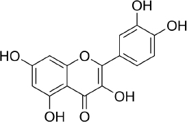
Quercetin (C15H10O7,Figure 2) is a flavonoid in fruits and vegetables. It has unique biological properties that can improve physical and mental health and reduce the risk of infection [90]. Quercetin has diverse biological activities, such as antioxidant [91,92], antidiabetic [93], cardioprotective [94], anti-obesity [95] antihypertension [96], anti-Alzheimer [97], antitumor [98] and anti-hyperpigmentation [99].

Figure 2. Quercetin
ADMET-12-2442-g002.jpg

The antioxidant properties of quercetin occur due to its ability to capture free radicals and bind transition metal ions [100]. The presence of the 3',4'-diphenolic group in quercetin allows this flavonoid to be effective in superoxide anion scavenging activity [101].

Research shows that quercetin will be produced in white blood cells and the liver shortly after consuming vegetables rich in quercetin [102], where quercetin is able to prevent damage to red blood cell membranes due to smoking [103]. Several studies have shown that quercetin has hepatoprotective activity in mice induced with a high-fat diet [104], reducing oxidative stress caused by hyperglycemia and diabetes by modulating carcinogenic signalling pathways [105].

Several studies have shown the antidiabetic mechanism of quercetin in vitro and in vivo through increasing pancreatic islet regeneration and possibly increasing insulin release in STZ diabetic rats [106], reducing oxidative stress and protecting pancreatic β-cells [107], improving liver and kidney function by restoring cell proliferation through inhibiting CDKN1A gene expression [108], increasing adiponectin secretion by PPAR-γ independent mechanism, and reducing fasting glucose and HbA1c through decreasing intestinal maltose activity [109]. In their research, Ahmet et al. showed that quercetin accelerated wound healing in diabetic and non-diabetic mice through the mechanism of reducing proinflammatory cytokines [110], inhibited the secretion of cytochrome P450 2E1 (CYP2E1) during the development of diabetes, thereby preventing oxidative damage in the liver [111] and also provided neuroprotective effects through the mechanism of preventing acetylcholinesterase (AChE) activity in the brains of diabetic mice [112]. In the case of anti-obesity, quercetin is reported to have the ability to stimulate hepatic mitochondrial oxidative metabolism through the induction of heme oxygenase-1 (HO-1) in the nuclear factor-related erythroid factor 2, Nrf-2 pathway [113]. In studies of cardioprotective activity, quercetin was reported to work in post-traumatic reversal reactions of cardiac dysfunction by reducing cardiomyocyte apoptosis, thereby suppressing the increase in tumour necrosis factor (TNF) alpha, reactive oxygen species (ROS) and Ca2+ production [114], as well as an alternative treatment for ischemia-reperfusion injury (IRI) by inducing blood vessel dilation through inhibition of endothelin-1 receptors, increased stimulation of nitric oxide (NO) and activation of calcium channels [115]. For antihypertensive activity, quercetin works to inhibit the activity of the cytochrome P450 (CYP) 4A enzyme and the soluble epoxide hydrolase (sHE) enzyme, the two main enzymes of arachidonic acid metabolism in the kidneys which regulate blood pressure [116]. In in silico research using molecular docking, quercetin was able to bind angiotensin-converting-enzyme (ACE), an enzyme responsible for regulating blood pressure with an optimal binding energy of −35.564 kJ/mol [117].

Having a neuroprotective activity, quercetin works to inhibit cholinesterase (ChE) activity, thereby restoring the balance of acetylcholinesterase (AChE) and butyrylcholinesterase (BChE) in brain tissue [118]. Studies in mouse models of Alzheimer's showed that quercetin reduced extracellular β-amyloidosis plaques, tauopathy, astrogliosis and microgliosis in the hippocampus and amygdala of mice, which are characteristic of Alzheimer's [119]. Quercetin induces apoptosis through activation of the mitochondrial pathway (caspase cascade) and by inhibiting signals in the human hepatoma cell line - HepG2 [120], disrupts the Akt/PKB pathway by inhibiting the proliferation process and induces apoptosis [121], increases TNF-related cytotoxicity apoptosis-inducing ligand (TRAIL-anticancer drug) by activating caspases and inhibiting Akt phosphorylation [122] and induces the apoptotic pathway in MCF-7 cells [123]. Structurally, the mechanism of action of quercetin is the inhibition of the DNA topoisomerase I and II enzymes, which play a role in cleavage in the proliferate phase, through substitution of the keto group C-4 and substitution of the hydroxyl groups of ring A and ring B at positions C-3, C-5, C-7, C3' and C4' [124].

Studies also reported the ability of quercetin to inhibit tyrosinase, an enzyme that plays a role in the synthesis of melanin [125]. Controlling tyrosine activity will lead to the treatment of hyperpigmentation disorders in mammals and enzymatic browning of fruits and fungi [126]. An in vitro study showed that quercetin could significantly inhibit both the monophenolase and diphenolase activity of tyrosinase and inhibited the formation of dopaquinone in a reversible competitive manner with an IC50 value of (30.8±7.4) 4mol/L [125] and (44.5±1.3) 3mol/L [127]. In silico studies through molecular docking suggested the inhibition activity of quercetin due to the catechol structure (3’,4’ dihydroxy groups in B ring) of quercetin that chelated copper in the active site of tyrosinase resulting in the blocking access of the substrate L-DOPA. The report showed the non-hydrogen bonding interaction with various amino acid residues, including Gly281, Ser282, Met280, His263, Phe292, Val283, His61, Ala286, His85, His259, Phe264, Asn260, Met257 and Val248 [127], while Fan et al. [128] reported this bonding occurs on Cys83 and His85. Another study by Park et al. [129] reported that quercetin-7--O-α-L-rhamnoside, a quercetin glycoside, inhibits tyrosinase activity and melanogenesis in α-MSH plus IBMX-stimulated B16F10 melanoma cells. Docking simulation revealed hydrogen bonding of this flavonoid with amino acid residues His85, His244, Thr261, and Gly281 of tyrosinase.

Despite its diverse biological activities, the use of quercetin is limited by its low level of solubility, which affects its bioavailability [130]. Several studies have been carried out to increase the bioavailability of this compound, including the combination of quercetin with insulin, which can increase bioavailability by 20 % [131], encapsulating quercetin with lecithin-chitosan nanoparticles [132], encapsulating with lipid nanoparticles [133], using rice bran protein as an emulsifier [134], co-crystallization with nicotinamide [135], and by attaching a sugar group to the 3-OH position of quercetin in order to increase the whitening effect [136].

Apigenin

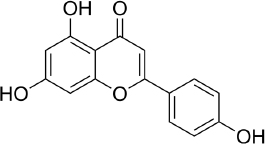
Apigenin (APG, 4′,5,7-trihydroxyflavone - C15H10O5,Figure 3) attracted attention for the first time in the 1960s when this compound was found to suppress the release of histamine from cells of white basophils and exhibited bronchial dilating effects on the lungs [137]. A number of studies show the ability of these flavones to inhibit and stop cell proliferation in several types of cancer, such as pancreatic, colon, liver, blood, lung, prostate, breast, thyroid, skin and neck [138-140]. In addition, apigenin is also reported to have antioxidant [141], and anti-inflammation [142].

Figure 3. Apigenin
ADMET-12-2442-g003.jpg

Currently, the development of research on apigenin therapy is being focused on its use to reduce chemotherapy resistance of various anticancer drugs by targeting several signalling pathways at the cell/molecular level [143]. Research on the effect of apigenin at the cellular level on prostate cancer using transformed human prostate epithelial cells and various prostate cancer cells (RWPE-1 cells and prostate cancer LNCaP, PC-3 and DU145 cells) in vivo and in vitro found that apigenin accumulates in the nuclear matrix and binds to DNA, thereby reducing oxidative DNA damage and apoptosis [144].

Another mechanism was also reported by Sukhla and Gupta [145], where apigenin accumulated in cells has the potential to interfere with androgen receptor signalling and inhibit androgen-responsive genes. In breast cancer research, apigenin was reported to be able to block the development of progestin-dependent BT-474 breast cancer cell (BCC) xenograft tumours in female mice [146]. In addition, apigenin is able to reduce cell proliferation through modulation of mitogen-activated protein kinase (MAPK), phosphoinositide 3-kinase (PI3K-Akt) [147,148]. This MAPK modulation is also key to the anti-inflammatory activity of apigenin [149], while Kang et al. [150] demonstrated apigenin's regulation of the production of TNF-α, IL-6, IL-8, and GM-CSF in HMC-1 cells. A molecular docking study shows the interaction between apigenin and some genes related to papillary thyroid carcinoma with the binding energy range from −31.4636 to −18.7025 kJ/mol [151].

Zhang et al. [152] reported the whitening effect of apigenin through in vitro testing using melanin production and tyrosinase activity assays. They propose that the higher rate of inhibitory activity is due to the presence of 7 and 4’ hydroxyl groups in this flavonoid. An in vitro test was also reported by Karaoglan et al.[153], where apigenin showed a tendency to inhibit tyrosinase activity by 49.36±0.24 %. Molecular docking simulations showed a hydrogen bond between the hydroxyl group of the benzopyran ring and the carbonyl group of Met280, as well as a hydrophobic interaction with residues of Val248, Phe264, Met280, Val283, Ala286, and Phe292. Additionally, polar interactions were observed with His61, Hid85, Ser282, His263, Asn260, and His259. However, an in vivo study report by Chauhan et al. [154] using a hydroquinone-induced vitiligo mouse model found that apigenin significantly prevented vitiligo by acting as an anti-inflammatory, increasing tyrosine, and reducing the expression of non-phosphorylated P38 mitogen-activated protein kinases (p38MAPK). The activation of the p38MAPK pathway, which resulted in an increase in melanogenesis in B16 cells, was also reported by Ye et al. [155], who suggested apigenin for hypopigmentation disorder treatment. On the other hand, the glycoside of apigenin, apigenin-6-C-glucoside, tends to suppress melanin synthesis via the down-regulation of intracellular tyrosinase signalling due to the presence of the hydroxyl group at the A and B rings [156].

Scutellarin

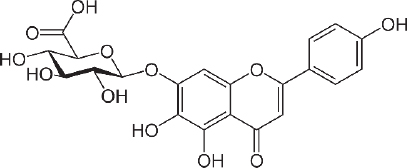
Scutellarin (7-O-β-D -glucuronide - C21H18O12 -Figure 4) was first investigated for drug development in the late 1970s when it was isolated from the Chinese herbal plant, Erigeron breviscapus [157]. This flavonoid is studied for the treatment of heart disease, stroke and diabetes complications because of its ability to relax blood vessels and its anti-inflammatory, antimicrobial, anticoagulation, antioxidant properties and myocardial protection [158,159]. The scutellarin relaxing effect was studied for preventing SARS CoV-2 using molecular docking by Chen and Du [160] and found that scutellarin was able to interact with angiotensin-converting enzyme 2 (ACE2), the host receptor of SARS CoV-2.

Figure 4. Scutellarin
ADMET-12-2442-g004.jpg

The binding energy to ACE2 is estimated at --62.3415 kJ/mol, with binding sites at Glu495, Unk957, and Arg482. For the treatment of stroke, scutellarin was reported to have therapeutic effects on cerebral ischemia by activating the astrocytic Janus kinase 2/signal transducer and activator of transcription 3 (JAK2/STAT3) signalling pathway [161], and by reducing the infarct cerebral tissue area in middle cerebral artery occlusion (MCAO) rats [162].

Luo et al. [163] reported that scutellarin, which has anti-inflammatory activity, can provide protection against hyperglycaemia induced by vascular inflammation. Furthermore, Long et al. [164], who conducted research on mice, reported that scutellarin was able to inhibit damage to apoptotic cells and disruption of mouse testes morphology due to hyperglycaemia. Su et al. [165] also reported the ability of scutellarin to inhibit protein kinase translocation by in vivo and in vitro studies, making it possible to treat complications due to diabetes. Wang et al. [166] reported the protective effect of scutellarin on intervertebral disc degeneration (IVDD) by reducing the amount of ROS, alleviating mitochondrial damage, and decreasing the expression levels of apoptosis-related biomarkers.

The antitumor activity of scutellarin was reported through inhibiting proliferation and inducing apoptosis of HepG2 cells in liver cancer [167], attenuating the development of fibrosarcoma and inhibiting cancer cell metastasis [168], and inhibiting the invasive potential of melanoma cell lines by suppressing the EMT and angiogenesis through the PI3K/Akt/mTOR signalling pathway [169]. Scutellarin was also reported to significantly reduce multiple myeloma xenograft tumour burden in nude mice [170]. Research on white mice reported the protective effect of scutellarin against acute cardiac toxicity due to the use of the drug doxorubicin, one of the most frequently used cancer drugs [171]. Besides that, scutellarin was also able to protect against the disruption of blood flow to the heart [172].

Scutellarin also reports the capability to inhibit cellular tyrosinase enzymes, leading to decreased melanin production with no cytotoxicity effect [173]. Using in vitro and computational simulation, Chen et al. [174] reported scutellarin inhibited tyrosinase activity in a competitive manner with an IC50 of 91 μM and predicted that scutellarin was mainly bound with tyrosinase via Arg268 residue.

Hispidulin

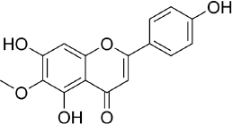
Hispidulin (4',5,7-trihydroxy-6-methoxyflavone, C16H12O6,Figure 5) is one of the flavonoids that is an active ingredient in Chinese medicine and is reported to have anticancer, anti-inflammatory, and antioxidant activities [175]. The anticancer activity of hispidulin is due to its ability to inhibit the proliferation and metastasis of hepatocellular carcinoma (HCC) cells by activating PPAR-γ [176], suppressing allergic inflammatory reactions by reducing the release of histamine and inflammatory cytokines such as TNF-α and interleukin-4 [177]. Hispidulin is also reported to activate AMP-activated protein kinase (AMPK), thereby suppressing eukaryotic initiation factor 4E-binding protein (4E-BP1) via the rapamycin (mTOR) pathway in glioblastoma multiforme (GBM) cells, one of the most common and deadly types of brain cancer [178]. The same mechanism was also reported in relation to ovarian cancer, where activation of AMPK increased the sensitivity of TNF-related apoptosis-inducing ligand (TRAIL), thereby reducing the protein expression of MCL-1, a group of antiapoptotic proteins [179]. In pancreatic cancer, the target of hispidulin is the vascular endothelial growth factor (VEGF) receptor 2 mediated PI3K/Akt/mTOR signalling pathway in endothelial cells, thereby suppressing pancreatic tumor cell growth and angiogenesis [180]. In studies of hepatoblastoma cancer cells hispidulin was reported to induce apoptosis through mitochondrial dysfunction and inhibition of the PI3K/Akt signalling pathway [181].

Figure 5. Hispidulin
ADMET-12-2442-g005.jpg

The anti-inflammatory effect of hispidulin in neuroinflammation was reported by its capability to increase dopamine levels in the prefrontal cortex of phencyclidine-treated mice and reverse social withdrawal in schizophrenia-1 mutant mice [182].

In an in vivo study using epilepsy-gerbil models, hispidulin was shown to reduce seizure suffering with the same effect as diazepam, a drug for anxiety disorders [183]. Hispidulin was also reported to relieve intractable motor tic disorder in a mouse model for hyperdopaminergic states, a condition found in patients with schizophrenia and obsessive-compulsive disorder [184]. The anti-seizure effect of hispidulin was reported to be achieved through suppressing the inflammatory process and activating the mitogen-activated protein kinases A in kainic acid-induced rats [185].

The antidiabetic activity of hispidulin was reported by stimulating glucagon-like peptide-1 secretion and suppressing hepatic glucose production [186]. Hispidulin is also reported to have potential as a therapy in diabetic retinopathy because of its ability to improve high glucose-induced proliferation by reducing the expression of protein kinase, phosphorylated extracellular regulated kinase and VEGF-A, and inhibiting mRNA levels of TNF-α [187]. Molecular docking in this study shows that hispidulin has the highest affinity with VEGF-A and the second highest with TNF-α compared to other modelled compounds. Hispidulin forms hydrogen bonds with Cys5, Asp6 and Glu12 in VEGF-A and Tyr227 and Tyr195 on TNF-α.

Salvigenin

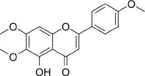
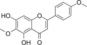
Salvigenin (5-hydroxy-6,7-bis(trideuteriomethoxy)-2-[4-(trideuteriomethoxy) phenyl] chromen-4-one, C18H16O6,Figure 6) is a derivative of apigenin with different biological activities and reported antioxidant, anticancer and antidiabetic activities [188]. In vitro studies using human neuroblastoma SH-SY5Y cells showed that salvigenin protected cells from H2O2-induced oxidative stress [189], while in vivo study using atrazine-induced rats showed that salvigenin protected the liver tissues via regulating antioxidant, anti-inflammatory and anti-apoptotic [190]. Uydes-Dogan et al. [191] reported a vasorelaxation effect on rat aortic rings related to antioxidant activity, thus enabling the use of this flavonoid in cardiovascular disease treatment.

Figure 6. Salvigenin
ADMET-12-2442-g006.jpg

Research conducted by Noori et al. [192] in female mice showed the antitumor activity of salvigenin through modulation of cytokine production, where there was a decrease in the production of interleukin 4 and an increase in the production of interferon γ. In liver cancer, salvigenin was reported to have the ability to reduce the proliferation, migration and invasion of hepatocellular HCC cells and suppress cell glycosides and chemoresistance by modulating the PI3K/AKT/GSK-3β pathway [193]. Anticancer activity was also reported in relation to oral squamous cell carcinoma, where molecular docking studies showed the binding energy of salvigenin with AKT1 was-33.0536 kJ/mol on Gly294 and Lys179 [194]. The combination of salvigenin with doxorubicin (DOXO), a treatment for chemotherapy patients, was reported to reduce DOXO toxicity through the mechanism of increasing Bax/Bcl-2 ratio, caspase-3 expression and PARP cleavage [195].

Salvigenin was reported to increase insulin secretion and reduce HbA1c, and at the same time was able to influence the lipid profile by reducing triglycerides, total cholesterol, and HDL in diabetic rats [196]. An in vitro test using hepatic HuH7 cells shows the capability of salvigenin to inhibit lipogenesis and stimulate mitochondrial functionality [197]. Molecular docking simulation showed that the antidiabetic mechanism of salvigenin is by forming two hydrogen bonds at Lys169 and Glu443 with α-glucokinase [198].

Acacetin

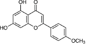
Acacetin (5,7-Dihydroxy-4'-methoxyflavone, C16H12O5,Figure 7) is an apigenin derivative but has different activities from apigenin. Several studies have reported the antioxidant, anti-inflammatory, and anti-cancer activities of this flavonoid [199]. A review by Semwal et al. [200]found that there was commercial health supplements based on acacetin with more than 1000 patents related to acacetin as appetite suppression, treatment for prostate cancer, anti-allergy and anti-inflammatory activities. A test on type 2 diabetic mice models shows that acacetin could improve blood glucose and lipid metabolism and liver and kidney dysfunction, where this potential was related to the antioxidant and anti-inflammatory activity of this flavonoid [201]. Using RINm5F cells, Wang et al. [202] reported the mechanism of acacetin against lipo-toxicity in pancreatic β-cells. This mechanism involves reducing oxidative stress by scavenging intracellular ROS, upregulating endogenous antioxidant enzymes, diminishing sub-G1 DNA fraction in cells exposed to free fatty acid (FFA), and decreasing endoplasmic reticulum stress by mitigating the overload of intracellular Ca2+ and reducing pro-apoptotic protein expression in FFA - stimulated cells. Han et al. [203] reported that acacetin has the capability to attenuate diabetes-accelerated atherosclerosis by protecting vascular endothelial cells from injury induced by hyperglycaemia. This protection is achieved by preserving mitochondrial function through Sirt1-mediated activation of Sirt3/AMPK/PGC-1α signalling molecules.

Figure 7. Acacetin
ADMET-12-2442-g007.jpg

Research related to the anti-inflammatory activity of acacetin has reported the anti-neuroinflammatory effects in Parkinson's disease mouse models. Acacetin was found to protect dopaminergic cells and inhibit the production of inflammatory trigger factors such as nitric oxide, prostaglandin E2, and TNF - α [204]. In the context of dental inflammation, acacetin has shown the ability to suppress inflammation by regulating autophagy and glycogen synthase kinase 3β (GSK-3β) signalling in human periodontal ligament cells [205].

The anticancer activity of acacetin is reported by its ability to inhibit cell growth and induce apoptosis in gastric carcinoma cells [206], inhibit the migration of MDA-MB-231 and T47D cells in breast cancer [207], inhibit the activity of signal transducer and activator of transcription 3 (STAT3) in prostate cancer [208], inhibit the invasion and migration of A549 cells in lung cancer through inhibiting the phosphorylation of Jun N-terminal kinase 1 and 2 (JNK1/2), reduction of activator protein-1 (AP-1) and nuclear factor kappa B - NF-κB [209]. Zhang et al. [210] reported that acacetin can induce cell cycle arrest in the G2/M phase, apoptosis, and autophagy in breast cancer cells, resulting in downregulation of PI3Kγ-p110 and the disruption of the PI3K/AKT-mammalian target signalling pathway of rapamycin (mTOR). Molecular docking in this study shows that acacetin forms hydrogen bonds with PI3Kγ via Ser806, Ala885, and Val882, and hydrophobic interactions with Lys833 and Asp964. The PI3K/AKT/mTOR pathway has become a “hot spot” of molecular biomarker-based/targeted therapy because research in several types of cancer, such as breast, liver, colorectal, prostate, and gastric cancers, shows the presence of irregularities in this pathway [211].

Pectolinaringenin

Pectolinaringenin (5,7-dihydroxy-4′,6-dimethoxy-flavone, C17H14O6,Figure 8) was first isolated from the Linaria vulgaris plant more than 100 years ago and has since been isolated as the main component in herbal plants in various countries [212]. Pectolinarigenin has been reported to have various biological activities, such as antioxidant, anticancer, anti-inflammatory, antidiabetic and treatment for various brain-related illnesses [213].

Figure 8. Pectolinaringenin
ADMET-12-2442-g008.jpg

Shiraiwa et al. [214] reported the antioxidant activity of pectolinarigenin through in vitro inhibition tests using the 2,20-Azobis (2-amidinopropane) dihydrochloride (AAPH) assay. Furthermore, using HepG2 cancer cells, the study reported that pectolinarigenin-induced antioxidant enzymes, heme oxygenase-1, NAD(P)H:quinone oxidoreductase 1, and aldo-keto reductase family 1 member B10. The induction mechanism for these enzymes is through nuclear accumulation of Nrf2 which increases transcriptional activity mediated by the antioxidant response element (ARE) and suppresses Nrf2 degradation through modification of Kelch-like ECH-associated protein 1 (Keap-1). Research by Pang et al. [215] using SH-SY5Y neuronal cells showed that pectolinarin, a glycoside of pectolinarigenin can scavenge hydroxyl and nitric oxide radicals, increase cell viability, reduce ROS production and lactate dehydrogenase release (LDH). Therefore, it is promoted to be applied for the treatment of oxidative stress-related neurodegenerative diseases.

The activity of pectolinaringen in inhibiting several types of cancer, including liver cancer, has been reported. Research using HCC cells, SMMC7721 and PLC5, has shown that this flavonoid can suppress the proliferation of HCC cells by inducing cell apoptosis and cell cycle arrest. It also reduces migration and invasion of HCC cells and deactivates the PI3K/AKT/mTOR/ ERK signalling pathway [216]. Research on nasopharyngeal cancer using C666-1 cells have shown that pectolinaringen can induce apoptosis in C666-1 cells through the mitochondrial-related apoptotic pathway and ROS-induced apoptotic pathway [217]. The western blot test in this study showed an increase in cleavage caspase 3 and 9 levels, indicating that the caspase inhibitor (z-VADfmk) significantly prevented the increase of apoptotic cells. Zhou et al. [218] reported the potential of pectolinaringen as a treatment for pancreatic cancer using human pancreatic cancer cells (Patu 8988 and BxPC-3). Pectolinaringen induces apoptosis and reduces the phosphorylation of signal transducer and activator of transcription 3 (STAT-3). In breast cancer, in vitro assays of MCF-7 cancer cells demonstrated antiproliferative activity of pectolinaringen by inducing apoptosis and downregulation of B-cell lymphoma 2 (Bcl2) expression [219]. Furthermore, research on gastric cancer cells using AGS and MKN28 cells showed the ability of pectolinaringen to inhibit the viability of human gastric cancer cells via the AKT/PI3K/mTOR pathway [220]. This inhibitory mechanism begins with the stimulation of intracellular protein ubiquitination and proteasome degradation of proteins, including caspase-3/-7 and AKT. This is followed by Beclin-1-independent autophagy and subsequent caspase-dependent apoptosis, leading to gastric cancer cells' death.

The anti-melanogenic activity of pectolinarigenin was reported through in vitro tests using melan-A cells, where pectolinarigenin was able to inhibit melanogenesis by inhibiting the protein expression of microphthalmia-associated transcription factor (MITF), tyrosinase, tyrosinase-related protein (TRP)-1, and TRP-2, which play a role in synthesis thereby reducing melanin synthesis [221]. In line with its anti-melanogenic ability, Deng et al. [222] conducted in vitro research using A375 and CHL-1 cells and found that pectolinarigenin was able to inhibit cell viability, proliferation, invasion and migration and induce apoptosis through the apoptotic ROS-mitochondrial pathway.

Hesperetin and hesperidin

Hesperetin (3',5,7-Trihydroxy-4'-methoxyflavanone, C16H14O6,Figure 9a) together with its glycoside, hesperidin ((2S)-3',5-Dihydroxy-4′-methoxy-7- [α-L- rhamnopyranosyl- (1→6)-β-D-glucopyranosyloxy] flavan-4-one, C28H34O15,Figure 9b) has diverse biological activities, such as antioxidant, anti-inflammatory, neuroprotective, anticancer, cardiovascular protection and antidiabetes [223-225]. The antioxidant activity of hesperetin and hesperidin occurs through direct radical scavenging and augmenting cellular antioxidant defense [226]. The direct radical scavenging pathway plays an important role in protecting the body's DNA, proteins and other tissues. Hesperetin and hesperidin are reported to protect tissue damage from exposure to toxic compounds, such as hydrogen peroxide, 1,2-dimethylhydrazine, benzo(a)pyrene and peroxynitrite [227,228]. In the second pathway, hesperidin is reported to play a role in increasing cellular defence through the ability to upregulate the protein levels of nuclear factor erythroid 2-related factor 2 (Nrf-2) [229]and induction of heme oxidase-1 through extracellular signal-regulated protein kinase [ERK)/Nrf2 signalling [230].

Figure 9. Hesperetin (a), hesperidin (b)
ADMET-12-2442-g009.jpg

In connection with this antioxidant ability, studies report the inhibitory effect of hesperidin and hespertin on the formation of advanced glycation end products (AGEs), the end products of the Maillard reaction [231]. AGEs cause damage to extracellular proteins, thereby contributing to diabetes complications such as cataracts, nephropathy, vasculopathy, proliferative retinopathy, and atherosclerosis [232]. The study conducted by Shi et al. [233] on streptozotocin-induced diabetic rats showed that hesperidin suppressed blood retina breakdown and increased retinal thickness, reduced blood glucose, aldose reductase activity, retinal levels of TNF-α, ICAM-1, VEGF, IL-1β, and AGEs, as well as reduced the level of plasma malondialdehyde (MDA) and increased SOD activity. Using computational molecular docking, Gong et al. [234] reported the α-glucosidase inhibition activity of hesperetin, where two hesperetin rings interact with several residues near the active site of the enzyme, such as Lys155, Asn241, Glu304, Pro309, Phe311, and Arg312. An in vivo study by Akiyama et al. [235]also reported the capability of hesperidin glycosides to reduce blood glucose levels in diabetic rats by altering the activities of glucose-regulating enzymes and lowering the serum and liver lipid levels.

The ability of hesperetin in cancer treatment has been reported in several studies. Yang et al. [236] reported the ability of hesperetin as an inhibitor of the transforming growth factor β (TGF-β) signaling pathway, where this inhibition will block the metastatic properties of cancer cells. Through testing on male Wistar rats induced by 1,2-dimethylhydrazine (DMH) as an agent of colon carcinogenesis, Arangathan and Nalini [237] reported that hesperetin was able to reduce tumour multiplicity, tumour incidence and burden of DMH-induced colorectal tumorigenesis. Histological observations showed that the administration of hesperetin in experimental conditions affected colon carcinogenesis at every stage. The group that was given hesperetin continuously showed significant changes compared to the group that was only given hesperetin at the initiation and post-initiation stages. Using the same DMH induction method in male Wistar rats, Nalini et al. [238] reported the efficacy of hesperetin as a chemo-preventive agent. It was found to inhibit cell proliferation markers, angiogenic growth factors (VEGF, EGF, bFGF), COX-2 mRNA expression and induce apoptosis. In order to find out the effect of hesperetin on colon cancer apoptosis, the HT-29 human colon adenocarcinoma cell line was used, where hesperetin was able to inhibit the proliferation of HT-29 cells by inducing apoptosis through the Bax-dependent mitochondrial pathway, which involves an oxidant/antioxidant imbalance [239].

In breast cancer research, in vitro tests using MCF-7 cells showed that hesperetin inhibited cell proliferation, induced cell cycle arrest at the G1 phase, and induced apoptosis [240]. This study also reports the regulation of cyclin-dependent kinases-4 (CDK-4) and p21Cip1, which may participate in the inhibitory mechanism. In vivo study reported by Ye et al. [241] in a mouse model that was injected with MCF-7aro cells, where hesperetin inhibited the activity of the aromatase enzyme (estrogen synthetase) and suppressed the proliferation of the MCF-7 breast cancer cell line. Aromatase is a key enzyme in the conversion of androgens to estrogen, and exposure to both endogenous and exogenous estrogens has been linked to the initiation and promotion of hormone-dependent diseases such as breast cancer [242]. Measurements of mouse blood plasma showed a decrease in plasma estrogen and messenger RNA expression of the estrogen-responsive gene pS2 in mouse models [241]. This research shows that the mechanism of hesperetin in preventing cell growth is through down-regulating the expression of cyclin D1, CDK4 and Bcl-xL and up-regulating p57Kip2 expression.

The skin protective activity of hesperetin tested using murine B16-F10 melanoma cells shows the ability of hesperetin to stimulate melanogenesis through the activation of MAPK, phosphorylation of cAMP-responsive element binding protein (CREB), and glycogen synthase kinase-3b [243]. Using inhibition kinetics and computational simulation, Si et al. [244] reported that hesperetin inhibited tyrosinase in a competitive manner and predicted that putative hesperetin-binding residues include Met280, His61, His85, and His259. Another report by Hong et al. [245] using multivariate curve resolution-alternate least squares (MCR-ALS) analysis suggested that tyrosinase interacted with hesperetin and formed a tyrosinase-hesperetin complex. Molecular docking showed that hesperetin entered the hydrophobic cavity of tyrosinase PPO and bound near the dinuclear copper active centre. It interacted with Val283, Phe264, His85, Asn260, Val248, and His263 via hydrophobic interactions, formed hydrogen bonds with Met280, His89, and His259 residues, and also interacted with Phe292, His61, Phe90, Glu256, His244, Asn260, Phe264, and Gly281 via van der Waals forces.

In research related to SARS-CoV-2, hesperidin and hesperetin were reported to be able to prevent the binding of the virus to angiotensin-converting enzyme 2 (ACE2) in host cells, inhibit viral replication after penetration into host cells, and prevent and counteract excessive proinflammatory reactions from the immune system [246]. Molecular docking studies on 3 SARS-CoV-2 target proteins, namely SARS-CoV-2 Mpro, SARS-CoV-2 PLpro and SARS-CoV-2 spike glycoprotein, show that the ligand binding affinity score for hesperidin is 24.27, -41.84 and -33.89 kJ/mol, respectively [247]. Another molecular docking carried out on 24 SARS-CoV-2 proteins reported that hesperidin exhibited high binding affinity to 2 target proteins for preventing the virus RNA synthesis and replication (3CLpro and Helicase) and was the only natural compound in the simulation that could target the binding interface between spike and ACE2 through the formation of a hydrogen bond at Tyr440 [248].

Luteolin

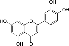
Luteolin (3,4,5,7-tetrahydroxy flavone, C15H10O6,Figure 10) is a natural flavonoid widely isolated from traditional Chinese medicine plants used for treating hypertension, inflammatory disorders, and cancer [249]. Luteolin is reported to have various biological activities such as antioxidant, antidiabetic, neuroprotective, antiallergy, and anticancer [250-252].

Figure 10. Luteolin
ADMET-12-2442-g010.jpg

Luteolin's antioxidant activity is due to the presence of the 1,4-pyrone moiety group [253] and its ability to donate H atoms with low energy [254]. In in vivo study using rat hepatocytes cells induced by tert-butyl hydroperoxide (tBHP), the antioxidant mechanism of luteolin is through up-regulating antioxidant enzyme gene transcription through up-regulating protein heme oxygenase-1, glutamate cysteine ligase, and glutamate-cysteine expression ligase modifier subunit via the extracellular signal-regulated protein kinase 2/nuclear factor erythroid 2-related factor 2 (ERK2/Nrf2) pathway [255].

Antidiabetic activity of luteolin was reported through in vitro inhibition tests of the α-glucosidase and α-amylase enzymes [256]. Molecular docking of the α-glucosidase enzyme shows a hydrogen bond between the hydroxyl group of luteolin and Asp285 on H27 and H33 [257], while research by Rekha et al. [198] shows a bond energy of -30.9616 kJ/mol with the glucokinase receptor on Ser151, Asp20 and Thr228.

In neuroprotective research, the administration of luteolin improved the learning and memory abilities of Alzheimer's disease model mice. This was followed by the inhibition of neuroinflammation and a decrease in the expression of endoplasmic reticulum stress markers in brain tissue [258]. Research conducted on subarachnoid haemorrhage model mice demonstrated that luteolin was able to repair oxidative damage by increasing the expression of nuclear factor-erythroid 2-related factor 2 (Nrf2) and downregulation of the activation of inflammatory nod-like receptor pyrin domain-containing 3 (NLRP3) [259]. In cases of intracerebral haemorrhage caused by brain injury, luteolin was reported to prevent the activation and infiltration of microglia and reduce the release of proinflammatory factors (IL-6, IL-1β, TNF-α). Additionally, it inhibited the activation of the TLR4/TRAF6/NF-κB signalling pathway [260].

Schomber et al. [261] reported an in vivo study of luteolin's ability to inhibit the growth of xenografted melanoma tumours in a mouse model induced by tumour cells BRAF-mt A375 and BRAF-wt WM3211 cells. Tumour cells shrank after administration of luteolin, while in vitro studies showed luteolin plays a role in the interaction pathway between cells (extracellular matrix), oncogenic pathway, and immune response signalling pathway. In relation to the melanogenesis pathway, an in silico study reported the capability of luteolin to inhibit the tyrosinase enzyme by establishing a binding between four hydrogen bonds to Tyr65, Lys79, Cys83, and Glu322 [128]. Molecular docking simulation also shows that the hydroxyl groups of the B ring of luteolin will bind to Asn81 and Cys83, and the HPLC and UPLC-MS analyses explained that luteolin acted as a substrate or a suicide inhibitor [262].

Using human hepatoma cell lines, HepG2, HLF, and HAK-1B, as well as the human neuroblastoma cell line IMR-32, Selvendiran et al. [263] reported the protective ability of luteolin against cancer through the mechanism of increasing CD95 (cluster of differentiation 95) expression in neoplastic cells in vivo and in vitro, promoting phosphorylation of signal transducer and activator of transcription 3 (STAT3) through a ubiquitination-dependent process; and was able to significantly inhibit the growth of human HCC xenografts in nude mice. In esophageal cancer research using human ESCC cell lines (EC1 and KYSE450 ESCC), it was shown that luteolin was able to induce apoptosis and caspase-3 activation as well as induce cell cycle arrest at the G2/M phase, where in vivo tests on model mice injected with EC1 showed a decrease in mass tumours in the luteolin treatment group [264].

Nepetin

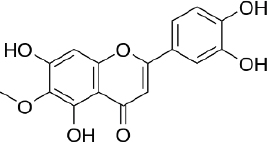
Nepetin (Eupafolin, 6- methoxy-5,7,3′,4′-tetrahydroxy-flavone, C16H12O7,Figure 11) is a flavonoid that was isolated for the first time from Eupatorium perfoliatum L., which is widely used in traditional Chinese medicines and Indian tribes [265]. Research shows nepetin has anti-inflammatory, antioxidant and antitumor properties. The anti-inflammatory mechanism of nepetin is to reduce the release of inflammatory mediators (iNOS, COX-2, and NO) and proinflammatory cytokines (IL-6 and TNF-α) in RAW264.7 macrophages induced by lipopolysaccharide [266]. The research also reported inhibition of phosphorylation of p38 MAPK, ERK1/2, JNK, AKT, as well as p65 nuclear translocation of p65 and c-fos.

Figure 11. Nepetin
ADMET-12-2442-g011.jpg

The antitumor activities of nepetin have been investigated and have revealed significant inhibitory potential on VEGF - induced cell proliferation [267]. Liu et al. [268] reported that nepetin inhibited the proliferation of prostate cancer cells by binding to PI3-Akt and attenuating its kinase activity. In a cervical cancer study using human cervical adenocarcinoma cells, nepetin was found to induce apoptosis mediated by caspase-dependent pathways involving caspases-3, -9, and -8, which are initiated by the B-cell Lymphoma-2 -dependent loss of mitochondrial membrane potential (ΔΨm) [269]. Chen and Cheng [270] reported the antitumor activity of nepetin in the human non-Hodgkin lymphoma cell line, OCI-LY-3, through inducing cell apoptosis. The study also revealed the ability of nepetin to suppress the Akt/mTOR signalling pathway and promote autophagy.

Xu et al. [271]reported the capability of nepetin to inhibit the aggregation of human islet amyloid polypeptide (hIAPP). The research shows the average number of hydrogen bonds between hIAPP22-28 octamer and nepetin increased from 28 to 71, with possible binding sites occurs in Asn22 and Ser28. The hIAPP is the peptide produced by pancreatic β cells in the islet of Langerhans, and the increase of this peptide is associated with type 2 diabetes mellitus [272]. The antidiabetic activity of nepetin was also reported through the inhibition of α-amylase, which was reported to be strong compared to other flavonoids such as scutellarin, apigenin and hispidulin, due to the adjacent position of the dihydroxyl group on the B-ring [273]. The molecular docking simulation of this study shows that nepetin formed four hydrogen bonds with Gln63 and Asp197.

The protective effect of nepetin on the skin was reported by Huey-Ko et al. [274], where nepetin was able to decrease cellular melanin content and tyrosinase activity on B16F10 melanoma cells. Inhibition of melanin production occurs through the reduction of phospho-cAMP response element-binding protein and microphthalmia-associated transcription factor (MITF), downregulation of tyrosinase synthesis and TRP expression, and induction of phosphorylation of ERK1/2 and p38 MAPK.

Conclusions

The isolation and identification of flavonoids from the Clerodendrum genus have revealed a variety of compounds using various methods. Several factors determine the flavonoid content in Clerodendrum genus, such as the plant material part, the extraction techniques, and the solvent combination. Isolated flavonoids also show significant biological activity, highlighting the antioxidant, anti-inflammatory agents, antimicrobials, antidiabetic, anticancer, anti-tyrosinase, and neuroprotective agents.

Challenges and perspective

Flavonoids isolated from the Clerodendrum genus hold potential benefits for promoting health, but there are several challenges to pursuing their full potential. Future research should focus on optimizing the extraction and isolation techniques, establishing structure-activity relationships, and synthesizing derivatives to enhance the bioavailability and bioactivity of the flavonoids. As research continues to develop the phytochemistry and bioactivities of the Clerodendrum genus, there are also opportunities to translate these findings into practical applications, such as the development of functional foods, nutraceuticals, pharmaceuticals, and cosmeceuticals.
Acknowledgements

The authors would like to express profound appreciation to all members of the Research Center for Molecular Biotechnology and Bioinformatics - Universitas Padjadjaran for their generous support and for fully funding support by Universitas Padjajaran through Hibah Review Article Number 2338/UN6.3.1/PT.00/2024.

Notes

[1] Conflicts of interest Declaration of interest statement: The authors declared no potential conflicts of interest with respect to the research, authorship, and/or publication of this article.

[2] Funding: This work was supported by Hibah Review Artikel Universitas Padjadjaran Number 2338/UN6.3.1/ /PT.00/2024

References

[1] 

Erukainure O.L.; Oke O. V.; Ajiboye A.J.; Okafor O.Y.. Nutritional qualities and phytochemical constituents of Clerodendrum volubile, a tropical non-conventional vegetable. International Food Research Journal 18 (2011) 1393-1399. http://www.ifrj.upm.edu.my/18%20(04)%202011/(27)IFRJ-2011-009.pdf

[2] 

Shrivastava N.; Patel T.. Clerodendrum and Heathcare: An Overview. Medicinal and Aromatic Plant Science and Biotechnology ©2007 Global Science Books 1 (2007) 142-150. http://www.globalsciencebooks.info/Online/GSBOnline/images/0706/MAPSB_1(1)/MAPSB_1(1)142-150o.pdf

[3] 

Kar P.; Goyal A.K.; Das A.P.; Sen A.. Antioxidant and pharmaceutical potential of Clerodendrum L.: An overview. International Journal of Green Pharmacy 8 (2014). 210-216. https://www.greenpharmacy.info/index.php/ijgp/article/view/415

[4] 

Yadav A.; Kuamr V.P.; Chand T.; Bora R.H.. Ethno-medicinal knowledge of Clerodendrum L. among different tribes of Nambor reserve forest, Assam, India. Journal of Pharmacognosy and Phytochemistry 7 (2018). 1567-1570. https://www.phytojournal.com/archives/2018/vol7issue5/PartAA/7-5-235-900.pdf

[5] 

Rueda R.M.. The Genus Clerodendrum (Verbenaceae) in Mesoamerica. Annals of the Missouri Botanical Garden 80 (1993) 870-890. https://doi.org/10.2307/2399934 https://doi.org/10.2307/2399934

[6] 

Hamilton V.. Mabberley’s Plant-Book: A Portable Dictionary of Plants, their Classification and Uses (4th edition). Reference Reviews 32 (2018) 28-29. https://doi.org/10.1108/RR-12-2017-0259 https://doi.org/10.1108/RR-12-2017-0259

[7] 

Leeratiwong C.; Chantaranothai P.; Paton A.J.. A Synopsis of the Genus Clerodendrum L. (Lamiaceae ) in Thailand. Tropical Natural History 11 (2011) 177-211. https://www.thaiscience.info/journals/Article/TNAH/10801631.pdf

[8] 

Guang-Wei L.; Katsuyuki M.; Tokihito Y.; Kenjiro Y.. Effects of extract from Clerodendron trichotomum on blood pressure and renal function in rats and dogs. Journal of Ethnopharmacology 42 (1994) 77-82. https://doi.org/10.1016/0378-8741(94)90100-7 https://doi.org/10.1016/0378-8741(94)90100-7

[9] 

Patel J.J.; Acharya S.R.; Acharya N.S.. Clerodendrum serratum (L.) Moon. - A review on traditional uses, phytochemistry and pharmacological activities. Journal of Ethnopharmacology 154 (2014) 268-285. https://doi.org/10.1016/j.jep.2014.03.071 https://doi.org/10.1016/j.jep.2014.03.071

[10] 

Solapure P.; Dr. Pradeep ; Mundugaru R.; Hegde P.L.. Comparative anti-inflammatory activity of Clerodendrum serratum (Linn) Moon and Solanum xanthocarpum Schrad and Wendl in wistar ablino rats. The Journal of Phytopharmacology 5 (2016) 38-44. http://dx.doi.org/10.31254/phyto.2016.5201 https://doi.org/10.31254/phyto.2016.5201

[11] 

Rabiul H.; Subhasish M.; Sinha S.; Roy M.G.; Sinha D.; Gupta S.. Hepatoprotective activity of clerodendron inerme against paracetamol induced hepatic injury in rats for pharmaceutical product. International Journal of Drug Development and Research 3 (2011) 118-126. https://www.scholarscentral.com/pdfs/109115/hepatoprotective-activity-of-clerodendron-inerme-againstparacetamol-induced-hepatic-injury-in-rats-for-pharmaceutical-product.pdf

[12] 

Payum T.. Phytoconstituents and proximate composition of clerodendrum colebrookianum walp.: A widely used anti high blood pressure medicinal food plant in eastern himalayas. Pharmacognosy Journal 12 (2020) 1534-1540. http://dx.doi.org/10.5530/pj.2020.12.210 https://doi.org/10.5530/pj.2020.12.210

[13] 

Kar M.K.; Swain T.R.; Mishra S.K.. Antidiabetic activity of clerodendrum philippinum schauer leaves in streptozotocin induced diabetic rats. International Journal of Pharmacy and Pharmaceutical Sciences 7 (2015) 386-389. https://journals.innovareacademics.in/index.php/ijpps/article/view/7428

[14] 

Kuźma Ł.; Gomulski J.. Biologically Active Diterpenoids in the Clerodendrum Genus—A Review. International Journal of Molecular Sciences 23 (2022) 11001. https://doi.org/10.3390/ijms231911001 https://doi.org/10.3390/ijms231911001

[15] 

Wang J.H.; Luan F.; He X.D.; Wang Y.; Li M.X.. Traditional uses and pharmacological properties of Clerodendrum phytochemicals. Journal of Traditional and Complementary Medicine 8 (2018) 24-38. https://doi.org/10.1016/j.jtcme.2017.04.001 https://doi.org/10.1016/j.jtcme.2017.04.001

[16] 

Santos E.L.; Maia B.H.L.N.S.; Ferriani A.P.; Teixeira S.D.. Flavonoids: Classification, Biosynthesis and Chemical Ecology. in: Flavonoids - From Biosynthesis to Human Health, IntechOpen Limited, London, UK, 2017, pp. 1-16. http://dx.doi.org/10.5772/67861 https://doi.org/10.5772/67861

[17] 

Havsteen B.H.. The biochemistry and medical significance of the flavonoids. Pharmacology and Therapeutics 96 (2002) 67-202. https://doi.org/10.1016/s0163-7258(02)00298-x https://doi.org/10.1016/s0163-7258(02)00298-x

[18] 

Dixon R.A.; Pasinetti G.M.. Flavonoids and isoflavonoids: From plant biology to agriculture and neuroscience. Plant Physiology 154 (2010) 453-457. https://doi.org/10.1104/pp.110.161430 https://doi.org/10.1104/pp.110.161430

[19] 

Dias M.C.; Pinto D.C.G.A.; Silva A.M.S.. Plant flavonoids: Chemical characteristics and biological activity. Molecules 26 (2021) 5377. https://doi.org/10.3390/molecules26175377 https://doi.org/10.3390/molecules26175377

[20] 

Feng W.; Hao Z.; Li M.. Isolation and Structure Identification of Flavonoids. in: Flavonoids - From Biosynthesis to Human Health, IntechOpen Limited, London, UK, pp. 17-43. http://dx.doi.org/10.5772/67810 https://doi.org/10.5772/67810

[21] 

Chen S.; Wang X.; Cheng Y.; Gao H.; Chen X.. A Review of Classification, Biosynthesis, Biological Activities and Potential Applications of Flavonoids. Molecules 28 (2023) 4982. https://doi.org/10.3390/molecules28134982 https://doi.org/10.3390/molecules28134982

[22] 

Panche A.N.; Diwan A.D.; Chandra S.R.. Flavonoids: An overview. Journal of Nutritional Science 5 (2016) e47. https://doi.org/10.1017/jns.2016.41 https://doi.org/10.1017/jns.2016.41

[23] 

Liu W.; Cui X.; Zhong Y.; Ma R.; Liu B.; Xia Y.. Phenolic metabolites as therapeutic in inflammation and neoplasms: Molecular pathways explaining their efficacy. Pharmacological Research 193 (2023) 106812. https://doi.org/10.1016/j.phrs.2023.106812 https://doi.org/10.1016/j.phrs.2023.106812

[24] 

Prasanth K.G.; Anandbabu A.; Venkatanarayanan R.; Dineshkumar B.; Sankar V.. HPTLC Technique: Determination of flavonoid from Clerodendrum viscosum vent roots. Der Pharma Chemica 4 (2012) 926-929. https://www.derpharmachemica.com/pharma-chemica/hptlc-technique-determination-of-flavonoid-from-clerodendrum-viscosum-vent-roots.pdf

[25] 

Zhou J.; Yang Q.; Zhu X.; Lin T.; Hao D.; Xu J.. Antioxidant activities of clerodendrum cyrtophyllum turcz leaf extracts and their major components. PLoS ONE 15 (2020) e0234435. https://doi.org/10.1371/journal.pone.0234435 https://doi.org/10.1371/journal.pone.0234435

[26] 

Chaves J.O.; de Souza M.C.; da Silva L.C.; Lachos-Perez D.; Torres-Mayanga P.C.; da F. Machado A.P.; Forster-Carneiro T.; Vázquez-Espinosa M.; González-de-Peredo A.V.; Barbero G.F.; Rostagno M.A.. Extraction of Flavonoids From Natural Sources Using Modern Techniques. Frontiers in Chemistry 8 (2020) 1-25. https://doi.org/10.3389/fchem.2020.507887 https://doi.org/10.3389/fchem.2020.507887

[27] 

Magozwi D.K.; Dinala M.; Mokwana N.; Siwe-Noundou X.; Krause R.W.M.; Sonopo M.; McGaw L.J.; Augustyn W.A.; Tembu V.J.. Flavonoids from the genus euphorbia: Isolation, structure, pharmacological activities and structure-activity relationships. Pharmaceuticals 14 (2021) 428. https://doi.org/10.3390/ph14050428 https://doi.org/10.3390/ph14050428

[28] 

Li W.; Zhang X.; Wang S.; Gao X.; Zhang X.. Research Progress on Extraction and Detection Technologies of Flavonoid Compounds in Foods. Foods 13 (2024) 628. https://doi.org/10.3390/foods13040628 https://doi.org/10.3390/foods13040628

[29] 

Prashith Kekuda T.R.; Sudharshan S.J.. Ethnobotanical uses, phytochemistry and biological activities of Clerodendrum paniculatum L. (Lamiaceae): A comprehensive review. Journal of Drug Delivery and Therapeutics 8 (2018) 28-34. https://jddtonline.info/index.php/jddt/article/view/1930

[30] 

Leena P.N.; Aleykutty N.A.. Isolation and Spectral identification of Quercetin fron the alcoholic root extract of Clerodendrum paniculatum. International Journal of Pharma Sciences and Research 7 (2016) 47-50. http://www.ijpsr.info/docs/IJPSR16-07-01-005.pdf

[31] 

Kopilakkal R.; Chanda K.; Balamurali M.M.. Hepatoprotective and Antioxidant Capacity of Clerodendrum paniculatum Flower Extracts against Carbon Tetrachloride-Induced Hepatotoxicity in Rats. ACS Omega 6 (2021) 26489-26498. https://doi.org/10.1021/acsomega.1c03722 https://doi.org/10.1021/acsomega.1c03722

[32] 

Pertiwi D.; Sitorus P.; Hafiz I.; Satria D.. Analysis of Component and Antibacterial Activity of Ethanol Extract and Etyl Acetate Fraction of Pagoda (Clerodendrum paniculatum L.) Leaves against Pseudomonas aeruginosa and MRSA. Research Journal of Pharmacy and Technology 15 (2022) 3047-3050. https://doi.org/10.52711/0974-360X.2022.00509 https://doi.org/10.52711/0974-360X.2022.00509

[33] 

Hafiz I.; Rosidah, Silalahi J.. Antioxidant and anti-inflammatory activity of pagoda leaves (Clerodendrum paniculatum l.) ethanolic extract in white male rats (Rattus novergicus). International Journal of PharmTech Research 9 (2016) 165-170. https://sphinxsai.com/2016/ph_vol9_no5/1/(165-170)V9N5PT.pdf

[34] 

Hegde N.P.; Hungund B.S.. Phytochemical profiling of Clerodendrum paniculatum leaf extracts: GC-MS, LC-MS analysis and comparative evaluation of antimicrobial, antioxidant & cytotoxic effects. Natural Product Research (2022) 2957-2964. https://doi.org/10.1080/14786419.2022.2140339 https://doi.org/10.1080/14786419.2022.2140339

[35] 

Venkatesh S, Aswani K, Asheena Asharaf V V, Anjitha P, Suresh A, Babu G. Anti-diabetic activity of Clerodendrum paniculatum leaves by In-vitro, In-vivo and Ex-vivo methods. GSC Biological and Pharmaceutical Sciences 16 (2021) 211-218. https://doi.org/10.30574/gscbps.2021.16.1.0210 https://doi.org/10.30574/gscbps.2021.16.1.0210

[36] 

Varghese S.; Kannappan P.; Kanakasabapathi D.; Madathil S.R.; Perumalsamy M.. Antidiabetic and antilipidemic effect of Clerodendrum paniculatum flower ethanolic extract. An in vivo investigation in Albino Wistar rats. Biocatalysis and Agricultural Biotechnology 36 (2021) 102095. https://doi.org/10.1016/j.bcab.2021.102095 https://doi.org/10.1016/j.bcab.2021.102095

[37] 

Priyanka K.; Kuppast I.; Ramesh B.; Gururaj S.; Annegowda H.. Screening of aerial parts of the plant Clerodendrum paniculatum Linn for anti-anxiety activity. GSC Biological and Pharmaceutical Sciences 8 (2019) 46-50. http://dx.doi.org/10.30574/gscbps.2019.8.1.0159 https://doi.org/10.30574/gscbps.2019.8.1.0159

[38] 

Sundaraganapathy S.; L.eena P.N.. Development and Assessment of Anti Cancer Activity of Phytosome Using Isolated Compound from Clerodendron Paniculatum Linn Root Extract. International Journal of Pharma Research and Health Sciences 4 (2016) 1399-1402. https://pharmahealthsciences.net/pdfs/volume4-issue52016/11.vol4-issue5-2016-MS-15331.pdf

[39] 

Barman M.; Barman A.; Ray S.. Clerodendrum inerme (L.) Gaertn.: a critical review on current progress in traditional uses, phytochemistry, pharmacological aspects and toxicity. Phytochemistry Reviews (2024). http://dx.doi.org/10.1007/s11101-024-09934-y https://doi.org/10.1007/s11101-024-09934-y

[40] 

Huang W.J.; Lee H.J.; Chen H.L.; Fan P.C.; Ku Y.L.; Chiou L.C.. Hispidulin, a constituent of Clerodendrum inerme that remitted motor tics, alleviated methamphetamine-induced hyperlocomotion without motor impairment in mice. Journal of Ethnopharmacology 166 (2015) 18-22. https://doi.org/10.1016/j.jep.2015.03.001 https://doi.org/10.1016/j.jep.2015.03.001

[41] 

Achari B.; Chaudhuri C.; Saha C.R.; Dutta P.K.; Pakrashi S.C.. A clerodane diterpene and other constituents of Clerodendron inerme. Phytochemistry 29 (1990) 3671-3673. https://doi.org/10.1016/0031-9422(90)85302-V https://doi.org/10.1016/0031-9422(90)85302-V

[42] 

Ibrahim S.R.M.; Alshali K.Z.; Fouad M.A.; Elkhayat E.S.; Al Haidari R.A.; Mohamed G.A.. Chemical constituents and biological investigations of the aerial parts of Egyptian Clerodendrum inerme. Bulletin of Faculty of Pharmacy, Cairo University 52 (2014) 165-170. https://doi.org/10.1016/j.bfopcu.2014.05.002 https://doi.org/10.1016/j.bfopcu.2014.05.002

[43] 

Yankanchi Y.S.; Koli S.A.. Anti-inflammatory and Analgesic activity of mature leaves methanol extract of clerodendrum inerme L. (Gaertn). Journal of Pharmaceutical Sciences and Research 2 (2010) 782-785. https://www.europub.co.uk/articles/anti-inflammatory-and-analgesic-activity-of-mature-leaves-methanol-extract-of-clerodendrum-inerme-l-gaertn-A-155215

[44] 

Nindatu M.; Noya F.; Taihuttu Y.. Efektivitas antimalaria rebusan tanaman lamburung meit (Clerondrum inerme Linn) pada penderita malaria di daerah pelayanan puskesmas Kairatu Barat, Kabupaten Seram Barat, Maluku. Molucca Medica (2018) 11-19. http://dx.doi.org/10.30598/molmed.2018.v11.i2.11 https://doi.org/10.30598/molmed.2018.v11.i2.11

[45] 

Khan S.A.; Rasool N.; Riaz M.; Nadeem R.; Rashid U.; Rizwan K.; Zubair M.; Bukhari I.H.; Gulzar T.. Evaluation of antioxidant and cytotoxicity studies of clerodendrum inerme. Asian Journal of Chemistry 25 (2013) 7457-7462. http://dx.doi.org/10.14233/ajchem.2013.14831 https://doi.org/10.14233/ajchem.2013.14831

[46] 

Fan P.C.; Huang W.J.; Chiou L.C.. Intractable chronic motor tics dramatically respond to clerodendrum inerme (L) gaertn. Journal of Child Neurology 24 (2009) 887-890. https://doi.org/10.1177/0883073808331088 https://doi.org/10.1177/0883073808331088

[47] 

Chen H.L.; Lee H.J.; Huang W.J.; Chou J.F.; Fan P.C.; Du J.C.; Ku Y.L.; Chiou L.C.. Clerodendrum inerme leaf extract alleviates animal behaviors, hyperlocomotion, and prepulse inhibition disruptions, mimicking tourette syndrome and schizophrenia. Evidence-Based Complementary and Alternative Medicine (2012) 284301. https://doi.org/10.1155/2012/284301 https://doi.org/10.1155/2012/284301

[48] 

Chowdhary Y.. Chemical Composition of Clerodendrum Phlomidis: A Review. Asian Journal of Research in Pharmaceutical Sciences 12:2 (2022) 133-136. https://doi.org/10.52711/2231-5659.2022.00022 https://doi.org/10.52711/2231-5659.2022.00022

[49] 

Rajamanoharan P.R.S.. An ethno botanical survey of medicinal plants in Sillalai, Jaffna, Northern Province, Sri Lanka. International Journal of Herbal Medicine 1:4 (2014) 22-30. https://www.cabidigitallibrary.org/doi/pdf/10.5555/20143146451

[50] 

Santosh Kumar J.; Krishna Chaitanya M.; Semotiuk A.J.; Krishna V.. Indigenous knowledge of medicinal plants used by ethnic communities of South India. Ethnobotany Research and Applications 18 (2019) 1-112. http://dx.doi.org/10.32859/era.18.4.1-112 https://doi.org/10.32859/era.18.4.1-112

[51] 

Mohan Maruga Raja M.K.; Mishra S.H.. Comprehensive review of Clerodendrum phlomidis: A traditionally used bitter. Journal of Chinese Integrative Medicine 8 (2010) 510-524. http://dx.doi.org/10.3736/jcim20100602 https://doi.org/10.3736/jcim20100602

[52] 

Muthu C.; Reegan A.D.; Kingsley S.; Ignacimuthu S.. Larvicidal activity of pectolinaringenin from Clerodendrum phlomidis L. against Culex quinquefasciatus Say and Aedes aegypti L. (Diptera: Culicidae). Parasitology Research 111 (2012) 1059-1065. https://doi.org/10.1007/s00436-012-2932-8 https://doi.org/10.1007/s00436-012-2932-8

[53] 

Bharitkar Y.P.; Hazra A.; Shah S.; Saha S.; Matoori A.K.; Mondal N.B.. New flavonoid glycosides and other chemical constituents from Clerodendrum phlomidis leaves: Isolation and characterisation. Natural Product Research 29 (2015) 1850-1856. https://doi.org/10.1080/14786419.2015.1009457 https://doi.org/10.1080/14786419.2015.1009457

[54] 

Yadav A.K.; Thakur J.P.; Agrawal J.; Saikia D.; Pal A.; Gupta M.M.. Bioactive chemical constituents from the root of Clerodendrum phlomidis. Medicinal Chemistry Research 24 (2015) 1112-1118. https://doi.org/10.1007/s00044-014-1191-x https://doi.org/10.1007/s00044-014-1191-x

[55] 

Vaghasiya Y.; Chanda S. V.. Screening of methanol and acetone extracts of fourteen Indian medicinal plants for antimicrobial activity. Turkish Journal of Biology 31 (2007) 243-248. https://journals.tubitak.gov.tr/cgi/viewcontent.cgi?article=2053&context=biology

[56] 

Dhanabal S.P.; Marugaraja M.K.M.; Suresh B.. Antidiabetic activity of Clerodendron phlomoidis leaf extract in alloxan-induced diabetic rats. Indian Journal of Pharmaceutical Sciences 70 (2008) 841-844. https://pmc.ncbi.nlm.nih.gov/articles/PMC3040893/pdf/IJPhS-70-841.pdf

[57] 

Chidrawar V.R.; Patel K.N.; Chitme H.R.; Shiromwar S.S.. Pre-clinical evolutionary study of Clerodendrum phlomidis as an anti-obesity agent against high fat diet induced C57BL/6J mice. Asian Pacific Journal of Tropical Biomedicine 2 (2012) S1509-1519. https://doi.org/10.1016/S2221-1691(12)60446-8 https://doi.org/10.1016/S2221-1691(12)60446-8

[58] 

Bhangale J.; Patel R. V, Jat R.; Bhangale J.O.. Antiarthritic activity of crude extract of Clerodendrum phlomidis (L.) leaves in FCA induced arthritis in rats. World Journal of Pharmaceutical Research 9 (2020) 715-729. https://www.researchgate.net/publication/348693789_ANTIARTHRITIC_ACTIVITY_OF_CRUDE_EXTRACT_OF_CLERODENDRUM_PHLOMIDIS_L_LEAVES_IN_FCA_INDUCED_ARTHRITIS_IN_RATS

[59] 

Gokani R.H.; Lahiri S.K.; Santani D.D.; Shah M.B.. Evaluation of immunomodulatory activity of Clerodendrum phlomidis and Premna integrifolia root. International Journal of Pharmacology 3 (2007) 352-356. https://doi.org/10.3923/ijp.2007.352.356 https://doi.org/10.3923/ijp.2007.352.356

[60] 

Joshi H.; Megeri K.. Antiamnesic evaluation of C. phlomidis Linn. bark extract in mice. Revista Brasileira de Ciencias Farmaceuticas/Brazilian Journal of Pharmaceutical Sciences 44 (2008) 717-725. https://doi.org/10.1590/S1516-93322008000400019 https://doi.org/10.1590/S1516-93322008000400019

[61] 

Rani S.; Ahamed N.; Rajaram S.; Saluja R.; Thenmozhi S.; Murugesan T.. Anti-diarrhoeal evaluation of Clerodendrum phlomidis Linn. leaf extract in rats. Journal of Ethnopharmacology 68 (1999) 315-319. https://doi.org/10.1016/S0378-8741(99)00103-8 https://doi.org/10.1016/S0378-8741(99)00103-8

[62] 

Brimson J.M.; Onlamoon N.; Tencomnao T.; Thitilertdecha P.. Clerodendrum petasites S. Moore: The therapeutic potential of phytochemicals, hispidulin, vanillic acid, verbascoside, and apigenin. Biomedicine and Pharmacotherapy 118 (2019) 1-6. https://doi.org/10.1016/j.biopha.2019.109319 https://doi.org/10.1016/j.biopha.2019.109319

[63] 

Singharachai C.; Palanuvej C.; Kiyohara H.; Yamada H.; Ruangrungsi N.. Pharmacognostic specification of five root species in Thai traditional medicine remedy: Ben-Cha-Lo-Ka-Wi-Chian. Pharmacognosy Journal 3 (2011) 1-11. https://doi.org/10.5530/pj.2011.21.1 https://doi.org/10.5530/pj.2011.21.1

[64] 

Panthong A.; Kanjanapothi D.; Taylor W.C.. Ethnobotanical review of medicinal plants from thai traditional books, Part I: Plants with anti-inflammatory, anti-asthmatic and antihypertensive properties. Journal of Ethnopharmacology 18 (1986) 213-228. https://doi.org/10.1016/0378-8741(86)90001-2 https://doi.org/10.1016/0378-8741(86)90001-2

[65] 

Hazekamp A.; Verpoorte R.; Panthong A.. Isolation of a bronchodilator flavonoid from the Thai medicinal plant Clerodendrum petasites. Journal of Ethnopharmacology 78 (2001) 45-49. https://doi.org/10.1016/S0378-8741(01)00320-8 https://doi.org/10.1016/S0378-8741(01)00320-8

[66] 

Hasriadi, Jongchanapong A.; Thongphichai W.; Dasuni Wasana P.W.; Sukrong S.; Suttisri R.; Amnuoypol S.; Towiwat P.. Antinociceptive efficacy of Clerodendrum petasites S. Moore, a Thai medicinal plant, and its CNS safety profiles. Journal of Traditional and Complementary Medicine 13 (2023) 81-92. https://doi.org/10.1016/j.jtcme.2022.11.001 https://doi.org/10.1016/j.jtcme.2022.11.001

[67] 

Arjsri P.; Srisawad K.; Mapoung S.; Semmarath W.; Thippraphan P.; Umsumarng S.; Yodkeeree S.; Dejkriengkraikul P.. Hesperetin from Root Extract of Clerodendrum petasites S. Moore Inhibits SARS-CoV-2 Spike Protein S1 Subunit-Induced NLRP3 Inflammasome in A549 Lung Cells via Modulation of the Akt/MAPK/AP-1 Pathway. International Journal of Molecular Sciences 23 (2022) 10346. https://doi.org/10.3390/ijms231810346 https://doi.org/10.3390/ijms231810346

[68] 

Kwuansawat T.; Putalun W.; Tassaneeyakul W.; Mahakunakorn P.. Phytochemical constituents and anti-inflammatory activities of Clerodendrum petasites. International Journal of Health Sciences (2022) 3497-3510. https://doi.org/10.53730/ijhs.v6nS1.5542 https://doi.org/10.53730/ijhs.v6nS1.5542

[69] 

Thitilertdecha P.; Guy R.H.; Rowan M.G.. Characterisation of polyphenolic compounds in Clerodendrum petasites S. Moore and their potential for topical delivery through the skin. Journal of Ethnopharmacology 154 (2014) 400-407. https://doi.org/10.1016/j.jep.2014.04.021 https://doi.org/10.1016/j.jep.2014.04.021

[70] 

Panthong A.; Kanjanapothi D.; Taesotikul T.; Wongcome T.; Reutrakul V.. Anti-inflammatory and antipyretic properties of Clerodendrum petasites S. Moore. Journal of Ethnopharmacology 85 (2003) 151-156. https://doi.org/10.1016/S0378-8741(02)00368-9 https://doi.org/10.1016/S0378-8741(02)00368-9

[71] 

Thitilertdecha P.; Rowan M.G.; Guy R.H.. Topical formulation and dermal delivery of active phenolic compounds in the Thai medicinal plant - Clerodendrum petasites S. Moore. International Journal of Pharmaceutics 478 (2015) 39-45. https://doi.org/10.1016/j.ijpharm.2014.11.004 https://doi.org/10.1016/j.ijpharm.2014.11.004

[72] 

Ajao A.A.; Oseni O.M.; Oladipo O.T.; Adams Y.A.; Mukaila Y.O.; Ajao A.A.. Clerodendrum volubile P. Beauv (Lamiaceae), an underutilized indigenous vegetable of utmost nutritive and pharmacological importance. Beni-Suef University Journal of Basic and Applied Sciences 7 (2018) 606-611. https://doi.org/10.1016/j.bjbas.2018.07.003 https://doi.org/10.1016/j.bjbas.2018.07.003

[73] 

Okaiyeto K.; Falade A.O.; Oguntibeju O.O.. Traditional uses, nutritional and pharmacological potentials of clerodendrum volubile. Plants 10 (2021) 1893. https://doi.org/10.3390/plants10091893 https://doi.org/10.3390/plants10091893

[74] 

Erukainure O.L.; Mesaik M.A.; Atolani O.; Muhammad A.; Chukwuma C.I.; Islam M.S.. Pectolinarigenin from the leaves of Clerodendrum volubile shows potent immunomodulatory activity by inhibiting T − cell proliferation and modulating respiratory oxidative burst in phagocytes. Biomedicine and Pharmacotherapy 93 (2017) 529-535. https://doi.org/10.1016/j.biopha.2017.06.060 https://doi.org/10.1016/j.biopha.2017.06.060

[75] 

Ugbaja R.N.; Akinhanmi T.F.; James A.S.; Ugwor E.I.; Babalola A.A.; Ezenandu E.O.; Ugbaja V.C.; Emmanuel E.A.. Flavonoid-rich fractions from Clerodendrum volubile and Vernonia amygdalina extenuates arsenic-invoked hepato-renal toxicity via augmentation of the antioxidant system in rats. Clinical Nutrition Open Science 35 (2021) 12-25. https://doi.org/10.1016/j.nutos.2020.12.003 https://doi.org/10.1016/j.nutos.2020.12.003

[76] 

Erukainure O.L.; Zaruwa M.Z.; Choudhary M.I.; Naqvi S.A.; Ashraf N.; Hafizur R.M.; Muhammad A.; Ebuehi O.A.T.; Elemo G.N.. Dietary fatty acids from leaves of Clerodendrum volubile induce cell cycle arrest, downregulate matrix metalloproteinase-9 expression, and modulate redox status in human breast cancer. Nutrition and Cancer 68 (2016) 634-645. https://doi.org/10.1080/01635581.2016.1156714 https://doi.org/10.1080/01635581.2016.1156714

[77] 

Erukainure O.L.; Narainpersad N.; Singh M.; Olakunle S.; Islam M.S.. Clerodendrum volubile inhibits key enzymes linked to type 2 diabetes but induces cytotoxicity in human embryonic kidney (HEK293) cells via exacerbated oxidative stress and proinflammation. Biomedicine and Pharmacotherapy 106 (2018) 1144-1152. https://doi.org/10.1016/j.biopha.2018.07.013 https://doi.org/10.1016/j.biopha.2018.07.013

[78] 

Saheed A.; Olufunke G.; Gideon Olorundare, Deeba N S.; Hasan M.; Albrecht R.; Mamoru K.. Cytotoxic Potentials of Clerodendrum Volubile against Prostate Cancer Cells and Its Possible Proteomic Targets. Journal of Clinical Nutrition and Food Science 2 (2019) 46-53. https://www.researchgate.net/publication/339075343_Cytotoxic_Potentials_of_Clerodendrum_Volubile_against_Prostate_Cancer_Cells_and_Its_Possible_Proteomic_Targets

[79] 

Ogunwa T.H.; Adeyelu T.T.; Fasimoye R.Y.; Oyewale M.B.; Ademoye T.A.; Ilesanmi O.C.; Awe O.B.; Ajiboye S.A.; Oloye B.O.; Sholanke D.R.. Phytochemical evaluation and in vitro antioxidant status of Clerodendrum volubile (an indigenous medicinal plant). Pakistan Journal of Pharmaceutical Research 2 (2016) 77-88. https://journals.indexcopernicus.com/api/file/viewByFileId/120019

[80] 

Adefegha S.A.; Oboh G.. Antioxidant and inhibitory properties of Clerodendrum volubile leaf extracts on key enzymes relevant to non-insulin dependent diabetes mellitus and hypertension. Journal of Taibah University for Science 10 (2016) 521-533. https://doi.org/10.1016/j.jtusci.2015.10.008 https://doi.org/10.1016/j.jtusci.2015.10.008

[81] 

Verma R.K.; Paraidathathu T.. Herbal medicines used in the traditional Indian medicinal system as a therapeutic treatment option for overweight and obesity management. International Journal of Pharmacy and Pharmaceutical Sciences 6 (2014) 40-47. https://www.researchgate.net/publication/260677281_Herbal_medicines_used_in_the_traditional_Indian_medicinal_system_as_a_therapeutic_treatment_option_for_overweight_and_obesity_management_A_review

[82] 

Jadeja R.N.; Thounaojam M.C.; Singh T.B.; Devkar R. V.; Ramachandran A. V.. Traditional uses, phytochemistry and pharmacology of Clerodendron glandulosum Coleb - a review. Asian Pacific Journal of Tropical Medicine 5 (2012) 1-6. https://doi.org/10.1016/S1995-7645(11)60236-8 https://doi.org/10.1016/S1995-7645(11)60236-8

[83] 

Deb P.K.; Khound P.; Bhattacharjee S.; Choudhury P.; Sarma H.; Devi R.; Sarkar B.. Variation in chemical constituents, in-vitro bioactivity and toxicity profile among different parts of Clerodendrum glandulosum Lindl. (C. colebrookianum Walp.). South African Journal of Botany 140 (2021) 50-61. https://doi.org/10.1016/j.sajb.2021.03.023 https://doi.org/10.1016/j.sajb.2021.03.023

[84] 

Khound P.; Deb P.K.; Bhattacharjee S.; Medina K.D.; Sarma P.P.; Sarkar B.; Devi R.. Phenolic enriched fraction of Clerodendrum glandulosum Lindl. leaf extract ameliorates hyperglycemia and oxidative stress in streptozotocin-nicotinamide induced diabetic rats. Journal of Ayurveda and Integrative Medicine 15 (2024) 100906. https://doi.org/10.1016/j.jaim.2024.100906 https://doi.org/10.1016/j.jaim.2024.100906

[85] 

Jadeja R.N.; Thounaojam M.C.; Ansarullah, Patel V.B.; Devkar R. V.; Ramachandran A. V.. Protective effect of Clerodendron glandulosum extract against experimentally induced metabolic syndrome in rats. Pharmaceutical Biology 48 (2010) 1312-1319. https://doi.org/10.3109/13880201003739304 https://doi.org/10.3109/13880201003739304

[86] 

Jadeja R.N.; Thounaojam M.C.; Ramani U. V.; Devkar R. V.; Ramachandran A. V.. Anti-obesity potential of Clerodendron glandulosum.Coleb leaf aqueous extract. Journal of Ethnopharmacology 135 (2011) 338-343. https://doi.org/10.1016/j.jep.2011.03.020 https://doi.org/10.1016/j.jep.2011.03.020

[87] 

Jadeja R.; Thounaojam M.; Ansarullah, Ramachandran A. V.; Devkar R.. Phytochemical constituents and free radical scavenging activity of Clerodendron glandulosum.Coleb methanolic extract. Journal of Complementary and Integrative Medicine 6 (2009). https://doi.org/10.2202/1553-3840.1226. https://doi.org/10.2202/1553-3840.1226

[88] 

Jadeja R.N.; Thounaojam M.C.; Ansarullah, Devkar R. V.; Ramachandran A. V.. Clerodendron glandulosum Coleb., Verbenaceae, ameliorates high fat diet-induced alteration in lipid and cholesterol metabolism in rats. Revista Brasileira de Farmacognosia 20 (2010) 117-123. http://dx.doi.org/10.1590/S0102-695X2010000100023 https://doi.org/10.1590/S0102-695X2010000100023

[89] 

Jadeja R.N.; Thounaojam M.C.; Ansarullah, Devkar R. V.; Ramachandran A. V.. A preliminary study on hypolipidemic effect of aqueous leaf extract of Clerodendron glandulosum.Coleb. International Journal of Green Pharmacy 3 (2009) 285-289. https://www.greenpharmacy.info/index.php/ijgp/article/view/102

[90] 

Davis J.M.; Murphy E.A.; Carmichael M.D.. Effects of the dietary flavonoid quercetin upon performance and health. Current Sports Medicine Reports 8 (2009) 206-213. http://dx.doi.org/10.1249/JSR.0b013e3181ae8959 https://doi.org/10.1249/JSR.0b013e3181ae8959

[91] 

Bustos P.S.; Deza-Ponzio R.; Páez P.L.; Albesa I.; Cabrera J.L.; Virgolini M.B.; Ortega M.G.. Protective effect of quercetin in gentamicin-induced oxidative stress in vitro and in vivo in blood cells. Effect on gentamicin antimicrobial activity. Environmental Toxicology and Pharmacology 48 (2016) 253-264. https://doi.org/10.1016/j.etap.2016.11.004 https://doi.org/10.1016/j.etap.2016.11.004

[92] 

Amorati R.; Baschieri A.; Cowden A.; Valgimigli L.. The antioxidant activity of quercetin in water solution. Biomimetics 2 (2017) 9. https://doi.org/10.3390/biomimetics2030009 https://doi.org/10.3390/biomimetics2030009

[93] 

Kim J.H.; Kang M.J.; Choi H.N.; Jeong S.M.; Lee Y.M.; Kim J.I.. Quercetin attenuates fasting and postprandial hyperglycemia in animal models of diabetes mellitus. Nutrition Research and Practice 5 (2011) 107-111. https://doi.org/10.4162/nrp.2011.5.2.107 https://doi.org/10.4162/nrp.2011.5.2.107

[94] 

Bhat I.U.H.; Bhat R.. Quercetin: A bioactive compound imparting cardiovascular and neuroprotective benefits: Scope for exploring fresh produce, their wastes, and by-products. Biology 10 (2021) 586. https://doi.org/10.3390/biology10070586 https://doi.org/10.3390/biology10070586

[95] 

Chen S.; Jiang H.; Wu X.; Fang J.. Therapeutic Effects of Quercetin on Inflammation, Obesity, and Type 2 Diabetes. Mediators of Inflammation 2016 (2016) 340637. https://doi.org/10.1155/2016/9340637 https://doi.org/10.1155/2016/9340637

[96] 

Serban M.C.; Sahebkar A.; Zanchetti A.; Mikhailidis D.P.; Howard G.; Antal D.; Andrica F.; Ahmed A.; Aronow W.S.; Muntner P.; Lip G.Y.H.; Graham I.; Wong N.; Rysz J.; Banach M.. Effects of Quercetin on Blood Pressure: A Systematic Review and Meta-Analysis of Randomized Controlled Trials. Journal of the American Heart Association 5 (2016). https://doi.org/10.1161/jaha.115.002713. https://doi.org/10.1161/jaha.115.002713

[97] 

Szwajgier D.; Borowiec K.; Zapp J.. Activity-guided purification reveals quercetin as the most efficient cholinesterase inhibitor in wild strawberry (Fragaria vesca L.) and apricot (Prunus armeniaca L.) fruit extract. Emirates Journal of Food and Agriculture 31 (2019) 386-394. https://doi.org/10.9755/ejfa.2019.v31.i5.1957 https://doi.org/10.9755/ejfa.2019.v31.i5.1957

[98] 

Rauf A.; Imran M.; Khan I.A.; ur-Rehman M.; Gilani S.A.; Mehmood Z.; Mubarak M.S.. Anticancer potential of quercetin: A comprehensive review. Phytotherapy Research 32 (2018) 2109-2130. https://doi.org/10.1002/ptr.6155 https://doi.org/10.1002/ptr.6155

[99] 

El-Nashar H.A.S.; Gamal El-Din M.I.; Hritcu L.; Eldahshan O.A.. Insights on the inhibitory power of flavonoids on tyrosinase activity: A survey from 2016 to 2021. Molecules 26 (2021) 7546. https://doi.org/10.3390/molecules26247546 https://doi.org/10.3390/molecules26247546

[100] 

Salem Alrawaiq N.; Abdullah A.. A review of flavonoid quercetin: Metabolism, Bioactivity and antioxidant properties. International Journal of PharmTech Research 6 (2014) 933-941. https://sphinxsai.com/2014/phvolpt3/1/(933-941)Jul-Aug14.pdf

[101] 

Yuting Chen, Rongliang Zheng, Zhongjian Jia, Yong Ju. Flavonoids as superoxide scavengers and antioxidants. Free Radical Biology and Medicine 9 (1990) 19-21. https://doi.org/10.1016/0891-5849(90)90045-K https://doi.org/10.1016/0891-5849(90)90045-K

[102] 

Manach C.; Morand C.; Texier O.; Favier M.L.; Agullo G.; Demigne C.; Regerat F.; Remesy C.. Quercetin metabolites in plasma of rats fed diets containing rutin or quercetin. Journal of Nutrition 125 (1995) 1911-1922. https://doi.org/10.1093/jn/125.7.1911 https://doi.org/10.1093/jn/125.7.1911

[103] 

Begum A.N.; Terao J.. Protective effect of quercetin against cigarette tar extract-induced impairment of erythrocyte deformability. Journal of Nutritional Biochemistry 13 (2002) 265-272. https://doi.org/10.1016/S0955-2863(01)00219-4 https://doi.org/10.1016/S0955-2863(01)00219-4

[104] 

Mariee A.D.; Abd-Allah G.M.; El-Beshbishy H.A.. Protective effect of dietary flavonoid quercetin against lipemic-oxidative hepatic injury in hypercholesterolemic rats. Pharmaceutical Biology 50 (2012) 265-272. https://doi.org/10.1016/S0955-2863(01)00219-4 https://doi.org/10.1016/S0955-2863(01)00219-4

[105] 

Yarahmadi A.; Khademi F.; Mostafavi-Pour Z.; Zal F.. In-Vitro Analysis of Glucose and Quercetin Effects on m-TOR and Nrf-2 Expression in HepG2 Cell Line (Diabetes and Cancer Connection). Nutrition and Cancer 70 (2018) 770-775. https://doi.org/10.1080/01635581.2018.1470654 https://doi.org/10.1080/01635581.2018.1470654

[106] 

Vessal M.; Hemmati M.; Vasei M.. Antidiabetic effects of quercetin in streptozocin-induced diabetic rats. Comparative Biochemistry and Physiology - C Toxicology and Pharmacology 135 (2003) 357-364. https://doi.org/10.1016/S1532-0456(03)00140-6 https://doi.org/10.1016/S1532-0456(03)00140-6

[107] 

Coskun O.; Kanter M.; Korkmaz A.; Oter S.. Quercetin, a flavonoid antioxidant, prevents and protects streptozotocin-induced oxidative stress and β-cell damage in rat pancreas. Pharmacological Research 51 (2005) 117-123. https://doi.org/10.1016/j.phrs.2004.06.002 https://doi.org/10.1016/j.phrs.2004.06.002

[108] 

Kobori M.; Masumoto S.; Akimoto Y.; Takahashi Y.. Dietary quercetin alleviates diabetic symptoms and reduces streptozotocin-induced disturbance of hepatic gene expression in mice. Molecular Nutrition and Food Research 53 (2009) 859-868. https://doi.org/10.1002/mnfr.200800310 https://doi.org/10.1002/mnfr.200800310

[109] 

Wein S.; Behm N.; Petersen R.K.; Kristiansen K.; Wolffram S.. Quercetin enhances adiponectin secretion by a PPAR-γ independent mechanism. European Journal of Pharmaceutical Sciences 41 (2010) 16-22. https://doi.org/10.1016/j.ejps.2010.05.004 https://doi.org/10.1016/j.ejps.2010.05.004

[110] 

Ahmed O.M.; Mohamed T.; Moustafa H.; Hamdy H.; Ahmed R.R.; Aboud E.. Quercetin and low level laser therapy promote wound healing process in diabetic rats via structural reorganization and modulatory effects on inflammation and oxidative stress. Biomedicine and Pharmacotherapy 101 (2018) 58-73. https://doi.org/10.1016/j.biopha.2018.02.040 https://doi.org/10.1016/j.biopha.2018.02.040

[111] 

Maksymchuk O.; Shysh A.; Rosohatska I.; Chashchyn M.. Quercetin prevents type 1 diabetic liver damage through inhibition of CYP2E1. Pharmacological Reports 69 (2017) 1386-1392. https://doi.org/10.1016/j.pharep.2017.05.020 https://doi.org/10.1016/j.pharep.2017.05.020

[112] 

Maciel R.M.; Carvalho F.B.; Olabiyi A.A.; Schmatz R.; Gutierres J.M.; Stefanello N.; Zanini D.; Rosa M.M.; Andrade C.M.; Rubin M.A.; Schetinger M.R.; Morsch V.M.; Danesi C.C.; Lopes S.T.A.. Neuroprotective effects of quercetin on memory and anxiogenic-like behavior in diabetic rats: Role of ectonucleotidases and acetylcholinesterase activities. Biomedicine and Pharmacotherapy 84 (2016) 559-568. https://doi.org/10.1016/j.biopha.2016.09.069 https://doi.org/10.1016/j.biopha.2016.09.069

[113] 

Kim C.S.; Kwon Y.; Choe S.Y.; Hong S.M.; Yoo H.; Goto T.; Kawada T.; Choi H.S.; Joe Y.; Chung H.T.; Yu R.. Quercetin reduces obesity-induced hepatosteatosis by enhancing mitochondrial oxidative metabolism via heme oxygenase-1. Nutrition and Metabolism 12 (2015) 33. https://doi.org/10.1186/s12986-015-0030-5 https://doi.org/10.1186/s12986-015-0030-5

[114] 

Jing Z.; Wang Z.; Li X.; Li X.; Cao T.; Bi Y.; Zhou J.; Chen X.; Yu D.; Zhu L.; Li S.. Protective Effect of Quercetin on Posttraumatic Cardiac Injury. Scientific Reports 6 (2016) 30812. https://doi.org/10.1038/srep30812 https://doi.org/10.1038/srep30812

[115] 

Zhang Y.M.; Zhang Z.Y.; Wang R.X.. Protective Mechanisms of Quercetin Against Myocardial Ischemia Reperfusion Injury. Frontiers in Physiology 11 (2020) 30812. https://doi.org/10.1038/srep30812 https://doi.org/10.1038/srep30812

[116] 

Elbarbry F.; Abdelkawy K.; Moshirian N.; Abdel-Megied A.M.. The antihypertensive effect of quercetin in young spontaneously hypertensive rats; role of arachidonic acid metabolism. International Journal of Molecular Sciences 21 (2020) 6554. https://doi.org/10.3390/ijms21186554 https://doi.org/10.3390/ijms21186554

[117] 

Muhammad S.; Fatima N.. In silico analysis and molecular docking studies of potential angiotensin-converting enzyme inhibitor using quercetin glycosides. Pharmacognosy Magazine 11 (2015) S123-S126. https://pmc.ncbi.nlm.nih.gov/articles/PMC4461951/

[118] 

Orhan I.E.. Cholinesterase Inhibitory Potential of Quercetin towards Alzheimer’s Disease - A Promising Natural Molecule or Fashion of the Day? - A Narrowed Review. Current Neuropharmacology 19 (2020) 2205-2213. http://dx.doi.org/10.2174/1570159X18666201119153807 https://doi.org/10.2174/1570159X18666201119153807

[119] 

Sabogal-Guáqueta A.M.; Muñoz-Manco J.I.; Ramírez-Pineda J.R.; Lamprea-Rodriguez M.; Osorio E.; Cardona-Gómez G.P.. The flavonoid quercetin ameliorates Alzheimer’s disease pathology and protects cognitive and emotional function in aged triple transgenic Alzheimer’s disease model mice. Neuropharmacology 93 (2015) 134-145. https://doi.org/10.1016/j.neuropharm.2015.01.027 https://doi.org/10.1016/j.neuropharm.2015.01.027

[120] 

Granado-Serrano A.B.; Martín M.A.; Bravo L.; Goya L.; Ramos S.. Quercetin induces apoptosis via caspase activation, regulation of Bcl-2, and inhibition of PI-3-kinase/Akt and ERK pathways in a human hepatoma cell line (HepG2). Journal of Nutrition 136 (2006) 2715-2721. https://doi.org/10.1093/jn/136.11.2715 https://doi.org/10.1093/jn/136.11.2715

[121] 

Gulati N.; Laudet B.; Zohrabian V.M.; Murali R.; Jhanwar-Uniyal M.. The antiproliferative effect of Quercetin in cancer cells is mediated via inhibition of the PI3K-Akt/PKB pathway. Anticancer Research 26 (2006) 1177-1181. https://ar.iiarjournals.org/content/anticanres/26/2A/1177.full.pdf

[122] 

Kim Y.H.; Lee Y.J.. TRAIL apoptosis is enhanced by quercetin through Akt dephosphorylation. Journal of Cellular Biochemistry 100 (2007) 998-1009. https://doi.org/10.1002/jcb.21098 https://doi.org/10.1002/jcb.21098

[123] 

Wu Q.; Needs P.W.; Lu Y.; Kroon P.A.; Ren D.; Yang X.. Different antitumor effects of quercetin, quercetin-3′-sulfate and quercetin-3-glucuronide in human breast cancer MCF-7 cells. Food and Function 9 (2018) 1736-1746. https://doi.org/10.1039/C7FO01964E https://doi.org/10.1039/C7FO01964E

[124] 

Constantinou A.; Mehta R.; Runyan C.; Rao K.; Vaughan A.; Moon R.. Flavonoids as DNA topoisomerase antagonists and poisons: Structure-activity relationships. Journal of Natural Products 58 (1995) 217-225. https://doi.org/10.1021/np50116a009 https://doi.org/10.1021/np50116a009

[125] 

Fan M.; Zhang G.; Hu X.; Xu X.; Gong D.. Quercetin as a tyrosinase inhibitor: Inhibitory activity, conformational change and mechanism. Food Research International 100 (2017) 226-233. https://doi.org/10.1016/j.foodres.2017.07.010 https://doi.org/10.1016/j.foodres.2017.07.010

[126] 

Zolghadri S.; Bahrami A.; Hassan Khan M.T.; Munoz-Munoz J.; Garcia-Molina F.; Garcia-Canovas F.; Saboury A.A.. A comprehensive review on tyrosinase inhibitors. Journal of Enzyme Inhibition and Medicinal Chemistry 34 (2019) 279-309. https://doi.org/10.1080/14756366.2018.1545767 https://doi.org/10.1080/14756366.2018.1545767

[127] 

Jakimiuk K.; Sari S.; Milewski R.; Supuran C.T.; Şöhretoğlu D.; Tomczyk M.. Flavonoids as tyrosinase inhibitors in in silico and in vitro models: basic framework of SAR using a statistical modelling approach. Journal of Enzyme Inhibition and Medicinal Chemistry 37 (2022) 421-430. https://doi.org/10.1080/14756366.2021.2014832 https://doi.org/10.1080/14756366.2021.2014832

[128] 

Fan M.; Ding H.; Zhang G.; Hu X.; Gong D.. Relationships of dietary flavonoid structure with its tyrosinase inhibitory activity and affinity. LWT 107 (2019) 25-34. https://doi.org/10.1016/j.lwt.2019.02.076 https://doi.org/10.1016/j.lwt.2019.02.076

[129] 

Park S. A.; Jegal J.; Chung K.W.; Jung H.J.; Noh S.G.; Chung H.Y.; Ahn J.; Kim J.; Yang M.H.. Isolation of tyrosinase and melanogenesis inhibitory flavonoids from Juniperus chinensis fruits. Bioscience, Biotechnology and Biochemistry 82 (2018) 2041-2048. https://doi.org/10.1080/09168451.2018.1511367 https://doi.org/10.1080/09168451.2018.1511367

[130] 

Alizadeh S.R.; Ebrahimzadeh M.A.. Quercetin derivatives: Drug design, development, and biological activities, a review. European Journal of Medicinal Chemistry 229 (2022) 49-56. https://doi.org/10.1016/j.ejmech.2021.114068 https://doi.org/10.1016/j.ejmech.2021.114068

[131] 

Singh S.; Kushwah V.; Agrawal A.K.; Jain S.. Insulin- and quercetin-loaded liquid crystalline nanoparticles: Implications on oral bioavailability, antidiabetic and antioxidant efficacy. Nanomedicine 13 (2018) 521-537. https://doi.org/10.2217/nnm-2017-0278 https://doi.org/10.2217/nnm-2017-0278

[132] 

Valencia M.S.; Franco da Silva Júnior M.; Xavier Júnior F.H.; de Oliveira Veras B.; Fernanda de Oliveira Borba E.; Gonçalves da Silva T.; Xavier V.L.; Pessoa de Souza M.; das G. Carneiro-da-Cunha M.. Bioactivity and cytotoxicity of quercetin-loaded, lecithin-chitosan nanoparticles. Biocatalysis and Agricultural Biotechnology 31 (2021) 101879. https://doi.org/10.1016/j.bcab.2020.101879 https://doi.org/10.1016/j.bcab.2020.101879

[133] 

Pinheiro R.G.R.; Granja A.; Loureiro J.A.; Pereira M.C.; Pinheiro M.; Neves A.R.; Reis S.. Quercetin lipid nanoparticles functionalized with transferrin for Alzheimer’s disease. European Journal of Pharmaceutical Sciences 148 (2020) 105314. https://doi.org/10.1016/j.ejps.2020.105314 https://doi.org/10.1016/j.ejps.2020.105314

[134] 

Chen W.; Ju X.; Aluko R.E.; Zou Y.; Wang Z.; Liu M.; He R.. Rice bran protein-based nanoemulsion carrier for improving stability and bioavailability of quercetin. Food Hydrocolloids 108 (2020) 106042. https://doi.org/10.1016/j.foodhyd.2020.106042 https://doi.org/10.1016/j.foodhyd.2020.106042

[135] 

Wu N.; Zhang Y.; Ren J.; Zeng A.; Liu J.. Preparation of quercetin-nicotinamide co-crystals and their evaluation under in vivo and in vitro conditions. RSC Advances 10 (2020) 21852-21859. https://doi.org/10.1039/D0RA03324C https://doi.org/10.1039/D0RA03324C

[136] 

Choi M.H.; Yang S.H.; Kim D.S.; Kim N.D.; Shin H.J.; Liu K.. Novel quercetin derivative of 3,7-dioleylquercetin shows less toxicity and highly potent tyrosinase inhibition activity. International Journal of Molecular Sciences 22 (2021) 4264. https://doi.org/10.3390/ijms22084264 https://doi.org/10.3390/ijms22084264

[137] 

Kashyap D.; Sharma A.; Tuli H.S.; Sak K.; Garg V.K.; Buttar H.S.; Setzer W.N.; Sethi G.. Apigenin: A natural bioactive flavone-type molecule with promising therapeutic function. Journal of Functional Foods 48 (2018) 457-471. https://doi.org/10.1016/j.jff.2018.07.037 https://doi.org/10.1016/j.jff.2018.07.037

[138] 

Madunić J.; Madunić I.V.; Gajski G.; Popić J.; Garaj-Vrhovac V.. Apigenin: A dietary flavonoid with diverse anticancer properties. Cancer Letters 413 (2018) 11-22. https://doi.org/10.1016/j.canlet.2017.10.041 https://doi.org/10.1016/j.canlet.2017.10.041

[139] 

Huang C.; Wei Y.X.; Shen M.C.; Tu Y.H.; Wang C.C.; Huang H.C.. Chrysin, abundant in Morinda citrifolia fruit water-EtOAc extracts, combined with apigenin synergistically induced apoptosis and inhibited migration in human breast and liver cancer cells. Journal of Agricultural and Food Chemistry 64 (2016) 4235-4245. https://doi.org/10.1021/acs.jafc.6b00766 https://doi.org/10.1021/acs.jafc.6b00766

[140] 

Nabavi S.; Habtemariam S.; Daglia M.; Nabavi S.. Apigenin and Breast Cancers: From Chemistry to Medicine. Anti-Cancer Agents in Medicinal Chemistry 15 (2015) 728-735. http://dx.doi.org/10.2174/1871520615666150304120643 https://doi.org/10.2174/1871520615666150304120643

[141] 

Xu X.; Li M.; Chen W.; Yu H.; Yang Y.; Hang L.. Apigenin Attenuates Oxidative Injury in ARPE-19 Cells thorough Activation of Nrf2 Pathway. Oxidative Medicine and Cellular Longevity 2016 (2016) 378461. https://doi.org/10.1155/2016/4378461 https://doi.org/10.1155/2016/4378461

[142] 

Cai S.Q.; Tang Z.M.; Xiong C.; Wu F.F.; Zhao J.R.; Zhang Q.; Wang L.; Zhang X.N.; Zhao X.H.. The anti-inflammatory effects of apigenin and genistein on the rat intestinal epithelial (IEC-6) cells with TNF-α stimulation in response to heat treatment. Current Research in Food Science 5 (2022) 918-926. https://doi.org/10.1016/j.crfs.2022.05.011 https://doi.org/10.1016/j.crfs.2022.05.011

[143] 

Rahmani A.H.; Alsahli M.A.; Almatroudi A.; Almogbel M.A.; Khan A.A.; Anwar S.; Almatroodi S.A.. The Potential Role of Apigenin in Cancer Prevention and Treatment. Molecules 27 (2022) 6051. https://doi.org/10.3390/molecules27186051 https://doi.org/10.3390/molecules27186051

[144] 

Sharma H.; Kanwal R.; Bhaskaran N.; Gupta S.. Plant flavone apigenin binds to nucleic acid bases and reduces oxidative DNA damage in prostate epithelial cells. PLoS ONE 9 (2014) e91588. https://doi.org/10.1371/journal.pone.0091588 https://doi.org/10.1371/journal.pone.0091588

[145] 

Shukla S.; Gupta S.. Abstract 3804: Transcriptional repression of androgen receptor in human prostate cancer cells by plant flavone apigenin. Cancer Research 70 (2010) 3804. https://doi.org/10.1158/1538-7445.AM10-3804 https://doi.org/10.1158/1538-7445.AM10-3804

[146] 

Mafuvadze B.; Liang Y.; Besch-Williford C.; Zhang X.; Hyder S.M.. Apigenin Induces Apoptosis and Blocks Growth of Medroxyprogesterone Acetate-Dependent BT-474 Xenograft Tumors. Hormones and Cancer 3 (2012) 160-171. https://doi.org/10.1007/s12672-012-0114-x https://doi.org/10.1007/s12672-012-0114-x

[147] 

Wu P.S.; Yen J.H.; Kou M.C.; Wu M.J.. Luteolin and apigenin attenuate 4-hydroxy- 2-nonenal-mediated cell death through modulation of UPR, Nrf2-ARE and MAPK pathways in PC12 cells. PLoS ONE 10 (2015) e0130599. https://doi.org/10.1371/journal.pone.0130599 https://doi.org/10.1371/journal.pone.0130599

[148] 

Shukla S.; Gupta S.. Apigenin-induced cell cycle arrest is mediated by modulation of MAPK, PI3K-Akt, and loss of cyclin D1 associated retinoblastoma dephosphorylation in human prostate cancer cells. Cell Cycle 6 (2007) 1102-1114. https://doi.org/10.4161/cc.6.9.4146 https://doi.org/10.4161/cc.6.9.4146

[149] 

Patil R.H.; Babu R.L.; Naveen Kumar M.; Kiran Kumar K.M.; Hegde S.M.; Ramesh G.T.; Chidananda Sharma S.. Apigenin inhibits PMA-induced expression of pro-inflammatory cytokines and AP-1 factors in A549 cells. Molecular and Cellular Biochemistry 403 (2015) 95-106. https://doi.org/10.1007/s11010-015-2340-3 https://doi.org/10.1007/s11010-015-2340-3

[150] 

Kang O.H.; Lee J.H.; Kwon D.Y.. Apigenin inhibits release of inflammatory mediators by blocking the NF-κB activation pathways in the HMC-1 cells. Immunopharmacology and Immunotoxicology 33 (2011) 473-479. https://doi.org/10.3109/08923973.2010.538851 https://doi.org/10.3109/08923973.2010.538851

[151] 

Li D.; Wang L.; Jing Y.; Jiang B.; Zhao L.; Miao Y.; Xin S.; Ge C.. Exploring Molecular Targets and Mechanisms of Apigenin in the Treatment of Papillary Thyroid Carcinoma Based on Network Pharmacology and Molecular Docking Analysis. Natural Product Communications 17(17) (2022). https://doi.org/10.1177/1934578X221135435 https://doi.org/10.1177/1934578X221135435

[152] 

Zhang R.; Hu X.; Zhang B.; Wang Z.; Hao C.; Xin J.; Guo Q.. Whitening Activity of Constituents Isolated from the Trichosanthes Pulp. Evidence-Based Complementary and Alternative Medicine 2020 (2020) 582579. https://doi.org/10.1155/2020/2582579 https://doi.org/10.1155/2020/2582579

[153] 

Karaoğlan E.S.; Koca M.. Tyrosinase, cholinesterase inhibitory activity and molecular docking studies on apigenin and vitexin. İstanbul Journal of Pharmacy 50 (2020) 268-271. http://dx.doi.org/10.26650/IstanbulJPharm.2019.0076 https://doi.org/10.26650/IstanbulJPharm.2019.0076

[154] 

Chauhan K.; Goel F.; Singh S.. Apigenin protects melanocytes and improve tyrosinase activity in a hydroquinone induced vitiligo mouse model targeting P38 MAP kinase signaling: histopathology and immunohistochemistry analysis. Naunyn-Schmiedeberg’s Archives of Pharmacology (2023) 4859-4869. https://doi.org/10.1007/s00210-023-02917-4 https://doi.org/10.1007/s00210-023-02917-4

[155] 

Ye Y.; Wang H.; Chu J.H.; Chou G.X.; Yu Z.L.. Activation of p38 MAPK pathway contributes to the melanogenic property of apigenin in B16 cells. Experimental Dermatology 20 (2011) 755-757. https://doi.org/10.1111/j.1600-0625.2011.01297.x https://doi.org/10.1111/j.1600-0625.2011.01297.x

[156] 

Apaza Ticona L.; Thiebaut Estrada C.; Rumbero Sánchez Á. Inhibition of melanin production and tyrosinase activity by flavonoids isolated from Loranthus acutifolius. Natural Product Research 35 (2021) 4690-4693. https://doi.org/10.1080/14786419.2019.1709185 https://doi.org/10.1080/14786419.2019.1709185

[157] 

Wang L.; Ma Q.. Clinical benefits and pharmacology of scutellarin: A comprehensive review. Pharmacology and Therapeutics 190 (2018) 105-127. https://doi.org/10.1016/j.pharmthera.2018.05.006 https://doi.org/10.1016/j.pharmthera.2018.05.006

[158] 

Zhang G.H.; Wang Q.; Chen J.J.; Zhang X.M.; Tam S.C.; Zheng Y.T.. The anti-HIV-1 effect of scutellarin. Biochemical and Biophysical Research Communications 334 (2005) 812-816. https://doi.org/10.1016/j.bbrc.2005.06.166 https://doi.org/10.1016/j.bbrc.2005.06.166

[159] 

Wang D.; Wang L.; Gu J.; Yang H.; Liu N.; Lin Y.; Li X.; Shao C.. Scutellarin inhibits high glucose-induced and hypoxia-mimetic agent-induced angiogenic effects in human retinal endothelial cells through reactive oxygen species/hypoxia-inducible factor-1a/vascular endothelial growth factor pathway. Journal of Cardiovascular Pharmacology 64 (2014) 218-227. https://doi.org/10.1097/fjc.0000000000000109 https://doi.org/10.1097/fjc.0000000000000109

[160] 

Chen H.; Du Q.. Potential natural compounds for preventing SARS-CoV-2 (2019-nCoV) infection. Preprints (2020). http://dx.doi.org/10.20944/preprints202001.0358.v3 https://doi.org/10.20944/preprints202001.0358.v3

[161] 

Jia Q.Y.; Chen H.L.; Qi Z.; Zhang X.L.N.; Zheng L.Y.; Liu T.T.; Yuan Y.; Yang L.; Wu C.Y.. Network pharmacology to explore the mechanism of scutellarin in the treatment of brain ischaemia and experimental verification of JAK2/STAT3 signalling pathway. Scientific Reports 13 (2023) 7557 1-11. https://doi.org/10.1038/s41598-023-33156-5 https://doi.org/10.1038/s41598-023-33156-5

[162] 

Yuan Y.; Zha H.; Rangarajan P.; Ling E.A.; Wu C.. Anti-inflammatory effects of Edaravone and Scutellarin in activated microglia in experimentally induced ischemia injury in rats and in BV-2 microglia. BMC Neuroscience 15 (2014) 125. https://doi.org/10.1186/s12868-014-0125-3 https://doi.org/10.1186/s12868-014-0125-3

[163] 

Luo P.; Tan Z.H.; Zhang Z.F.; Zhang H.; Liu X.F.; Mo Z.J.. Scutellarin isolated from Erigeron multiradiatus inhibits high glucose-mediated vascular inflammation. Yakugaku Zasshi 128 (2008) 1293-1299. https://doi.org/10.1248/yakushi.128.1293 https://doi.org/10.1248/yakushi.128.1293

[164] 

Long L.; Wang J.; Lu X.; Xu Y.; Zheng S.; Luo C.; Li Y.. Protective effects of scutellarin on type II diabetes mellitus-induced testicular damages related to reactive oxygen species/Bcl-2/Bax and reactive oxygen species/microcirculation/staving pathway in diabetic rat. Journal of Diabetes Research 2015 (2015) 52530. https://doi.org/10.1155/2015/252530 https://doi.org/10.1155/2015/252530

[165] 

Su Y.; Liu W.; Ma L.; Liu X.; Liu Z.; Zhu B.. Scutellarin inhibits translocation of protein kinase C in diabetic thoracic aorta of the rat. Clinical and Experimental Pharmacology and Physiology 39 (2012) 136-140. https://doi.org/10.1111/j.1440-1681.2011.05645.x https://doi.org/10.1111/j.1440-1681.2011.05645.x

[166] 

Wang Z.; Zhang P.; Zhao Y.; Yu F.; Wang S.; Liu K.; Cheng X.; Shi J.; He Q.; Xia Y.; Cheng L.. Scutellarin Protects Against Mitochondrial Reactive Oxygen Species-Dependent NLRP3 Inflammasome Activation to Attenuate Intervertebral Disc Degeneration. Frontiers in Bioengineering and Biotechnology 10 (2022) 883118. https://doi.org/10.3389/fbioe.2022.883118 https://doi.org/10.3389/fbioe.2022.883118

[167] 

Xu H.; Zhang S.. Scutellarin-induced apoptosis in HepG2 hepatocellular carcinoma cells via a STAT3 pathway. Phytotherapy Research 27 (2013) 1524-1528. https://doi.org/10.1002/ptr.4892 https://doi.org/10.1002/ptr.4892

[168] 

Shi X.; Chen G.; Liu X.; Qiu Y.; Yang S.; Zhang Y.; Fang X.; Zhang C.; Liu X.. Scutellarein inhibits cancer cell metastasis in vitro and attenuates the development of fibrosarcoma in vivo. International Journal of Molecular Medicine 35 (2015) 31-38. https://doi.org/10.3892/ijmm.2014.1997 https://doi.org/10.3892/ijmm.2014.1997

[169] 

Li C.Y.; Wang Q.; Wang X.; Li G.; Shen S.; Wei X.. Scutellarin inhibits the invasive potential of malignant melanoma cells through the suppression epithelial-mesenchymal transition and angiogenesis via the PI3K/Akt/mTOR signaling pathway. European Journal of Pharmacology 858 (2019) 172463. https://doi.org/10.1016/j.ejphar.2019.172463 https://doi.org/10.1016/j.ejphar.2019.172463

[170] 

Shi L.; Wu Y.; liang LV D.; Feng L.. Scutellarein selectively targets multiple myeloma cells by increasing mitochondrial superoxide production and activating intrinsic apoptosis pathway. Biomedicine and Pharmacotherapy 109 (2019) 2109-2118. https://doi.org/10.1016/j.biopha.2018.09.024 https://doi.org/10.1016/j.biopha.2018.09.024

[171] 

Sun X.P.; Wan L.L.; Yang Q.J.; Huo Y.; Han Y.L.; Guo C.. Scutellarin protects against doxorubicin-induced acute cardiotoxicity and regulates its accumulation in the heart. Archives of Pharmacal Research 40 (2017) 875-883. https://doi.org/10.1007/s12272-017-0907-0 https://doi.org/10.1007/s12272-017-0907-0

[172] 

Lin L.L.; Liu A.J.; Liu J.G.; Yu X.H.; Qin L.P.; Su D.F.. Protective effects of scutellarin and breviscapine on brain and heart ischemia in rats. Journal of Cardiovascular Pharmacology 50 (2007) 327-332. https://doi.org/10.1097/fjc.0b013e3180cbd0e7 https://doi.org/10.1097/fjc.0b013e3180cbd0e7

[173] 

Dai L.; Gu L.; Maeda K.. Inhibitory effect and mechanism of scutellarin on melanogenesis. Cosmetics 8 (2021) 15. https://doi.org/10.3390/cosmetics8010015 https://doi.org/10.3390/cosmetics8010015

[174] 

Chen Q.; Shang C.; Han M.; Chen C.; Tang W.; Liu W.. Inhibitory mechanism of scutellarin on tyrosinase by kinetics, spectroscopy and molecular simulation. Spectrochimica Acta - Part A: Molecular and Biomolecular Spectroscopy 296 (2023) 122644. https://doi.org/10.1016/j.saa.2023.122644 https://doi.org/10.1016/j.saa.2023.122644

[175] 

Patel K.; Patel D.K.. Medicinal importance, pharmacological activities, and analytical aspects of hispidulin: A concise report. Journal of Traditional and Complementary Medicine 7 (2017) 360-366. https://doi.org/10.1016/j.jtcme.2016.11.003 https://doi.org/10.1016/j.jtcme.2016.11.003

[176] 

Han M.; Gao H.; Ju P.; quan Gao M.; ping Yuan Y.; hong Chen X.; li Liu K.; tao Han Y.; wu Han Z.. Hispidulin inhibits hepatocellular carcinoma growth and metastasis through AMPK and ERK signaling mediated activation of PPARγ. Biomedicine and Pharmacotherapy 103 (2018) 272-283. https://doi.org/10.1016/j.biopha.2018.04.014 https://doi.org/10.1016/j.biopha.2018.04.014

[177] 

Kim D.E.; jin Min K.; Kim M.J.; Kim S.H.; Kwon T.K.. Hispidulin inhibits mast cell-mediated allergic inflammation through down-regulation of histamine release and inflammatory cytokines. Molecules 24 (2019) 2131. 10.3390/molecules24112131. https://doi.org/10.3390/molecules24112131

[178] 

Lin Y.C.; Hung C.M.; Tsai J.C.; Lee J.C.; Chen Y.L.S.; Wei C.W.; Kao J.Y.; Der Way T.. Hispidulin potently inhibits human glioblastoma multiforme cells through activation of AMP-activated protein kinase (AMPK). Journal of Agricultural and Food Chemistry 58 (2010) 9511-9517. https://doi.org/10.1021/jf1019533 https://doi.org/10.1021/jf1019533

[179] 

Yang J.M.; Hung C.M.; Fu C.N.; Lee J.C.; Huang C.H.; Yang M.H.; Lin C.L.; Kao J.Y.; Der Way T.. Hispidulin sensitizes human ovarian cancer cells to TRAIL-induced apoptosis by AMPK activation leading to Mcl-1 block in translation. Journal of Agricultural and Food Chemistry 58 (2010) 10020-10026. https://doi.org/10.1021/jf102304g https://doi.org/10.1021/jf102304g

[180] 

He L.; Wu Y.; Lin L.; Wang J.; Wu Y.; Chen Y.; Yi Z.; Liu M.; Pang X.. Hispidulin, a small flavonoid molecule, suppresses the angiogenesis and growth of human pancreatic cancer by targeting vascular endothelial growth factor receptor 2-mediated PI3K/Akt/mTOR signaling pathway. Cancer Science 102 (2011) 219-225. https://doi.org/10.1111/j.1349-7006.2010.01778.x https://doi.org/10.1111/j.1349-7006.2010.01778.x

[181] 

Gao H.; Wang H.; Peng J.. Hispidulin Induces Apoptosis Through Mitochondrial Dysfunction and Inhibition of P13k/Akt Signalling Pathway in HepG2 Cancer Cells. Cell Biochemistry and Biophysics 69 (2014) 27-34. https://doi.org/10.1007/s12013-013-9762-x https://doi.org/10.1007/s12013-013-9762-x

[182] 

Mouri A.; Lee H.J.; Mamiya T.; Aoyama Y.; Matsumoto Y.; Kubota H.; Huang W.J.; Chiou L.C.; Nabeshima T.. Hispidulin attenuates the social withdrawal in isolated disrupted-in-schizophrenia-1 mutant and chronic phencyclidine-treated mice. British Journal of Pharmacology 177 (2020) 3210-3224. https://doi.org/10.1111/bph.15043 https://doi.org/10.1111/bph.15043

[183] 

Kavvadias D.; Sand P.; Youdim K.A.; Qaiser M.Z.; Rice-Evans C.; Baur R.; Sigel E.; Rausch W.D.; Riederer P.; Schreier P.. The flavone hispidulin, a benzodiazepine receptor ligand with positive allosteric properties, traverses the blood-brain barrier and exhibits anticonvulsive effects. British Journal of Pharmacology 142 (2004) 811-820. https://doi.org/10.1038/sj.bjp.0705828 https://doi.org/10.1038/sj.bjp.0705828

[184] 

Liao Y.H.; Lee H.J.; Huang W.J.; Fan P.C.; Chiou L.C.. Hispidulin alleviated methamphetamine-induced hyperlocomotion by acting at α6 subunit-containing GABAA receptors in the cerebellum. Psychopharmacology 233 (2016) 3187-3199. https://doi.org/10.1007/s00213-016-4365-z https://doi.org/10.1007/s00213-016-4365-z

[185] 

Lin T.Y.; Lu C.W.; Wang S.J.; Huang S.K.. Protective effect of hispidulin on kainic acid-induced seizures and neurotoxicity in rats. European Journal of Pharmacology 755 (2015) 6-15. https://doi.org/10.1016/j.ejphar.2015.02.041. https://doi.org/10.1016/j.ejphar.2015.02.041

[186] 

Wang Y.; Wang A.; Alkhalidy H.; Luo J.; Moomaw E.; Neilson A.P.; Liu D.. Flavone Hispidulin Stimulates Glucagon-Like Peptide-1 Secretion and Ameliorates Hyperglycemia in Streptozotocin-Induced Diabetic Mice. Molecular Nutrition and Food Research 64 (2020) 1900978. https://doi.org/10.1002/mnfr.201900978 https://doi.org/10.1002/mnfr.201900978

[187] 

Chen Y.; Sun J.; Zhang Z.; Liu X.; Wang Q.; Yu Y.. The potential effects and mechanisms of hispidulin in the treatment of diabetic retinopathy based on network pharmacology. BMC Complementary Medicine and Therapies 22 (2022) 141. https://doi.org/10.1186/s12906-022-03593-2 https://doi.org/10.1186/s12906-022-03593-2

[188] 

Patel D.K.. Therapeutic Benefit of Salvigenin Against Various forms of Human Disorders Including Cancerous Disorders: Medicinal Properties and Biological Application in the Modern Medicine. Current Chinese Science 1 (2021) 387-395. http://dx.doi.org/10.2174/2210298101666210224100246 https://doi.org/10.2174/2210298101666210224100246

[189] 

Rafatian G.; Khodagholi F.; Farimani M.M.; Abraki S.B.; Gardaneh M.. Increase of autophagy and attenuation of apoptosis by Salvigenin promote survival of SH-SY5Y cells following treatment with H2O2. Molecular and Cellular Biochemistry 371 (2012) 9-22. https://doi.org/10.1007/s11010-012-1416-6 https://doi.org/10.1007/s11010-012-1416-6

[190] 

Edwards H.; Javed K.; Yadev K.; Ara C.; Omer A.-M.. Therapeutic potential of salvigenin to combat atrazine induced liver toxicity in rats via regulating Nrf-2/Keap-1 and NF-κB pathway. Pesticide Biochemistry and Physiology 202 (2024) 105966. https://doi.org/10.1016/j.pestbp.2024.105966 https://doi.org/10.1016/j.pestbp.2024.105966

[191] 

Uydeş-Doǧan B.S.; Takir S.; Özdemir O.; Kolak U.; Topçu G.; Ulubelen A.. The comparison of the relaxant effects of two methoxylated flavones in rat aortic rings. Vascular Pharmacology 43 (2005) 220-226. https://doi.org/10.1016/j.vph.2005.07.002 https://doi.org/10.1016/j.vph.2005.07.002

[192] 

Noori S.; Hassan Z.M.; Yaghmaei B.; Dolatkhah M.. Antitumor and immunomodulatory effects of salvigenin on tumor bearing mice. Cellular Immunology 286 (2013) 16-21. https://doi.org/10.1016/j.cellimm.2013.10.005 https://doi.org/10.1016/j.cellimm.2013.10.005

[193] 

Shao H.; Chen J.; Li A.; Ma L.; Tang Y.; Chen H.; Chen Y.; Liu J.. Salvigenin Suppresses Hepatocellular Carcinoma Glycolysis and Chemoresistance Through Inactivating the PI3K/AKT/GSK-3β Pathway. Applied Biochemistry and Biotechnology (2023) 5217-5237. https://doi.org/10.1007/s12010-023-04511-z https://doi.org/10.1007/s12010-023-04511-z

[194] 

Hou F.; Liu Y.; Cheng Y.; Zhang N.; Yan W.; Zhang F.. Exploring the Mechanism of Scutellaria baicalensis Georgi Efficacy against Oral Squamous Cell Carcinoma Based on Network Pharmacology and Molecular Docking Analysis. Evidence-Based Complementary and Alternative Medicine 2021 (2021) 597586. https://doi.org/10.1155/2021/5597586 https://doi.org/10.1155/2021/5597586

[195] 

Sarvestani N.N.; Sepehri H.; Delphi L.; Farimani M.M.. Eupatorin and Salvigenin potentiate doxorubicin-induced apoptosis and cell cycle arrest in HT-29 and SW948 human colon cancer cells. Asian Pacific Journal of Cancer Prevention 19 (2018) 131-139. https://doi.org/10.22034/APJCP.2018.19.1.131 https://doi.org/10.22034/APJCP.2018.19.1.131

[196] 

Sadeghi H.; Mansourabadi A.; Rezvani M.; Ghobadi M.; Razavi N.; Bagheri M.. Salvigenin has Potential to Ameliorate Streptozotocin-induced Diabetes Mellitus and Heart Complications in Rats. British Journal of Medicine and Medical Research 15 (2016) 1-12. https://doi.org/10.9734/BJMMR/2016/25156 https://doi.org/10.9734/BJMMR/2016/25156

[197] 

Serino E.; Chahardoli A.; Badolati N.; Sirignano C.; Jalilian F.; Mojarrab M.; Farhangi Z.; Rigano D.; Stornaiuolo M.; Shokoohinia Y.; Taglialatela-Scafati O.. Salvigenin, a trimethoxylated flavone from achillea wilhelmsii c. Koch, exerts combined lipid-lowering and mitochondrial stimulatory effects. Antioxidants 10 (2021) 1042. https://doi.org/10.3390/antiox10071042 https://doi.org/10.3390/antiox10071042

[198] 

Vidhya Rekha U.. Molecular docking analysis of bioactive compounds from Plectranthus amboinicus with glucokinase. Bioinformation 18 (2022) 261-264. https://doi.org/10.6026/97320630018261 https://doi.org/10.6026/97320630018261

[199] 

Yao L.; Zhu S.; Liu W.; Manzoor Z.; Nisar M.F.; Li M.. A comprehensive review of pharmacological and Analytical Aspects of Acacetin. Natural Resources for Human Health 1 (2021) 8-18. https://doi.org/10.53365/nrfhh/141018 https://doi.org/10.53365/nrfhh/141018

[200] 

Semwal R.B.; Semwal D.K.; Combrinck S.; Trill J.; Gibbons S.; Viljoen A.. Acacetin—A simple flavone exhibiting diverse pharmacological activities. Phytochemistry Letters 32 (2019) 56-65. https://doi.org/10.1016/j.phytol.2019.04.021 https://doi.org/10.1016/j.phytol.2019.04.021

[201] 

Wang Y.; Liu L.; Ge M.; Cui J.; Dong X.; Shao Y.. Acacetin attenuates the pancreatic and hepatorenal dysfunction in type 2 diabetic rats induced by high-fat diet combined with streptozotocin. Journal of Natural Medicines 77 (2023) 446-454. https://doi.org/10.1007/s11418-022-01675-6 https://doi.org/10.1007/s11418-022-01675-6

[202] 

Wang N.; Gao Q.; Shi J.; Yulan C.; Ji W.; Sheng X.; Zhang R.. Acacetin antagonized lipotoxicity in pancreatic β-cells via ameliorating oxidative stress and endoplasmic reticulum stress. Molecular Biology Reports (2022) 8727-8740. https://doi.org/10.1007/s11033-022-07717-2 https://doi.org/10.1007/s11033-022-07717-2

[203] 

Han W.M.; Chen X.C.; Li G.R.; Wang Y.. Acacetin Protects Against High Glucose-Induced Endothelial Cells Injury by Preserving Mitochondrial Function via Activating Sirt1/Sirt3/AMPK Signals. Frontiers in Pharmacology 11 (2020) 607796. https://doi.org/10.3389/fphar.2020.607796 https://doi.org/10.3389/fphar.2020.607796

[204] 

Kim H.G.; Ju M.S.; Ha S.K.; Lee H.; Lee H.; Kim S.Y.; Oh M.S.. Acacetin protects dopaminergic cells against 1-methyl-4-phenyl-1,2,3,6- tetrahydropyridine-induced neuroinflammation in vitro and in vivo. Biological and Pharmaceutical Bulletin 35 (2012) 1287-1294. https://doi.org/10.1248/bpb.b12-00127 https://doi.org/10.1248/bpb.b12-00127

[205] 

Liu J.; Wang Y.G.; Yu S.Y.; Li C.E.; Kang S.M.. Protective effect of acacetin in human periodontal ligament cells via regulation of autophagy and inflammation. Pharmazie 75 (2020) 436-439. https://pubmed.ncbi.nlm.nih.gov/32797769/

[206] 

Pan M.H.; Lai C.S.; Hsu P.C.; Wang Y.J.. Acacetin induces apoptosis in human gastric carcinoma cells accompanied by activation of caspase cascades and production of reactive oxygen species. Journal of Agricultural and Food Chemistry 53 (2005) 620-630. https://doi.org/10.1021/jf048430m https://doi.org/10.1021/jf048430m

[207] 

Jones A.A.; Gehler S.. Acacetin and Pinostrobin Inhibit Malignant Breast Epithelial Cell Adhesion and Focal Adhesion Formation to Attenuate Cell Migration. Integrative Cancer Therapies 19 (2020). https://doi.org/10.1177/1534735420918945 https://doi.org/10.1177/1534735420918945

[208] 

Yun S.; Lee Y.J.; Choi J.; Kim N.D.; Han D.C.; Kwon B.M.. Acacetin inhibits the growth of stat3-activated du145 prostate cancer cells by directly binding to signal transducer and activator of transcription 3 (Stat3). Molecules 26 (2021) 6204. https://doi.org/10.3390/molecules26206204 https://doi.org/10.3390/molecules26206204

[209] 

Fong Y.; Shen K.H.; Chiang T.A.; Shih Y.W.. Acacetin inhibits TPA-induced MMP-2 and u-PA expressions of human lung cancer cells through inactivating JNK signaling pathway and reducing binding activities of NF-κB and AP-1. Journal of Food Science 75 (2010) H30-H38. https://doi.org/10.1111/j.1750-3841.2009.01438.x https://doi.org/10.1111/j.1750-3841.2009.01438.x

[210] 

Zhang H.W.; Hu J.J.; Fu R.Q.; Liu X.; Zhang Y.H.; Li J.; Liu L.; Li Y.N.; Deng Q.; Luo Q.S.; Ouyang Q.; Gao N.. Flavonoids inhibit cell proliferation and induce apoptosis and autophagy through downregulation of PI3Kγ mediated PI3K/AKT/mTOR/p70S6K/ULK signaling pathway in human breast cancer cells. Scientific Reports 8 (2018) 11255. https://doi.org/10.1038/s41598-018-29308-7 https://doi.org/10.1038/s41598-018-29308-7

[211] 

Zughaibi T.A.; Suhail M.; Tarique M.; Tabrez S.. Targeting pi3k/akt/mtor pathway by different flavonoids: A cancer chemopreventive approach. International Journal of Molecular Sciences 22 (2021) 12455. https://doi.org/10.3390/ijms222212455 https://doi.org/10.3390/ijms222212455

[212] 

Cheriet T.; Ben-Bachir B.; Thamri O.; Seghiri R.; Mancini I.. Isolation and biological properties of the natural flavonoids pectolinarin and pectolinarigenin—a review. Antibiotics 9 (2020) 417. https://doi.org/10.3390/antibiotics9070417 https://doi.org/10.3390/antibiotics9070417

[213] 

Patel N.; Kulshrestha R.; Bhat A.A.; Mishra R.; Singla N.; Gilhotra R.; Gupta G.. Pectolinarigenin and its derivatives: Bridging the gap between chemical properties and pharmacological applications. Pharmacological Research - Modern Chinese Medicine 10 (2024) 100378. https://doi.org/10.1016/j.prmcm.2024.100378 https://doi.org/10.1016/j.prmcm.2024.100378

[214] 

Shiraiwa M.; Kitakaze T.; Yamashita Y.; Ukawa Y.; Mukai K.; Ashida H.. Pectolinarigenin Induces Antioxidant Enzymes through Nrf2/ARE Pathway in HepG2 Cells. Antioxidants 11 (2022) 675. https://doi.org/10.3390/antiox11040675 https://doi.org/10.3390/antiox11040675

[215] 

Pang Q.Q.; Kim J.H.; Kim H.Y.; Kim J.H.; Cho E.J.. Protective Effects and Mechanisms of Pectolinarin against H2O2-Induced Oxidative Stress in SH-SY5Y Neuronal Cells. Molecules 28 (2023) 5826. https://doi.org/10.3390/molecules28155826 https://doi.org/10.3390/molecules28155826

[216] 

Wu T.; Dong X.; Yu D.; Shen Z.; Yu J.; Yan S.. Natural product pectolinarigenin inhibits proliferation, induces apoptosis, and causes G2/M phase arrest of HCC via PI3K/AKT/mTOR/ERK signaling pathway. OncoTargets and Therapy 11 (2018) 8633-8642. https://doi.org/10.2147/ott.s186186 https://doi.org/10.2147/ott.s186186

[217] 

Wang C.; Cheng Y.; Liu H.; Xu Y.; Peng H.; Lang J.; Liao J.; Liu H.; Liu H.; Fan J.. Pectolinarigenin Suppresses the Tumor Growth in Nasopharyngeal Carcinoma. Cellular Physiology and Biochemistry 39 (2016) 1795-1803. https://doi.org/10.1159/000447879 https://doi.org/10.1159/000447879

[218] 

Zhou B.; Hong Z.; Zheng H.; Chen M.; Shi L.; Zhao C.; Qian H.. Pectolinarigenin suppresses pancreatic cancer cell growth by inhibiting STAT3 Signaling. Natural Product Communications 12 (2017) 1861-1864. https://doi.org/10.1177/1934578X1701201212 https://doi.org/10.1177/1934578X1701201212

[219] 

Lu M.; Kong Q.; Xu X.; Lu H.; Lu Z.; Yu W.; Zuo B.; Su J.; Guo R.. Pectolinarigenin - A flavonoid compound from Cirsium Japonicum with potential anti-proliferation activity in MCF-7 breast cancer cell. Tropical Journal of Pharmaceutical Research 13 (2014) 225-228. https://doi.org/10.4314/tjpr.v13i2.9 https://doi.org/10.4314/tjpr.v13i2.9

[220] 

Lee H.J.; Saralamma V.V.G.; Kim S.M.; Ha S.E.; Raha S.; Lee W.S.; Kim E.H.; Lee S.J.; Heo J.D.; Kim G.S.. Pectolinarigenin induced cell cycle arrest, autophagy, and apoptosis in gastric cancer cell via PI3K/AKT/mTOR signaling pathway. Nutrients 10 (2018) 1043. https://doi.org/10.3390/nu10081043. https://doi.org/10.3390/nu10081043

[221] 

Lee S.; Lee D.H.; Kim J.C.; Um B.H.; Sung S.H.; Jeong L.S.; Kim Y.K.; Kim S.N.. Pectolinarigenin, an aglycone of pectolinarin, has more potent inhibitory activities on melanogenesis than pectolinarin. Biochemical and Biophysical Research Communications 493 (2017) 765-772. https://doi.org/10.1016/j.bbrc.2017.08.106 https://doi.org/10.1016/j.bbrc.2017.08.106

[222] 

Deng Y.; Zhang Q.; Li Y.; Wang L.; Yang S.; Chen X.; Gan C.; He F.; Ye T.; Yin W.. Pectolinarigenin inhibits cell viability, migration and invasion and induces apoptosis via a ROS-mitochondrial apoptotic pathway in melanoma cells. Oncology Letters 20 (2020) 116. https://doi.org/10.3892/ol.2020.11977 https://doi.org/10.3892/ol.2020.11977

[223] 

Choi S.S.; Lee S.H.; Lee K.A.. A Comparative Study of Hesperetin, Hesperidin and Hesperidin Glucoside: Antioxidant, Anti-Inflammatory, and Antibacterial Activities In Vitro. Antioxidants 11 (2022) 1618. https://doi.org/10.3390/antiox11081618 https://doi.org/10.3390/antiox11081618

[224] 

Najjar Khalilabad S.; Mirzaei A.; Askari V.R.; Mirzaei A.; Khademi R.; Baradaran Rahimi V.. How hesperidin and Hesperetin, as promising food Supplements, combat cardiovascular Diseases: A systematic review from bench to bed. Journal of Functional Foods 120 (2024) 106358. https://doi.org/10.1016/j.jff.2024.106358 https://doi.org/10.1016/j.jff.2024.106358

[225] 

Wdowiak K.; Walkowiak J.; Pietrzak R.; Bazan-Woźniak A.; Cielecka-Piontek J.. Bioavailability of Hesperidin and Its Aglycone Hesperetin—Compounds Found in Citrus Fruits as A Parameter Conditioning the Pro-Health Potential (Neuroprotective and Antidiabetic Activity)—Mini-Review. Nutrients 14 (2022) 2647. https://doi.org/10.3390/nu14132647 https://doi.org/10.3390/nu14132647

[226] 

Parhiz H.; Roohbakhsh A.; Soltani F.; Rezaee R.; Iranshahi M.. Antioxidant and anti-inflammatory properties of the citrus flavonoids hesperidin and hesperetin: An updated review of their molecular mechanisms and experimental models. Phytotherapy Research 29 (2015) 323-331. https://doi.org/10.1002/ptr.5256 https://doi.org/10.1002/ptr.5256

[227] 

Kim J.Y.; Jung K.J.; Choi J.S.; Chung H.Y.. Hesperetin: A potent antioxidant against peroxynitrite. Free Radical Research 38 (2004) 761-769. https://doi.org/10.1080/10715760410001713844 https://doi.org/10.1080/10715760410001713844

[228] 

Kamaraj S.; Anandakumar P.; Jagan S.; Ramakrishnan G.; Devaki T.. Modulatory effect of hesperidin on benzo(a)pyrene induced experimental lung carcinogenesis with reference to COX-2, MMP-2 and MMP-9. European Journal of Pharmacology 649 (2010) 320-327. https://doi.org/10.1016/j.ejphar.2010.09.017 https://doi.org/10.1016/j.ejphar.2010.09.017

[229] 

Elavarasan J.; Velusamy P.; Ganesan T.; Ramakrishnan S.K.; Rajasekaran D.; Periandavan K.. Hesperidin-mediated expression of Nrf2 and upregulation of antioxidant status in senescent rat heart. Journal of Pharmacy and Pharmacology 64 (2012) 1472-1482. https://doi.org/10.1111/j.2042-7158.2012.01512.x https://doi.org/10.1111/j.2042-7158.2012.01512.x

[230] 

Chen M.C.; Ye Y.I.Y.I.; Guang J.I.; Jian-Wen L.I.U.. Hesperidin upregulates heme oxygenase-1 to attenuate hydrogen peroxide-induced cell damage in hepatic L02 cells. Journal of Agricultural and Food Chemistry 58 (2010) 3330-3335. https://doi.org/10.1021/jf904549s https://doi.org/10.1021/jf904549s

[231] 

Li D.; Mitsuhashi S.; Ubukata M.. Protective effects of hesperidin derivatives and their stereoisomers against advanced glycation end-products formation. Pharmaceutical Biology 50 (2012) 1531-1535. https://doi.org/10.3109/13880209.2012.694106 https://doi.org/10.3109/13880209.2012.694106

[232] 

Vinson J.A.; Howard T.B.. Inhibition of protein glycation and advanced glycation end products by ascorbic acid and other vitamins and nutrients. Journal of Nutritional Biochemistry 7 (1996) 659-663. https://doi.org/10.1016/S0955-2863(96)00128-3 https://doi.org/10.1016/S0955-2863(96)00128-3

[233] 

Shi X.; Liao S.; Mi H.; Guo C.; Qi D.; Li F.; Zhang C.; Yang Z.. Hesperidin prevents retinal and plasma abnormalities in streptozotocin-induced diabetic rats. Molecules 17 (2012) 12868-12881. https://doi.org/10.3390/molecules171112868 https://doi.org/10.3390/molecules171112868

[234] 

Gong Y.; Qin X.Y.; Zhai Y.Y.; Hao H.; Lee J.; Park Y.D.. Inhibitory effect of hesperetin on α-glucosidase: Molecular dynamics simulation integrating inhibition kinetics. International Journal of Biological Macromolecules 101 (2017) 32-39. https://doi.org/10.1016/j.ijbiomac.2017.03.072 https://doi.org/10.1016/j.ijbiomac.2017.03.072

[235] 

Akiyama S.; Katsumata S.I.; Suzuki K.; Nakaya Y.; Ishimi Y.; Uehara M.. Hypoglycemic and hypolipidemic effects of hesperidin and cyclodextrin-clathrated hesperetin in Goto-Kakizaki rats with type 2 diabetes. Bioscience, Biotechnology and Biochemistry 73 (2009) 2779-2782. https://doi.org/10.1271/bbb.90576 https://doi.org/10.1271/bbb.90576

[236] 

Yang Y.; Wolfram J.; Shen H.; Fang X.; Ferrari M.. Hesperetin: An inhibitor of the transforming growth factor-β (TGF-β) signaling pathway. European Journal of Medicinal Chemistry 58 (2012) 390-395. https://doi.org/10.1016/j.ejmech.2012.10.028 https://doi.org/10.1016/j.ejmech.2012.10.028

[237] 

Aranganathan S.; Nalini N.. Efficacy of the potential chemopreventive agent, hesperetin (citrus flavanone), on 1,2-dimethylhydrazine induced colon carcinogenesis. Food and Chemical Toxicology 47 (2009) 2594-2600. https://doi.org/10.1016/j.fct.2009.07.019 https://doi.org/10.1016/j.fct.2009.07.019

[238] 

Nalini N.; Aranganathan S.; Kabalimurthy J.. Chemopreventive efficacy of hesperetin (citrus flavonone) against 1,2-dimethylhydrazine-induced rat colon carcinogenesis. Toxicology Mechanisms and Methods 22 (2012) 397-408. https://doi.org/10.3109/15376516.2012.673092 https://doi.org/10.3109/15376516.2012.673092

[239] 

Sivagami G.; Vinothkumar R.; Preethy C.P.; Riyasdeen A.; Akbarsha M.A.; Menon V.P.; Nalini N.. Role of hesperetin (a natural flavonoid) and its analogue on apoptosis in HT-29 human colon adenocarcinoma cell line - A comparative study. Food and Chemical Toxicology 50 (2012) 660-671. https://doi.org/10.1016/j.fct.2011.11.038 https://doi.org/10.1016/j.fct.2011.11.038

[240] 

Choi E.J.. Hesperetin induced G1-phase cell cycle arrest in human breast cancer MCF-7 cells: Involvement of CDK4 and p21. Nutrition and Cancer 59 (2007) 115-119. https://doi.org/10.1080/01635580701419030 https://doi.org/10.1080/01635580701419030

[241] 

Ye L.; Chan F.L.; Chen S.; Leung L.K.. The citrus flavonone hesperetin inhibits growth of aromatase-expressing MCF-7 tumor in ovariectomized athymic mice. Journal of Nutritional Biochemistry 23 (2012) 1230-1237. https://doi.org/10.1016/j.jnutbio.2011.07.003 https://doi.org/10.1016/j.jnutbio.2011.07.003

[242] 

van Meeuwen J.A.; Nijmeijer S.; Mutarapat T.; Ruchirawat S.; de Jong P.C.; Piersma A.H.; van den Berg M.. Aromatase inhibition by synthetic lactones and flavonoids in human placental microsomes and breast fibroblasts - A comparative study. Toxicology and Applied Pharmacology 228 (2008) 269-276. https://doi.org/10.1016/j.taap.2007.12.007 https://doi.org/10.1016/j.taap.2007.12.007

[243] 

Huang Y.C.; Liu K.C.; Chiou Y.L.. Melanogenesis of murine melanoma cells induced by hesperetin, a Citrus hydrolysate-derived flavonoid. Food and Chemical Toxicology 50 (2012) 653-659. https://doi.org/10.1016/j.fct.2012.01.012 https://doi.org/10.1016/j.fct.2012.01.012

[244] 

Si Y.X.; Wang Z.J.; Park D.; Chung H.Y.; Wang S.F.; Yan L.; Yang J.M.; Qian G.Y.; Yin S.J.; Park Y.D.. Effect of hesperetin on tyrosinase: Inhibition kinetics integrated computational simulation study. International Journal of Biological Macromolecules 50 (2012) 257-262. https://doi.org/10.1016/j.ijbiomac.2011.11.001 https://doi.org/10.1016/j.ijbiomac.2011.11.001

[245] 

Hong X.; Luo X.; Wang L.; Gong D.; Zhang G.. New Insights into the Inhibition of Hesperetin on Polyphenol Oxidase: Inhibitory Kinetics, Binding Characteristics, Conformational Change and Computational Simulation. Foods 12 (2023) 905. https://doi.org/10.3390/foods12040905 https://doi.org/10.3390/foods12040905

[246] 

Agrawal P.K.; Agrawal C.; Blunden G.. Pharmacological Significance of Hesperidin and Hesperetin, Two Citrus Flavonoids, as Promising Antiviral Compounds for Prophylaxis Against and Combating COVID-19. Natural Product Communications 16(16) (2021). https://doi.org/10.1177/1934578X211042540 https://doi.org/10.1177/1934578X211042540

[247] 

Tomic N.; Pojskic L.; Kalajdzic A.; Ramic J.; Kadric N.L.; Ikanovic T.; Maksimovic M.; Pojskic N.. Screening of Preferential Binding Affinity of Selected Natural Compounds to SARS-CoV-2 Proteins Using in Silico Methods. Eurasian Journal of Medicine and Oncology 4 (2020) 319-323. https://dx.doi.org/10.14744/ejmo.2020.72548 https://doi.org/10.14744/ejmo.2020.72548

[248] 

Wu C.; Liu Y.; Yang Y.; Zhang P.; Zhong W.; Wang Y.; Wang Q.; Xu Y.; Li M.; Li X.; Zheng M.; Chen L.; Li H.. Analysis of therapeutic targets for SARS-CoV-2 and discovery of potential drugs by computational methods. Acta Pharmaceutica Sinica B 10 (2020) 766-788. https://doi.org/10.1016/j.apsb.2020.02.008 https://doi.org/10.1016/j.apsb.2020.02.008

[249] 

Lin Y.; Shi R.; Wang X.; Shen H.-M.. Luteolin, a Flavonoid with Potential for Cancer Prevention and Therapy. Current Cancer Drug Targets 8 (2008) 634-646. http://dx.doi.org/10.2174/156800908786241050 https://doi.org/10.2174/156800908786241050

[250] 

Imran M.; Rauf A.; Abu-Izneid T.; Nadeem M.; Shariati M.A.; Khan I.A.; Imran A.; Orhan I.E.; Rizwan M.; Atif M.; Gondal T.A.; Mubarak M.S.. Luteolin, a flavonoid, as an anticancer agent: A review. Biomedicine and Pharmacotherapy 112 (2019) 108612. https://doi.org/10.1016/j.biopha.2019.108612 https://doi.org/10.1016/j.biopha.2019.108612

[251] 

Sangeetha R.. Luteolin in the management of type 2 diabetes mellitus. Current Research in Nutrition and Food Science 7 (2019) 393-398. http://dx.doi.org/10.12944/CRNFSJ.7.2.09 https://doi.org/10.12944/CRNFSJ.7.2.09

[252] 

Kempuraj D.; Thangavel R.; Kempuraj D.D.; Ahmed M.E.; Selvakumar G.P.; Raikwar S.P.; Zaheer S.A.; Iyer S.S.; Govindarajan R.; Chandrasekaran P.N.; Zaheer A.. Neuroprotective effects of flavone luteolin in neuroinflammation and neurotrauma. BioFactors 47 (2021) 190-197. https://doi.org/10.1002/biof.1687 https://doi.org/10.1002/biof.1687

[253] 

Leopoldini M.; Prieto Pitarch I.; Russo N.; Toscano M.. Structure, conformation, and electronic properties of apigenin, luteolin, and taxifolin antioxidants. A first principle theoretical study. Journal of Physical Chemistry A 108 (2004) 92-96. https://pubs.acs.org/doi/10.1021/jp035901j https://doi.org/10.1021/jp035901j

[254] 

Horváthová K.; Novotný L.; Tóthová D.; Vachálková A.. Determination of free radical scavenging activity of quercetin, rutin, luteolin and apigenin in H2O2-treated human ML cells K562. Neoplasma 51 (2004). 395-399. https://pubmed.ncbi.nlm.nih.gov/15640946/

[255] 

Huang C.S.; Lii C.K.; Lin A.H.; Yeh Y.W.; Yao H.T.; Li C.C.; Wang T.S.; Chen H.W.. Protection by chrysin, apigenin, and luteolin against oxidative stress is mediated by the Nrf2-dependent up-regulation of heme oxygenase 1 and glutamate cysteine ligase in rat primary hepatocytes. Archives of Toxicology 87 (2013) 167-178. https://doi.org/10.1007/s00204-012-0913-4 https://doi.org/10.1007/s00204-012-0913-4

[256] 

Kim J.S.; Kwon C.S.; Son K.H.. Inhibition of alpha-glucosidase and amylase by luteolin, a flavonoid. Bioscience, Biotechnology and Biochemistry 64 (2000) 2458-2461. https://doi.org/10.1271/bbb.64.2458 https://doi.org/10.1271/bbb.64.2458

[257] 

Ahmed D.; Kumar V.; Sharma M.; Verma A.. Target guided isolation, in-vitro antidiabetic, antioxidant activity and molecular docking studies of some flavonoids from Albizzia Lebbeck Benth. bark. BMC Complementary and Alternative Medicine 14 (2014) 155. https://doi.org/10.1186/1472-6882-14-155 https://doi.org/10.1186/1472-6882-14-155

[258] 

jian Kou J.; zhuo Shi J.; yang He Y.; jiao Hao J.; yu Zhang H.; mei Luo D.; ke Song J.; Yan Y.; mei Xie X.; hua Du G.; bin Pang X.. Luteolin alleviates cognitive impairment in Alzheimer’s disease mouse model via inhibiting endoplasmic reticulum stress-dependent neuroinflammation. Acta Pharmacologica Sinica 43 (2022) 840-849. https://doi.org/10.1038/s41401-021-00702-8 https://doi.org/10.1038/s41401-021-00702-8

[259] 

Zhang Z.H.; Liu J.Q.; Di Hu C.; Zhao X.T.; Qin F.Y.; Zhuang Z.; Zhang X.S.. Luteolin Confers Cerebroprotection after Subarachnoid Hemorrhage by Suppression of NLPR3 Inflammasome Activation through Nrf2-Dependent Pathway. Oxidative Medicine and Cellular Longevity 2021 (2021) 838101. https://doi.org/10.1155/2021/5838101 https://doi.org/10.1155/2021/5838101

[260] 

Yang Y.; Tan X.; Xu J.; Wang T.; Liang T.; Xu X.; Ma C.; Xu Z.; Wang W.; Li H.; Shen H.; Li X.; Dong W.; Chen G.. Luteolin alleviates neuroinflammation via downregulating the TLR4/TRAF6/NF-κB pathway after intracerebral hemorrhage. Biomedicine and Pharmacotherapy 126 (2020) 110044. https://doi.org/10.1016/j.biopha.2020.110044 https://doi.org/10.1016/j.biopha.2020.110044

[261] 

Schomberg J.; Wang Z.; Farhat A.; Guo K.L.; Xie J.; Zhou Z.; Liu J.; Kovacs B.; Liu-Smith F.. Luteolin inhibits melanoma growth in vitro and in vivo via regulating ECM and oncogenic pathways but not ROS. Biochemical Pharmacology 177 (2020) 114025. https://doi.org/10.1016/j.bcp.2020.114025 https://doi.org/10.1016/j.bcp.2020.114025

[262] 

Zhang L.; Zhao X.; Tao G.J.; Chen J.; Zheng Z.P.. Investigating the inhibitory activity and mechanism differences between norartocarpetin and luteolin for tyrosinase: A combinatory kinetic study and computational simulation analysis. Food Chemistry 223 (2017) 40-48. https://doi.org/10.1016/j.foodchem.2016.12.017 https://doi.org/10.1016/j.foodchem.2016.12.017

[263] 

Selvendiran K.; Koga H.; Ueno T.; Yoshida T.; Maeyama M.; Torimura T.; Yano H.; Kojiro M.; Sata M.. Luteolin promotes degradation in signal transducer and activator of transcription 3 in human hepatoma cells: An implication for the antitumor potential of flavonoids. Cancer Research 66 (2006) 4826-4834. https://doi.org/10.1158/0008-5472.can-05-4062 https://doi.org/10.1158/0008-5472.can-05-4062

[264] 

Chen P.; Zhang J.Y.; Sha B.B.; Ma Y.E.; Hu T.; Ma Y.C.; Sun H.; Shi J.X.; Dong Z.M.; Li P.. Luteolin inhibits cell proliferation and induces cell apoptosis via down-regulation of mitochondrial membrane potential in esophageal carcinoma cells EC1 and KYSE450. Oncotarget 8 (2017) 27471-27480. https://doi.org/10.18632/oncotarget.15832 https://doi.org/10.18632/oncotarget.15832

[265] 

Patel D.K.. Protective Role of Eupafolin against Tumor, Inflammation, Melanogenesis, Viral Disease and Renal Injury: Pharmacological and Analytical Aspects through Scientific Data Analysis. Current Chinese Science 2 (2022) 143-151. http://dx.doi.org/10.2174/2210298102666220302094321 https://doi.org/10.2174/2210298102666220302094321

[266] 

Chen C.C.; Lin M.W.; Liang C.J.; Wang S.H.. The anti-inflammatory effects and mechanisms of eupafolin in lipopolysaccharide-induced inflammatory responses in RAW264.7 Macrophages. PLoS ONE 11 (2016) e0158662. https://doi.org/10.1371/journal.pone.0158662 https://doi.org/10.1371/journal.pone.0158662

[267] 

Jiang H.; Wu D.; Xu D.; Yu H.; Zhao Z.; Ma D.; Jin J.. Eupafolin exhibits potent anti-angiogenic and antitumor activity in hepatocellular carcinoma. International Journal of Biological Sciences 13 (2017) 701-711. https://doi.org/10.7150/ijbs.17534 https://doi.org/10.7150/ijbs.17534

[268] 

Liu K.; Park C.; Chen H.; Hwang J.; Thimmegowda N.R.; Bae E.Y.; Lee K.W.; Kim H.G.; Liu H.; Soung N.K.; Peng C.; Jang J.H.; Kim K.E.; Ahn J.S.; Bode A.M.; Dong Z.; Kim B.Y.; Dong Z.. Eupafolin suppresses prostate cancer by targeting phosphatidylinositol 3-kinase-mediated Akt signaling. Molecular Carcinogenesis 54 (2015) 751-760. https://doi.org/10.1002/mc.22139 https://doi.org/10.1002/mc.22139

[269] 

Chung K.S.; Choi J.H.; Back N.I.; Choi M.S.; Kang E.K.; Chung H.G.; Jeong T.S.; Lee K.T.. Eupafolin, a flavonoid isolated from Artemisia princeps, induced apoptosis in human cervical adenocarcinoma HeLa cells. Molecular Nutrition and Food Research 54 (2010) 1318-1328. https://doi.org/10.1002/mnfr.200900305 https://doi.org/10.1002/mnfr.200900305

[270] 

Chen Z.; Cheng L.Q.. Eupafolin induces autophagy and apoptosis in B-cell non-Hodgkin lymphomas. Journal of Pharmacy and Pharmacology 73 (2021) 241-246. https://doi.org/10.1093/jpp/rgaa011 https://doi.org/10.1093/jpp/rgaa011

[271] 

Xu J.; Zheng T.; Zhao C.; Huang X.; Du W.. Resistance of nepetin and its analogs on the fibril formation of human islet amyloid polypeptide. International Journal of Biological Macromolecules 166 (2021) 435-447. https://doi.org/10.1016/j.ijbiomac.2020.10.202. https://doi.org/10.1016/j.ijbiomac.2020.10.202

[272] 

Alrouji M.; Al-Kuraishy H.M.; Al-Gareeb A.I.; Alexiou A.; Papadakis M.; Saad H.M.; Batiha G.E.S.. The potential role of human islet amyloid polypeptide in type 2 diabetes mellitus and Alzheimer’s diseases. Diabetology and Metabolic Syndrome 15 (2023) 101. https://doi.org/10.1186/s13098-023-01082-1 https://doi.org/10.1186/s13098-023-01082-1

[273] 

Wang X.; Yang J.; Li H.; Shi S.; Peng X.. Mechanistic study and synergistic effect on inhibition of α-amylase by structurally similar flavonoids. Journal of Molecular Liquids 360 (2022) 119485. https://doi.org/10.1016/j.molliq.2022.119485 https://doi.org/10.1016/j.molliq.2022.119485

[274] 

Ko H.H.; Chiang Y.C.; Tsai M.H.; Liang C.J.; Hsu L.F.; Li S.Y.; Wang M.C.; Yen F.L.; Lee C.W.. Eupafolin, a skin whitening flavonoid isolated from Phyla nodiflora, downregulated melanogenesis: Role of MAPK and Akt pathways. Journal of Ethnopharmacology 151 (2014) 386-393. https://doi.org/10.1016/j.jep.2013.10.054 https://doi.org/10.1016/j.jep.2013.10.054


This display is generated from NISO JATS XML with jats-html.xsl. The XSLT engine is libxslt.

accessibility

closePristupačnostrefresh

Ako želite spremiti trajne postavke, kliknite Spremi, ako ne - vaše će se postavke poništiti kad zatvorite preglednik.