
Introduction
The double-crowned tooth is among the suite of congenital anomalies of the single-rooted deciduous dentition. Double teeth are a developmental anomaly that describes adjacent teeth joined by the dentin or pulp and occur in two different ways: by division and by fusion (Brabant, 1967; Aguiló et al., 1999; Schuurs & van Loveren, 2000). The division (twinning or gemination) is an attempt to divide a germ (Aguiló et al., 1999; Scheid, 2007). The gemination results in two wholly separated crowns; or a large, incompletely separated crown having a single root canal. The anomalous tooth has a larger mesial-distal diameter than normal and is counted as one (Schuurs & van Loveren, 2000). Fusion is the union of two normally separated tooth germs. There may be a complete union to form one abnormally large tooth, a union of crowns, or a union of roots only. The tooth count reveals a missing tooth with the anomalous tooth is counted as one unless the fusion occurred with a supernumerary tooth (More & Tailor, 2012).
As a phenomenon, fused or geminated teeth are more common in the primary dentition and have a worldwide clinical frequency range of 0.5 - 4.1% (Scheid, 2007), ranging from 0.4% to 0.9% in the mandible (Schuurs & van Loveren, 2000). Most reports conclude that there is no sex preference for this anomaly (Santos et al., 2003). Fusion in the primary teeth more frequently occurs unilaterally in the mandibular central and lateral incisors, followed by lateral incisor and canines, whereas gemination mainly affects the primary maxillary lateral incisor (Aguiló et al., 1999; Santos et al., 2003; Benazzi et al., 2010). They may also contribute to aesthetic concerns, problems with space, occlusal disturbances, and delayed eruption of permanent successors (Gomes et al., 2014). It has been reported that approximately 50% of fused or geminated primary teeth result in abnormal permanent teeth like hypodontia; hence careful monitoring of the condition is recommended (Yuen et el., 1987). Although understanding how genetic, epigenetic, and environmental influences interplay during odontogenesis has been greatly advanced (Townsend et al., 2012), the etiology of these abnormalities remains unclear.
This variability in prevalence suggests a potential bioarchaeological utility as a discrete trait. However, tooth fusion is rare in archaeological populations and only a small number of cases are documented in Europe (Benazzi et al., 2010; Tritsaroli et al., 2018; Sperduti et al., 2021) and Iberian samples in particular (Silva & Silva, 2007).
This report aims to present a case of fusion of two adjacent primary teeth displayed by a non-adult dating to the last Islamic period of the Iberian Peninsula from the Late Medieval period to the Early Modern necropolis in central Almería (Spain) and make this data available for comparative analysis and illustrate the applications and the potential of microcomputed tomography (Micro-CT) in the analysis of tooth anatomy and root canal morphology in dental anomalies.
Materials and methods
The site of Macael Viejo dates from the Late Medieval to the Early Modern period (13th–16th century). Geographically located in the interior mountains at Southeast of Almeria (Spain), it lies 2.5 km from the current town of Macael (Fig.1). The foundation of Macael Viejo took place in the middle of the 13th century after the Christian conquest of the neighboring Kingdom of Murcia (Valladares & García, 2015). The population moved to the hardly accessible mountainous zone of Almeria. The site remained inhabited until the end of the War of the Alpujarra in 1571, an event that determined its abandonment and the deportation of its population (Valladares & García, 2015).
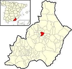
Figure 1. Location of Macael in the province of Almería (south-eastern Spain) (wikiwand.com) and photo of the necropolis of Macael Viejo.
The research carried out so far on this cemetery (2018-2021) allowed the recovery of 47 individuals. The organization of the maqbara is characterized by the alignments of graves of equivalent morphology and orientation, which determines the uniformity of the landscape and funerary rite, consistent with the Muslim religion.
During the analysis of an assemblage from Macael Viejo (Almeria, Spain), the remains of a non-adult (referred to as Individual 16) with the presence of double deciduous teeth (81 and 82) were registered. Individual 16 was in a nearly supine position, facing south/east orientation (Fig. 2). The skull and arms were not in their anatomical position. Preservation can be defined as a medium, with a Preservation Index of 40.9% (Safont et al., 2000). The skull was highly fragmented; therefore, its orientation could not be established. The legs were straight and parallel but slightly bent. The recovered joints were in close anatomical connection. No grave goods were associated with the burial. All the elements of the burial correspond to the Muslim rite.
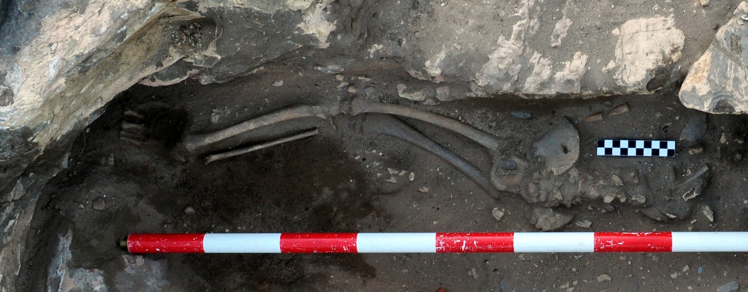
Figure 2. Remains of a non-adult individual 16 in the site of Macael Viejo, dated to the Late Medieval period to Early Modern periods (13th -16th century).
In the present work, it has been decided not to determine the sex since the secondary sexual characteristics have not yet been developed (Ferembach, Schwidetzky & Stlovkal, 1980). Standard anthropological methods for non-adult individuals were used for age estimation. These methods were based on dentition (AlQahtani, 2012), length of long bones (Maresh, 1970), and fusion of bones (Schaefer, 2008).
Regarding dental pathology, all teeth were examined under standardized lighting conditions by careful visual inspection. Dental wear and caries were recorded according to Hillson (2001). Possible linear enamel hypoplasia (LEH) and dental anomalies were recorded according to Goodman and Rose, (1990) and Ansari et al., (2019), respectively. The identification and characterization of deciduous double-crowned single-rooted teeth is based on the four descriptive categories reported by Aguiló et al. (1999). Fédération Dentaire Internationale (FDI) notation system was used.
Micro-CT is a non-invasive detection tool that employs radiation and digital X-ray detectors to capture images of a sample’s internal structure (on a micro-level) without damaging the sample. Micro-CT scan was performed in the Laboratory for Mineralized Tissue, Department for Anatomy at the University of Zagreb (Croatia). The teeth sample were scrutinized through 1076 Micro-CT (Bruker, Belgium) with the following parameters: voltage of 40 kV and electric current of 250 µA, corresponding to a resolution of 9 µm. The beam hardening was reduced by using a 0.025 mm thick titanium filter. The rotational step was set to 0.3° with a frame averaging set at 2. The acquired data were reconstructed using the NRecon software (Bruker, Belgium) through a dedicated GPU. Reconstructed data was visualized using CTAn and CTVox software (Bruker, Belgium).
Results
The estimated age of death based on dentition (AlQahtani, 2012) is given as the value of 5±1 years. Based on the length of preserved long bones (Maresh, 1970), the age was estimated to be 3.5 years, and the age based on the fusion of bones (Schaefer, 2008) corresponds to an interval between 4–5 years. Since dentition is the indicator that is least sensitive to changes (Prieto, 2008), it has been used as a reference method to estimate the age of non-adult individuals. Therefore, according to the method of AlQahtani (2012), it could be said that Individual 16 is a non-adult aged 5±1 years.
As we mentioned before, the skull was heavily damaged and very fragmented. Maxillary bone was not preserved, and the mandible was divided into 3 parts, with 3 teeth in sockets (Fig. 3). Apart from that, 13 loose teeth were also recovered, but none showed the sign of fusion.
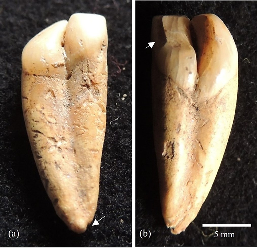
Figure 3. Heavily fragmented mandible with preserved teeth in situ.
The individual shows unilateral double-teeth among the primary mandibular central and right lateral incisors (81, 82) (Fig. 4). The double teeth reveal a bifid crown with a well-defined buccal and lingual groove that extends from the incisal edge to the apex of the root that appears separated at least 2 mm (Fig. 4). The lateral incisor was rotated to the mesial aspect forming a 90º angle on the distal aspect of the central incisor. Since no other anomaly was observed in the left mandibular quadrant, this represents a unilateral event. The central incisor was an interproximal dental wear facet on the mesial region equivalent to grade 1 on Hillson (2001), corresponding to dental ware limited to the enamel (Fig. 4).
 Figure 4. Primary maxillary teeth of Macael Viejo showing a fused central and lateral incisor (81, 82) on (a) buccal view and, (b) lingual view show an interproximal dental wear facet on the mesial region of central incisor (arrow). Observed the apex between the fused roots that appears separated at the last 2 mm (arrow).
Conventional X-ray examination revealed that the double tooth has two separate pulp chambers and root canals (Fig. 5), but obtaining more information about the union is impossible. Additionally, the Micro-CT images showed two separate canals leaving the pulp chamber, then joined to form one canal to the exiting site but still with a double form canal (Fig. 6).
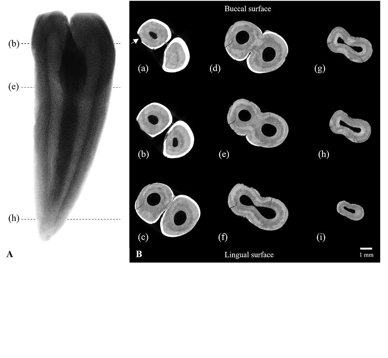
Figure 5. X-ray of primary maxillary teeth of Macael Viejo showing the partial fusion of crowns and roots.
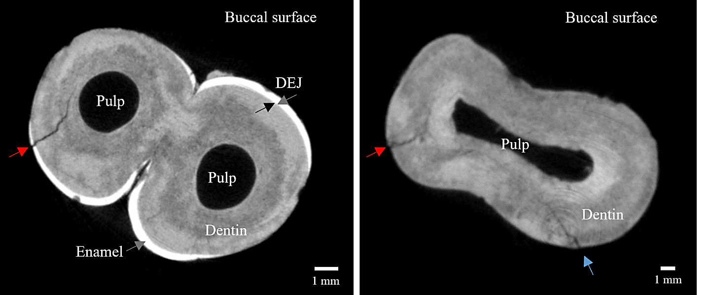
Figure 6. Representative cross-section of micro-CT data, (a-i) changes of the Micro-CT sequential images. (a) The arrow shows the interproximal dental wear facet on the mesial region of central incisor.
Micro-CT reveals the central incisor was an interproximal dental wear facet on the mesial region equivalent to grade 2 on Hillson (2001) which corresponds to the awareness that it is limited to the dentin on the occlusal and middle part of the crown (Fig. 5).
Additionally, the enamel thickness at the labial fused portion in the deciduous central/lateral incisor tended to decrease from the incisal edge towards the cervical region. Enamel thickness at the lingual fused portion tended to increase gradually from the incisal edge towards the cervical region (Fig. 5). Micro-CT also reveals the presence of incomplete radial microcracks. Originates in the enamel of the interproximal mesial wear facet and continues to the dentine microcrack without reaching the root canal (Fig. 5). These radial microcracks initiate from the base of flexing brittle enamel with in-dentine microcracks originating from the root wall without reaching the root canal (Fig. 5).
Macroscopic and microscopic techniques showed that the teeth are joined by crowns and along the root and fall within clearly distinct but joined roots, with two separate root canals at the last distal part of the root. Individual 16 corresponds to type-IV described by Aguiló et al. (1999). The Type IV anomaly is two fused crowns with two distinct but fused roots. The Micro-CT images (Fig. 6) show a difference from the conventional X-ray in interpreting the fusion of the root canals, which helps estimate the precise diagnosis.
Fused teeth usually show an increased risk for dental caries due to of conditions favoring dental plaque accumulation along the grooves between the crowns, but in this case, they were not recorded (Fig. 4).
No other bony or dental anomaly, such as supernumerary teeth, were detected in the skeletal remains of this child. The only condition observed in this individual was Linear Enamel Hypoplasia (LEH), visible in the permanent canine (2 well-defined lines), still without a formed root. LEH is a disruption in the enamel formation process that indicates the existence of a period of physiological stress experienced by the individual during the formation of the dental crown (Goodman and Rose, 1990). This process can last from the prenatal period up to 12 months in deciduous teeth, from birth to 7 years for permanent teeth (Hillson, 2008), and up to 16.5 years for third molars (AlQahtani et al., 2010), which makes it a good indicator of non-specific stress in childhood. These lines of hypoplasia tell us of at least two episodes of metabolic stress that this child survived, while the position of those lines indicates that those episodes occurred at the ages of 3.4 and 4.3 years (Reid & Dean, 2000).
Discussion
Because gemination and fusion can create teeth that appear morphologically and physiologically similar, it can be difficult to correctly diagnose the mechanism involved, necessitating reliance on tooth count (Mahendra et al., 2014). The use of Micro-CT was the key to reveal the fusion, the most probable cause of these double teeth cases. Most clinical literature agrees that there has to be a union of the dentine, and the process can result in a shared pulp chamber and root canal, or both elements can remain separate (Benazzi et al., 2010; Koszowski et al., 2014). In the clinical literature geminated teeth are usually not found in the mandible and are the minority crown form in the maxilla (Duncan and Helpin, 1987). Population differences have also been noted; the fusion of the mandibular incisors is evidently more prevalent in Spanish samples (Aguiló et al., 1999).
The primary teeth suffered more numerous and extensive subsurface microcracks. Microcracks occur when enamel surfaces contact very small hard particles. While the particles slide on the surface, a combined process of micro-cutting, cutting of enamel crystals at the level of rods and inter rods microstructures, and microcracking of both the surface and the subsurface takes place (Low et al., 2008). The microcracks readily formed in the deciduous enamel even at low loads, which indicates the poor fracture resistance of deciduous teeth by virtue of their lower hardness and fracture toughness, compared to the permanent tooth (Low et al., 2008).
LEH is often associated with early mortality (decreased life expectancy) and frequently is found in individuals who die at a young age (Šlaus, 2002). LEH points to physiological stress that this child survived at different stages of his/her life. Although not pointing to growth delay, the slight discrepancy between dental eruption and length of a preserved long bone shows that the child was at the lower limit of its age group, which can be connected to physiological stress. However, no correlation could be made between physiological stress and mentioned dental anomaly, as the lines of hypoplasia were noted on a permanent tooth, that starts its formation at 10 months, and the mentioned anomaly was noted on deciduous incisive, forming in utero.
Conclusion
Tooth fusion is a rare condition, and archaeological cases have only occasionally been reported. The present example adds to the documentation of a mandibular primary dental fusion of teeth abnormality in past populations. The presence of dental anomalies and correct diagnosis is always a challenge for a dental anthropologist. Therefore, morphological and radiological features should be carefully evaluated, and a description must be made. Micro-CT can provide new and accurate image data that have not been possible to obtain fully with conventional diagnostic methods and are essential for the definitive diagnosis of significant dental anomalies. Regular Micro-CT methodology application would further help understand and distinguish this phenomenon from others with similar characteristics.