Introduction
Multiple myeloma (MM) is a disseminated malignant tumor composed of monoclonal plasma cells. It accounts for about 8% of all hematological diseases and mostly occurs in elderly and it is more common in men than women (1.4:1) (1).
Revised International Myeloma Working Group (IMWG) diagnostic criteria for multiple myeloma include clonal bone marrow plasma cell ≥ 10% or biopsy - proven bony or extramedullary plasmacytoma and any one or more of the following myeloma defining events that are presented in theTable 1 (2).
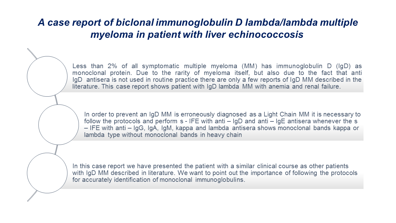
Our case report shows a monoclonal immunoglobulin D lambda type and monoclonal free light chains lambda type in a patient with anemia, renal insufficiency and clonal bone marrow plasma cell > 80%.
As a unique myeloma, immunoglobulin D (IgD) was first discovered in 1965. As a rare variant of the disease immunoglobulin D multiple myeloma (IgD MM) represents only 2% of all symptomatic myeloma. There is evidence that IgD MM is associated with high rates of kidney failure, amyloidosis, hypercalcemia and Bence Jones proteinuria (3-5).
There are two reasons why are just a few reports of IgD MM in literature, the first reason is its low concentration in plasma and the other one is the fact that antiserum for IgD is not used in routine practice. Because of its low concentration it can easily escape electrophoresis detection. Also, IgD MM is often misdiagnosed as light chain myeloma (LC MM) (3). In the case of IgD MM it is characterized by the high preponderance of lambda light chains over kappa light chains (4,6,7).
Case report
A 69 – year- old men with liver echinococcosis was hospitalized in the gastroenterology department. Patient reached out to the doctor in primary practice because of weakened appetite and loss of 10 kilograms in a last month. The doctor ordered laboratory tests and after receiving the results of anemia and renal insufficiency, he refers him to the hospital for further diagnostic and treatment. Patient general condition was poor and he was very weak. He has liver echinococcosis for the past 30 years and was operated in 2000 and 2005. After the second operation, lung echinococcosis was also verified. Until the current hospitalization the patient had no kidney problems. As a part of diagnostic processing, samples were referred to our laboratory for routine hematology, biochemistry and urine analysis. Also, in a diagnostic process hematologist examination was requested and he ordered among other analyses serum protein electrophoresis which showed presence of M protein. After this finding he requested measurement of free light chains (FLC) kappa (κ) and lambda (λ) type both, in serum and 24 h urine, β2-microglobulin and serum protein immunofixation. Patient gave his consent for publishing his laboratory data.
Result of laboratory tests showed normocytic anemia accompanied with serious kidney failure. Blood cell counts were performed on Sysmex XN 1000 (Sysmex Corporation, Kobe, Japan). Immunoglobulin analyses showed that concentrations of IgA and IgM are low and IgG is towards the lower limit of the reference interval. All biochemistry tests were performed on Beckman Coulter DxC 700 AU (Danaher Corporation, California, USA).
The serum concentration of kappa and lambda light chains showed an increase in lambda light chains and a decrease in κ/λ ratio. Laboratory results are presented inTable 2. In our case altered κ/λ ratio indicate excess production of clonal FLC by the proliferating plasma cell population. A high concentration of β2-microglobulin as well as a high erythrocytes sedimentation rate (ESR) was in a favor of multiple myeloma. Quantitative capillary photometry for ESR was performed on iSED (Alcor Scentific, Smithfield, USA) and β2-microglobulin was measured turbidimetrically on Alinity c analyzer (Abbott Laboratories, Chicago, USA).
Analysis of FLC in 24h urine showed Bence – Jones excretion with 7590 mg / 24h urine. Concentration of kappa and lambda light chain in both serum and urine was measured nephelometrically on BN II System (Siemens Healthcare, Marburg, Germany).
Serum protein electrophoresis was performed by capillary zone electrophoresis (CZE) method using Capillarys 3 Octa (Sebia, Lisses, France). It revealed a monoclonal peak (M – spike) in the gamma globulin fraction which is showed in theFigure 1.
In order to define the class and type of monoclonal protein, after electrophoresis serum protein immunofixation (s - IFE) was performed. Immunofixation (IF) is the gold standard method to confirm the presence of a monoclonal protein. Immunofixation is a two – step procedure consisting of electrophoresis on agarose and immunoprecipitation. In the first step proteins are separated electrophoretically into sharp bands on the gel and in the second step, a specific antiserum is added to each band for a molecule of individual class and type of immunoglobulin. If there is a monoclonal synthesized immunoglobulin it will make an insoluble complex with antisera which can be stained and proved (1).
Serum immunofixation was performed using agarose Hydragel 2/4 IF gels on Hydrasys 2 Scan (Sebia, Lisses, France). In the first step s-IFE was performed using the anti-IgG, IgA, IgM, total kappa and total lambda antisera. The result of the first s-IFE was the presence of two monoclonal lambda total light chain without corresponding heavy chain. Following the protocol we performed s-IFE using anti-IgD, anti-IgE, total kappa, total lambda, free kappa and free lambda antisera. Serum IFE with anti-IgE antisera, total kappa, total lambda, free kappa and free lambda antisera also showed two monoclonal bands lambda total light chain without corresponding heavy chain but there was also a band on free lambda chain, which has the same electrophoretic mobility as one band in total lambda. Visualization of the agarose gel with anti-IgD antisera showed monoclonal IgD band which has the same mobility in the electric field as one of the lambda bands. The second monoclonal lambda band has the same electrophoretic mobility as free lambda chain. The final result of s-IFE showed IgD lambda and free lambda monoclonal bands (Figure 1).
M spike was measured densimetrically using orthogonally way of measurement and its concentration was 21.60 g/L (Capillarys 3 Octa, Lisses, France). After receiving results from serum protein electrophoresis and s-IFE the bone marrow examination was ordered. The bone marrow examination was performed at pathology department. Result showed hypercellular bone marrow that is extensively diffusely infiltrated with cells that are only partially recognizable as mature and mildly atypical plasma cells and mostly blastoid cells in appearance.
For further diagnostic flow cytometry immunophenotyping is recommended.
Discussion
In this case report we presented patient with IgD lambda MM having anemia, renal failure and > 80% plasma cells in bone biopsy which meets IMWG criteria for MM diagnosis.
The rarity of IgD MM in our practice is shown by the fact that since we introduced s-IFE in our laboratory in September 2019 until today (October 2022) we had 457 requests for serum protein immunofixation and this is our first case of IgD MM. As for other types of myelomas we had: 20 IgG MM lambda type, 35 IgG MM kappa type, 4 IgM MM lambda type, 5 IgM MM kappa type, 7 IgA MM lambda type, 8 IgA kappa type, 7 LC MM lambda type and 4 LC MM kappa type.
In their study, Zagouri et al. states that more than 80% of IgD myeloma are lambda type, while only 38% of other myeloma have lambda light chains (8). The biological and pathogenetic features of the prevalence of lambda light chain in IgD MM are unclear. Also, most of the patients with IgD MM has a renal failure and significant Bence Jones proteinuria. The role of lambda light chain in kidney pathology is the subject of further research. Available data in the literature shows that patients with IgD MM have different clinical features than patients with other types of myelomas, and the exact reason for this cannot yet be determined with certainty (8). There are also reported a high – risk chromosomal abnormalities in patients with IgD MM but that is only on a small fraction of patients and therefore it cannot be assessed whether there are significant differences in these changes between patients with IgD MM compared to patients with another myeloma. Only a small percentage of patients with IgD MM had a detailed cytogenetics data. Current data cannot clearly say what has a greater influence on the clinical course and especially on the elevated risk of renal failure, whether it is specific cytogenetic abnormalities or tumor biology itself (8).
Kidney damage in multiple myeloma can be acute and chronic. Changes in the kidney tubules are dominant, while glomerulus and interstitial are much less affected. Kidney damage is caused by immunobiological activity of malignant clones, but also by deposits of immunoglobulin light chains that lead to obstruction of renal tubules and development of tubulointerstitial nephritis (9,10). Our patient, like other cases with IgD MM described in literature, had severe kidney failure. As the condition worsened, he had to undergo hemodialysis due to the high concentration of urea and creatinine.
Before development of the new drugs and autologous stem cell transplantation, survival of patients with IgD MM was shorter. With the new drugs and stem cell transplantation outcomes for these patients improved significantly (11,12). Bemelmans et al. in their study described the case of the patient with IgD MM with complete remission of more than eight years (13). Our patient general condition worsened so none of the treatments above has not started. Patient was treated with antibiotics, diuretics, electrolyte solutions and blood transfusions. Due to his previous lung problems, respiratory complications caused his death.
In order to prevent an IgD MM is erroneously diagnosed as a light chain MM it is necessary to follow the protocols and perform s-IFE with anti-IgD and anti-IgE antisera whenever the s-IFE with anti-IgG, IgA, IgM, kappa and lambda antisera shows monoclonal bands kappa or lambda type without monoclonal bands in heavy chain (14).
In conclusion, in this case report we have presented the patient with a similar clinical course as other patients with IgD MM described in literature. We want to point out the importance of following the protocols for accurately identification of monoclonal immunoglobulins.

