Skoči na glavni sadržaj

Izvorni znanstveni članak

https://doi.org/10.37427/botcro-2023-009

Placoneis modaomensis sp. nov. (Bacillariophyta; Cymbellaceae), a new species from Guangdong Province, China

Yu-Jie Li ; Yunnan University, School of Ecology and Environmental Science, Institute for Ecological Research and Pollution Control of Plateau Lakes, Kunming 650500, China
Ji-Shu Guo ; Yunnan University, School of Ecology and Environmental Science, Institute for Ecological Research and Pollution Control of Plateau Lakes, Kunming 650500, China
Hong-Ping Ni ; Yunnan University, School of Ecology and Environmental Science, Institute for Ecological Research and Pollution Control of Plateau Lakes, Kunming 650500, China
Ying-Yan Huang ; Ministry of Ecology and Environment, Administration for Pearl River Basin-South China Sea Ecology and Environment, Center for Ecology and Environment Monitoring and Research, Guangzhou, 510611, China
John Patrick Kociolek ; University of Colorado, Museum of Natural History and Department of Ecology and Evoluionary Biology, Boulder, Colorado-80309, USA
Yan-Ling Li ; Yunnan University, School of Ecology and Environmental Science, Institute for Ecological Research and Pollution Control of Plateau Lakes, Kunming 650500, China


Puni tekst: engleski pdf 1.321 Kb

str. 87-95

preuzimanja: 230

citiraj

Preuzmi JATS datoteku


Sažetak

One new species, Placoneis modaomensis, found in a freshwater environment from a tributary of the Pearl River, which lies in Modaomen Channel, Zhuhai County, Guangdong Province, China, is described based on light and scanning electron microscope observations. P. modaomensis sp. nov. has the morphological features that are typical for the genus, including external terminal raphe fissures curved to the opposite sides and areolae with internal volate occlusions. The new species is similar to P. amphibola (Cleve) E.J. Cox, P. amphiboliformis (Metzeltin, Lange-Bertalot & Soninkhishig) Vishnyakov, P. parvapolonica Lange-Bertalot & Wojtal, P. clementispronina Lange-Bertalot & Wojtal and P. nanoclementis Lange-Bertalot & Wojtal in the shape of the valves and in having coarse striae but it can be easily distinguished by the two main morphological characteristics: external central raphe endings bent in the opposite directions, and areolae covered by volate occlusions externally. The latter feature appears to be new for taxa assigned to the genus Placoneis. Data on the associated diatom flora and its ecology are also given. These findings increase our understanding about the morphology of Placoneis in general and the distribution of the genus in China.

Ključne riječi

diatoms; morphology; new species; Placoneis; taxonomy

Hrčak ID:

301963

URI

https://hrcak.srce.hr/301963

Datum izdavanja:

1.10.2023.

Posjeta: 625 *


Introduction

The genus Placoneis Mereschkowsky was erected by Mereschkowsky in 1903 for a group of species showing a single chloroplast with a central bridge and lateral lobes (Mereschkowsky 1903). With the shift to an emphasis on using frustular features to diagnose taxa (e.g., Hustedt 1930), species assigned to Placoneis were considered part of the large genus Navicula Bory (1822). Due to its structure of the chloroplast, Cox (1987) resurrected the genus Placoneis with P. gastrum (Ehrenberg) Mereschkowsky (Basionym: Pinnularia gastrum Ehrenberg) as the type species. Phylogenetic analysis of Placoneis based on morphological and molecular data showed that the genus was part of the Cymbellales, a group with valves that are asymmetrical to the apical and/or transapical axes, despite having symmetrical valves. Support for this phylogenetic placement includes the straight and expanded central raphe endings and more or less hooked distal ones, striae composed of rounded areolae which are internally closed by volae (tectulum), and a single chloroplast with a central bridge and lateral lobes extending under the valves (Cox 1987, 2003, 2004, Mann and Stickle 1995, Bruder and Medlin 2007). To date, more than 136 species are recognized to be part of this genus (Guiry and Guiry 2022).

The genus Placoneis has a relatively wide distribution range, including Europe (Cox 1987, Bruder and Medlin 2007, Levkov and Williams 2011, Kulikovskiy et al. 2016, Vishnyakov 2020), Asia (Mayama and Kawashima 1998, Metzeltin et al. 2009, Kulikovskiy et al. 2012, Pomazkina et al. 2019, Kezlya et al. 2020), North America (Johansen et al. 2004, Kociolek and Thomas 2010, Kociolek et al. 2014), South America (Metzeltin and Lange-Bertalot 1998, Metzeltin and Lange-Bertalot 2007, Straube et al. 2013, Maidana et al. 2017), West Africa (Fofana et al. 2014) and Antarctica (Zidarova et al. 2009). In China, research on Placoneis has focused on the discovery of newly recorded species for the country, such as P. prespanensis Levkov, Krstic & Nakov (Li et al. 2010), P. explanata (Hustedt) S. Mamaya (Liu et al. 2012), P. interglacialis (Hustedt) E.J. Cox (Liu et al. 2012), P. opportuna (Hustedt) Chudaev & Gololobova (Lin et al. 2018), P. anglophila var. signata (Hustedt) Lange-Bertalot (Liu et al. 2020). New Placoneis species have been proposed from China by Gong et al. (2013) but otherwise only few new species of this genus have been reported. In Guangdong Province the diversity of diatoms is relatively rich (Qiu et al. 2016, Lin et al. 2018), but previous studies have mainly focused on Navicula Bory, Nitzschia Hassall, Cymbella C. Agardh and Gomphonema Ehrenberg (Wang et al. 2018, 2021). Our understanding of Placoneis from Guangdong Province is very limited (Wang et al. 2021).

The purposes of our study are: (i) to describe a Placoneis species new to science from the tributary of the Pearl River: Modaomen Channel, Zhuhai County, Guangdong Province, China based on detailed morphological observation using both light and scanning electron microscopy, (ii) to discuss its characteristics compared to related species, and (iii) to provide ecological information on this new species.

Materials and methods

Modaomen is located in Zhuhai County, Guangdong Province, China, one of the eight major gates at the mouth of the Pearl River. The length of the Modaomen Channel is about 45 km, and the water depth varies from 5 to 13 m. The Channel is relatively straight and about 2200 m wide (Chen et al. 2014, Tong et al. 2018).

In 2021, samples containing Placoneis were collected from the Modaomen Channel (22°24′20″N, 113°36′25″E). Channel water pH and specific conductance were measured using a YSI 650 multi-parameter display system (650 MDS, YSI Incorporated 1700/1725 Brannum Lane, Yellow Springs, OH 45387 USA) with a 600XL probe. The diatom samples were further processed as described by Battarbee (1986). After several rinses in distilled water, the partially cleaned diatom material was air-dried onto cover slips and mounted onto slides using Naphrax. The sample and slides were deposited in the Herbarium of the Institute for Ecological Research and Pollution Control of Plateau Lakes, Yunnan University, Kunming, P.R. China (YUK). The isotype slides were stored in the Key Laboratory of Biodiversity of Aquatic Organisms, Harbin Normal University.

Morphological observations of specimens were made under oil immersion at 1000× magnification with light microscopy (LM) using an OLYMPUS BX51-DIC research microscope and a C5060 Olympus digital camera. At least 500 valves were identified and counted in each surface sediment sample. Cleaned material for scanning electron microscope (SEM) analysis was air-dried onto cover glasses, mounted onto stubs, and coated with 20 nm of Au (EMSCOP SC 500 sputter coater). Resulting stubs were examined in the LEO 1530 scanning electron microscope (SEM). Description of the new species follows the terminology provided by Round et al. (1990), Cox (2003), Metzeltin et al. (2009) and Lange-Bertalot and Wojtal (2014).

Results

Taxonomy

Division Bacillariophyta Haeckel 1878: 95

Class Bacillariophyceae Haeckel 1878 emend D.G. Mann in Round et al. 1990: 651

Subclass Bacillariophycidae D.G. Mann in Round et al. 1990: 125

Order Cymbellales D.G Mann in Round et al. 1990: 653

Family Cymbellaceae Greville 1833: 263, 409

Genus Placoneis Mereschkowsky 1903: 3

Placoneis modaomenensis Y.-L. Li sp. nov. Fig. 1A–H; Fig. 1C is the holotype
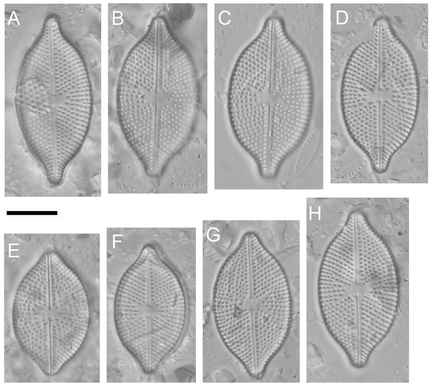
image1.png

Fig. 1. Placoneis modaomensis, Light Microscopy (LM), Differential Interference Contrast (DIC). A–H – valve views, showing size range and variability of the holotype population. Scale bar = 10 μm.

LM (Fig. 1): Valves elliptical to broadly elliptical, nearly symmetrical about the apical axis, with rostrate or rostrate-rounded apices. Length 25.0–32.5 μm, width 14.0–16.5 μm, length/width ratio 1.62–2.16, median 1.89 (n = 30). Raphe filiform, almost straight with slightly expanded, but not clearly deflected to any side. External central raphe ends slightly straight, no significant expansion or bending. External terminal raphe fissures hooked to opposite sides. Axial area narrow, linear. Central area transverse, irregular to bow-tie-shaped, rarely asymmetrical, occupying nearly 1/2 of the valve width. Isolated pore is absent from the central area. Striae radiate throughout, 12–14 in 10 μm. Areolae visible, 11–14 in 10 μm.

In SEM (Fig. 2 and Fig. 3): External raphe narrow, central raphe endings hooked opposite to each other (Fig. 2A, B, G, H, I). The terminal fissures curved, deflected in the opposite directions, extend onto the valve margin (Fig. 2C, D, E, F). Striae uniseriate, composed of round or elliptical areolae, extending to valve margin (Fig. 2A–F). Areolae covered by volate occlusions (Fig. 2G, H, I). Each areola has almost 1–4 siliceous protrusions internally, forming a variety of different shapes (Fig. 2A–I).
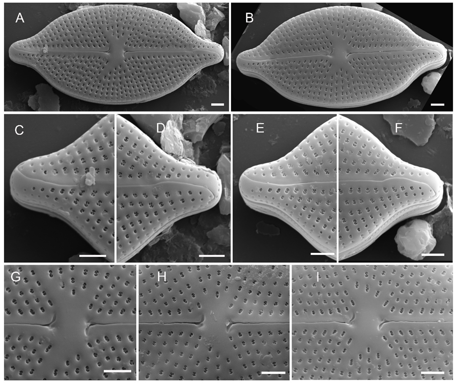
image2.png

Fig. 2. Placoneis modaomensis, Scanning Electron Microscope (SEM), external views. A, B – external view of an entire valve. C, D, E, F – valve apices, striae with elliptic/rounded areolae and hook-shaped terminal raphe fissures bent onto the valve margin. G, H, I – external view of valve center, the central raphe endings hooked in the opposite directions from each other, note volate occlusions. Scale bar = 2 μm.

Internal striae uniseriate, formed by elliptical areolae, separated by robust silica ribs (Fig. 3A–H). The raphe sternum clearly raised above the valve plane and expanded in the central area. Internal raphe straight, discontinuous with intermissio, lying in a prominent and raised raphe sternum. Central raphe endings straight, not expanded. Terminal raphe ends terminate as helictoglossae, offset from the raphe branch, bent slightly towards the valve margin (Fig. 3D, E, F). Striae are uniseriate, distinctly radiate, formed by round to elliptical shaped areolae (11–14 in 10 µm) and covered by dentate occlusions (Fig. 3G, H).
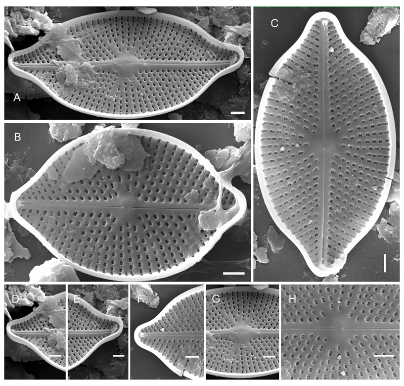
image3.png

Fig. 3. Placoneis modaomensis, Scanning Electron Microscope (SEM), internal views. A, B, C – internal view of entire valve. D, E, F – internal view of valve apex with terminal raphe ends offset and bent slightly towards the margin. G, H – internal detail of central area showing raised central part and areolae covered by volate occlusions. Scale bar = 2 μm.

Type: – CHINA. Guangdong Province: Zhuhai County, Modaomen Channel, GD1, 22°24′20″ N, 113°36′25″ E, elevation 0 m a.s.l., samples collected by Dr. Hong-Qu Tang, 26 th July 2021. Holotype MDM202172601 in Coll. Yan-Ling Li, Yunnan University, Kunming, China. Fig. 1C is of the holotype; Isotype YUNGL20220218, Harbin Normal University, Harbin, China)

Etymology:modaomensis, referring to the type locality from which the new species was obtained.

Associated diatom flora: Placoneis modaomensis is known from the Channel, situated at 0 m a.s.l. This species was associated with Amphora linearis F. Meister (1935: 97), Seminavis strigosa (Hustedt) Danielidis & Economou-Amilli (2003: 30), Aulacoseira granulata (Ehrenberg) Simonsen (1979: 58), Gomphonema parvulum (Kützing) Kützing (1849: 65), Navicula schroeteri F. Meister (1932: 38), Navicula viridula var. rostellata (Kützing) Cleve (1895: 15), Nitzschia clausii Hantzsch (1860: 40) and Nitzschia frustulum (Kützing) Grunow (1880: 98).

Ecology: The Modaomen Channel showed slightly alkaline conditions (pH 7.81), 29.8 °C of water temperature, 262 µS cm -1 of conductivity and 7.46 mg L -1 of dissolved oxygen (DO).

Discussion

Because Placoneis modaomenensis has symmetrical valve and radiate striae, it could easily be placed in one of four other genera: Paraplaconeis Kulikovskiy, Lange-Bertalot & Metzeltin (2012), Geissleria Lange-Bertalot & Metzeltin (1996), Rexlowea Kociolek & E.W.Thomas (2010) or Navicula Bory (1822). First, our new species resembles Paraplaconeis by symmetry of valve, but differs by the specific morphology of internal and external areolae patterns (Cox 1987, Cox 2003, Lange-Bertalot and Wojtal 2014). Second, P. modaomenensis is similar to the genus Geissleria on the basis of features observed with LM. These two genera are similar in terms of valve outline, but differ by the presence of the subpolar elongated areolae in the latter (Novais et al. 2013, Kulikovskiy et al. 2014). Third, P. modaomenensis is morphologically close to Rexlowea due to the valve outline, but these two genera are very distinct from one another by the arrangement, radiation and density of the striae (Kociolek and Thomas 2010). Fourth, P. modaomenensis can be confused with Navicula based on the valve symmetry. However, the former has exterior areolae covered by volate occlusions and internal areolae covered by dentate occlusions; while the latter areolae are all individually covered on the inside by a weakly convex hymen (Lange-Bertalot 2001, Li and Qi 2018). The characters found in Placoneis modaomensis are compared with those of morphologically most similar genera in Tab. 1.

While Placoneis seems easily placed as a member of Cymbellales, by virtue of its cytoplasmic features, its position in this lineage has been quite variable. Placoneis has been reported as outside the group of asymmetrical genera (Kociolek and Stoermer 1988, Nakov et al. 2014), deep within the cymbelloid lineage with some other genera naviculoid symmetry (Thomas et al. 2016, Kezlya et al. 2021) or in both positions depending upon the gene(s) used in the analysis (Bruder and Medlin 2007). Groupings within Placoneis are also enigmatic. Cox (1987) identified two groups within Placoneis, one with isolated pores, external terminal raphe fissures deflected in directions opposite one another and straight internal central raphe endings, while the other group has features of isolated pores absent, external terminal raphe fissures deflected in the same direction and recurved internal central raphe endings. Kociolek and Thomas (2010) noted some species from Colorado, USA, that did not conform to this organization of the genus. For example, P. fourtanierii Kociolek & Thomas (2010: 204) has external terminal raphe fissures that are deflected in opposite directions, but lack isolated pores and deflected internal raphe ends. P. coloradoensis Kociolek & Thomas (2010: 205) has external terminal raphe fissures that are deflected towards the same side, internal central raphe endings that are straight, but this species has isolated pores. In the case of P. modaomenensis, it has external terminal raphe fissures that curve in opposite sides, but has internal central raphe endings that are straight and no isolated pores. In addition, P. modaomenensis has areolae with volate occlusions positioned on the valve exterior which appears to be unique within Placoneis. Presence of central raphe endings turned opposite to one another in P. modaomenensis is shared with P. uruguayensis Metzeltin et al., a species without many features in common with other Placoneis species. The statement of Levkov et al. that “the full range of variation of morphology with the genus has been underestimated” (2007, p. 116) seems even more true now than when it was offered more than 15 years ago.

In terms of general valve shape and striae, P. modaomenensis shows some resemblance to P. amphibola (Cleve) E.J. Cox, P. amphiboliformis (Metzeltin, Lange-Bertalot and Soninkhishig) Vishnyakov, P. parvapolonica Lange-Bertalot & Wojtal, P. clementispronina Lange-Bertalot & Wojtal and P. nanoclementis Lange-Bertalot & Wojtal. However, P. modaomenensis differs strikingly from these species that are morphologically similar with respect to two features by the following: 1) external central raphe endings are bent in the opposite directions, and 2) both internal and external areolae are covered by volate occlusions.

Tab. 1. Morphological comparison of Placoneis to the most similar genera.

Placoneis modaomensis sp. nov. Placoneis Paraplaconeis Geissleria Rexlowea Navicula
Reference This studyCox 1987, Cox 2003, Lange-Bertalot and Wojtal 2014Kulikovskiy et al. 2012, Lange-Bertalot and Wojtal 2014Novais et al. 2013, Kulikovskiy et al. 2014Kociolek and Thomas 2010Lange-Bertalot 2001, Li and Qi 2018
Areolae Being covered by volate occlusions externally, being covered by dentate occlusions internallyBeing closed internally by volaeOpen internally and occluded externallyStraight slit-like pores openings without hymens, presence of the subpolar elongated areolaeLarge and more coarsely arranged puncta on the marginal valveBeing all individually covered on the inside by a weakly convex hymen
Striae Uniseriate, radiate throughout, extending to valve marginUsually uniseriate but sometimes biseriate, radiate near the centre of the valve, more parallel or convergent, at the apicesUniseriate only on the valve mantle, consistently biseriate on the valve faceRadiate throughout, the proximal three ones wider spaced than the othersRadiate, with the 1 st and or 2 nd striae at the apices parallel or, rarely, convergent Uniseriate, rarely biseriate, parallel, radiate or convergent
Chloroplast Single, asymmetrical chloroplast, central portion more or less along the apical axis of the cell, from which lobes extend under the valvesSingle, large plastid is divided into two plates lying one against each valve, connected by a broad columnTwo plastids, located on each side of the apical axis, and each plastid contains an elongated, slender protein nucleus
Raphe system The external central raphe ends slightly straight, the terminal fissures deflected in the opposite directions; the internal central raphe endings straight, terminal raphe ends terminate as helictoglossaeThe external central raphe endings are straight and slightly expanded; the internal endings usually deflected towards the secondary side, occasionally hooked in opposite directionsThe external raphe slit together with terminal fissures often curving primarily to opposite sides but finally to the same sideThe external raphe is almost straight; the internal raphe is not deflected between the central nodule and helictoglossa, internal rib has never been observedThe external proximal ends swollen, external distal ends hookedThe external terminal fissures are deflected to the secondary side; the internal central raphe ends are not deflected

The characters found in P. modaomenensis are compared with other closely-related species of the genus in Tab. 2.

Based on valve outline, the new species is similar to P. amphibola, P. amphiboliformis, P. parvapolonica, P. clementispronina and P. nanoclementis, and P. modaomenensis sp. nov. most resembles P. parvapolonica based on its more convex valve margin and the structure of raphe and areola. However, they can be easily separated from one another. For example, in terms of the central raphe endings, the central raphe endings of the new species hooked in the opposite directions from each other while they do not clearly deflect to any side in P. parvapolonica. Comparing the shape of the areolae, the exterior areolae come in a variety of shapes and internal areolae are mostly round to elliptical in the new species, but P. parvapolonica differs in having areolae whose exterior and internal openings are nearly circular to slightly elongated in shape. Taking into consideration the occlusion of the areolae, the exterior and internal areolae are covered by volate and dentate occlusions respectively in the P. modaomenensis sp. nov., while there is an absence of occlusions in exterior and internal areolae in P. parvapolonica. Besides, the size range of P. modaomensis is larger than P. parvapolonica (16–20 × 8.0–9.5 μm). Finally, the stria and areola density are much lower compared to P. parvapolonica (stria: 14–15 μm, areola: 40–44 in 10 μm). In summary, these differences are sufficient to justify the description of P. modaomensis as an independent species.

Placoneis species are prevalent in freshwater bodies, including alkaline waters, mesotrophic and oligotrophic conditions (Cox 1987, Fujita and Ohtsuka 2005, Bruder and Medlin 2007, Kezlya et al. 2021). Of the five species similar to our new species, P. amphibola has a nordic-alpine distribution in Europe, and also occurs in freshwater fossil deposits (Cox 2003). Except for P. amphibola, the relevant information of nutrition, pH, and conductivity are not recorded, but other species have certain similarities or differences with our species. In terms of pH, P. parvapolonica, P. clementispronina and P. nanoclementis were found in alkaline waters, but P. amphiboliformis was known from acidic rivers (Metzeltin et al. 2009, Lange-Bertalot and Wojtal 2014). From the nutritional level, P. clementispronina and P. nanoclementis were distributed in eutrophic waters, and P. parvapolonica was found in mesotrophic waters (Metzeltin et al. 2009, Lange-Bertalot and Wojtal 2014). As for the conductivity, P. parvapolonica and P. nanoclementis are found predominantly in waters with moderate to high conductivity, whereas P. amphiboliformis was discovered rivers with low conductivity (Metzeltin et al. 2009, Lange-Bertalot and Wojtal 2014). Here, the new species of Placoneis was collected from alkaline waters with moderately high conductivity, which is the most similar to P. parvapolonica and P. nanoclementis.

In short, the discovery of this new species promotes the understanding of morphological features and ecological distribution about the genus Placoneis, and contributes to our understanding of diatom diversity, especially in Guangdong Province.

Tab. 2. Morphological comparison of the currently described species in Placoneis.

P. modaomensis sp. nov P. parvapolonica P. amphibola P. amphiboliformis P. clementispronina P. nanoclementis
Reference This studyLange-Bertalot and Wojtal 2014Cox 2003Metzeltin et al. 2009Lange-Bertalot and Wojtal 2014Lange-Bertalot and Wojtal 2014
Valve shape Elliptical to broadly elliptical and the valve margin more convexBroadly ellipticalLinear-elliptic to linear-lanceolateEllipticalElliptical-lanceolateElliptical-lanceolate to elliptical
Central area Bow-tie, transverseExtended transapicallyBow-tie, transverseNot givenVariable in sizeExtended transapically, variable in shape
Isolated pores AbsentTwoNot givenNot givenTwoTwo
Valve length (μm) 25.0–32.516–2037–7534–6320–4014–23
Valve width (μm) 14.0–16.58.0–9.522–2718–2410–148.0–8.6

Number of

striae in 10 μm

12–1414–157–87–811–1412–13
Number of areolae in 10 μm 11–1440–44Not given10–1238–4140–44
External terminal raphe fissures Undulate, deflected in the opposite directions, extend to valve marginTurning first to opposite sides, finally turning to the same side as at the opposite poleDeflected towards one sideNot givenWeakly sinuous subapically and deflected to opposite sides, at one of the poles curving back weakly to the same sideShortly curved to apparently opposite sides
External central raphe end-ings Hooked in the opposite directions from each otherDistinctly expanded central pores, not clearly deflected to any sideExpandedNot givenExpanded but hardly deflected central poresDistinctly expanded central pores, not clearly deflected to any side
Areolae Uniseriate, exterior areolae covered by volate occlusions and formed a variety of different shapes, internal areolae are mostly round to elliptical covered by dentate occlusionsUniseriate, circular to slightly elongate apicallyNot givenRegularly elongated transapicallySmall, circular to weakly elongated apicallySlightly elongated apically

Acknowledgments

This work was supported by the projects of National Sciences and Foundation of China (Grant No. 42172206 and 41877296). We thank Ms. Yulan Luo for her help with scanning electron microscopy observations.

References


Battarbee, R. W., 1986. Diatom analysis. In: Berglund, B. E. (ed.), , editor. Handbook of holocene palaeoecology & palaeohydrology. p. 527–570. John Wiley and Sons Ltd.,; Chichester, UK.:


Bory de Saint-Vincent, J. B .G. M., 1822;Bacillariées. Dictionnaire Classique d'Histoire naturelle. 2:127–129


Bruder, K., Medlin, L. K., 2007;Molecular assessment of phylogenetic relationships in selected species/genera in the naviculoid diatoms (Bacillariophyta). I. The genus Placoneis. Nova Hedwigia. 8534:331–352. https://doi.org/10.1127/0029–5035/2007/0085–0331


Chen, W. H., Zou H. Z., Dong Y. J., 2014;Hydrodynamic of saltwater intrusion in the Modaomen waterway. Advances in Water Science. 25(5):713–723. [In Chinese with English abstract]. https://doi.org/10.14042/j.cnki.32.1309.2014.05.003


Cox, E. J., 1987;Placoneis Mereschkowsky: the re-evaluation of a diatom genus originally characterized by its chloroplast type. Diatom Research. 2(2):145–157. https://doi.org/10.1080/0269249X.1987.9704994


Cox, E. J., 2003;Placoneis Mereschkowsky (Bacillariophyta) revisited: resolution of several typification and nomenclatural problems, including the generitype. Botanical Journal of the Linnean Society. 141(1):53–83. https://doi.org/10.1046/j.1095-8339.2003.00115.x


Cox, E. J., 2004;Pore occlusions in raphid diatoms - a reassessment of their structure and terminology, with particular reference to members of the Cymbellales. Diatom: the Japanese journal of diatomology. 20:33–46. https://doi.org/10.11464/diatom1985.20.0_33


Fofana, C. A. K., Sow, E. H., Taylor, J., Ector, L., Van de Vijver, B., 2014;Placoneis cocquytiae a new raphid diatom (Bacillariophyceae) from the Senegal River (Senegal, West Africa). Phytotaxa. 161(2):139–147. https://doi.org/10.11646/phytotaxa.161.2.5


Fujita, Y., Ohtsuka, T., 2005;Diatoms from paddy fields in northern Laos. Diatom. 21:71–89. https://doi.org/10.11464/diatom1985.21.0_71


Gong, Z. J., Li, Y. L., Metzeltin, D., Lange-Bertalot, H., 2013;New species of Cymbella and Placoneis (Bacillariophyta) from late Pleistocene fossil, China. Phytotaxa. 150(1):29–40. https://doi.org/10.11646/phytotaxa.150.1.2


Guiry, M. D., Guiry, G. M., 2022. AlgaeBase. World-wide electronic publication,. National University of Ireland,; Galway.: Retrieved on May 5, 2022 from. https://www.algaebase.org


Hantzsch, C. A., 1860;Neue Bacillarien: Nitzschia vivax var. elongata, Cymatopleura nobilis. Hedwigia. 2:1–40


Hustedt, F., 1930. Bacillariophyta (Diatomeae). In: Pascher, A., (ed.), , editor. Die Süsswasser-Flora Mitteleuropas. 10:Gustav Fischer,; Jena.:


Johansen J. R., Lowe, R., Gómez, S. R., Kociolek, J. P., Makosky, S. A., 2004;New algal species records for the Great Smoky Mountains National Park, U.S.A., with an annotated checklist of all reported algal species for the park. Algological Studies. 111(1):17–44. https://doi.org/10.1127/1864–1318/2004/0111–0017


Kezlya, E., Glushchenko, A., Maltsev, Y., Gusev, S., Kuznetsov, A., Kociolek, J. P., Kulikovskiy, M., 2020;Placoneis cattiensis sp. nov. — a new, diatom (Bacillariophyceae: Cymbellales) soil species from Cát Tiên National Park (Vietnam). Phytotaxa. 460(4):237–248. https://doi.org/10.11646/phytotaxa.460.4.1


Kezlya, E. M., Glushchenko, A. M., Maltsev, Y. I., Gusev, E. S., Genkal, S., Kociolek, J. P., Kulikovskiy, M. S., 2021;Three new species of Placoneis Mereschkowsky (Bacillariophyceae: Cymbellales) with comments on cryptic diversity in the P. elginensis-Group. Water. 13(22):3276. https://doi.org/10.3390/w13223276


Kociolek, J. P., Stoermer, E. F., 1988;A preliminary investigation of the phylogenetic relationships of the freshwater, apical pore field-bearing cymbelloid and gomphonemoid diatoms (Bacillariophyceae). Journal of Phycology. 24(3):377–385. https://doi.org/10.1111/j.1529-8817.1988.tb04480.x


Kociolek, J. P., Thomas, E. W., 2010;Taxonomy and ultrastructure of five naviculoid diatoms (class Bacillariophyceae) from the Rocky Mountains of Colorado (USA), with the description of a new genus and four new species. Nova Hedwigia. 9012:195–214. https://doi.org/10.1127/0029–5035/2010/0090–0195


Kociolek, J. P., Laslandes, B., Bennett, D., Thomas, E., Brady, M., Graeff, C., 2014. Diatoms of the United States, 1. Taxonomy, ultrastructure and descriptions of new species and other rarely reported taxa from lake sediments in the western U.S.A. Cambridge University Press,; Cambridge.:


Kulikovskiy, M. S., Lange-Bertalot, H., Metzeltin, D., Witkowski, A., 2012;Lake Baikal: Hotspot of endemic diatoms I. Iconographia Diatomologica. 23:1–607


Kulikovskiy, M., Gusev, E., Andreeva, S., Annenkova, N., 2014;Phylogenetic position of the diatom genus Geissleria Lange-Bertalot & Metzeltin and description of two new species from Siberian mountain lakes. Phytotaxa. 177(5):249–260. https://doi.org/10.11646/phytotaxa.177.5.1


Kulikovskiy, M., Glushchenko, A., Genkal, S. I., Kuznetsova, I., 2016. Identification book of diatoms from Russia. Filigran, Yaroslavl [in Russian].


Kützing, F. T., 1849. Species algarum. Friedrich Arnold Brockhaus,; Leipzig.:


Lange-Bertalot, H., 2001;Navicula sensu stricto, 10 genera separated from Navicula sensu lato, Frustulia. Diatoms of Europe. 2:1–526


Lange-Bertalot, H., Wojtal, A. Z., 2014;Diversity in species complexes of Placoneis clementis (Grunow) Cox and Paraplaconeis placentula (Ehrenberg) Kulikovskiy, Lange-Bertalot & Metzeltin. Beihefte zur Nova Hedwigia. 143:403–420. https://doi.org/10.1127/1438–9134/2014/021


Levkov, Z., Williams, D. M., 2011;Fifteen new diatom (Bacillariophyta) species from Lake Ohrid, Macedonia. Phytotaxa. 30:1–41. https://doi.org/10.11646/phytotaxa.30.1.1


Li, J. Y., Qi, Y. Z., 2018. Bacillariophyta, Naviaulaceae (III).In: Flora Algarum Sinicarum Aquae Dulcis. Science Press,; Tomus, XXIII.: [In Chinese with English abstract].


Li, Y. L., Gong, Z. J., Wang, C.C., Shen, J., 2010;New species and new records of diatoms from Lake Fuxian, China. Journal of Systematics and Evolution. 48(1):65–72. https://doi.org/10.1111/j.1759–6831.2009.00059.x


Lin, X. R., Rioual, P., Bai, Z. J., Peng, W., Sun, M. J., Huang, X. Z., 2018;The recent diatom flora in Lake Kanas, Xinjiang: unusual species and new records in China. Acta Hydrobiologica Sinica. 42(3):641–654. [In Chinese with English abstract]. https://doi.org/10.7541/2018.080


Liu, Y., Fan, Y. W., Wang, Q. X., 2012;Newly recorded species in Cymbellaceae and Gomphonemataceae from Great Xing’an Mountains, China. Acta Hydrobiologica Sinica. 36:496–508. [In Chinese with English abstract].


Liu, Z. X., Liu, B., Quan, S. J., Long, J. Y., Mo, W. H., 2020;Two biraphid diatom species reported from China for the first time-Prestauroneis tumida and Placoneis anglophila var. signata. Acta Botanica Boreali-Occidentalia Sinica. 40:1784–1791. [In Chinese with English abstract].


Maidana, N. I., Aponte, G. A., Fey, M., Schäbitz, F., Morales, E. A., 2017;Cyclostepanos salsae and Placoneis patagonica, two new diatoms (Bacillariophyta) from Laguna Cháltel in southern Patagonia, Argentina. Nova Hedwigia Beiheft. 146:89–102. https://doi.org/10.1127/1438–9134/2017/089


Mann, D. G., Stickle, A. J., 1995;Sexual reproduction and systematics of Placoneis (Bacillariophyta). Phycologia. 34(1):74–86. https://doi.org/10.2216/i0031-8884-34-1-74.1


Mayama, S., Kawashima, A., 1998;New combinations for some taxa of Navicula and Stauroneis, and an avowed substitute for a taxon of Eunotia. Diatom. 14:69–71. https://doi.org/10.11464/diatom1985.14.0_69


Meister, F., 1932. Kieselalgen aus Asien. Gebrüder Borntraeger Verlag,; Berlin.:


Meister, F., 1935;Seltene und neue Kieselalgen I. Bericht der Schweizerischen Botanischen Gesellschaft. 44:88–108


Mereschkowsky, C., 1903;Über Placoneis, ein neues Diatomeen-Genus. Beihefte zum Botanischen Centralblatt. 15:1–30


Metzeltin, D., Lange-Bertalot, H., 1998;Tropical diatoms of South America I: About 700 predominantly rarely known or new taxa representative of the neotropical flora. Iconographia Diatomologica. 5:3–695


Metzeltin, D., Lange-Bertalot, H., 2007;Tropical Diatoms of South America II. Special remarks on biogeography disjunction. Iconographia Diatomologica. 18:1–877


Metzeltin D., Lange-Bertalot, H., Soninkhishig, N., 2009;Diatoms in Mongolia. Iconographia Diatomologica. 20:3–686


Nakov, T., Ruck, E. C., Galachyants, Y., Spaulding, S. A., Theriot, E. C., 2014;Molecular phylogeny of the Cymbellales (Bacillariophyceae, Heterokontophyta) with a comparison of models for accommodating rate variation across sites. Phycologia. 53(4):359–373. https://doi.org/10.2216/14-002.1


Novais, M. H., Wetzel, C. E., Van de Vijver, B., Morais, M. M., Hoffmann, L., Ector, L., 2013;New species and combinations in the genus Geissleria (Bacillariophyceae). Cryptogamie Algologie. 34(2):117–148. https://doi.org/10.7872/crya.v34.iss2.2013.117


Pomazkina, G. V., Rodionova, E. V., Sherbakova, T. A., 2019;Validation of 123 names of new diatom taxa from Lake Baikal. Limnology and Freshwater Biology. 1:181–198. https://doi.org/10.31951/2658–3518–2019–A–1–181


Qiu, L. C., Wei, G. F., Li, X. J., Shi, L. S., Lin, S. Z., Han, B. P., 2016;Species diversity and temporal-spatial distribution of benthic diatoms in Jianjiang River, Guangdong Province. Journal of Tropical and Subtropical Botany. 24:197–207. [In Chinese with English abstract]. https://doi.org/10.11926/j.issn.1005-3395.2016.02.011


Round, F. E., Crawford, R. M., Mann, D. G., 1990. The diatoms. biology and morphology of the genera. Cambridge University Press,; Cambridge.:


Simonsen, R., 1979;The diatom system: ideas on phylogeny. Bacillaria. 2:9–71


Straube, A., Tremarin, P. I., de Castro-Pires, E. C., Marquardt, G. C., Veiga Ludwig, T. A., 2013;Morphology, ultrastructure and distribution of Placoneis itamoemae sp. nov. (Cymbellaceae) from Brazil. Phytotaxa. 76(3):55–62. https://doi.org/10.11646/phytotaxa.76.3.13


Thomas, E. W., Stepanek, J. G., Kociolek, J. P., 2016;Historical and current perspectives on the systematics of the ‘enigmatic’diatom genus Rhoicosphenia (Bacillariophyta), with single and multi-molecular marker and morphological analyses and discussion on the monophyly of ‘monoraphid’diatoms. PloS One. 11(4):0152797. https://doi.org/10.1371/journal.pone.0152797


Tong, C. F., Li, L., Meng, Y. Q., Wang, B., 2018;Analysis of stratification-mixing mechanism during spring tide of dry season in the Modaomen waterway. Hydro-Science and Engineering. 48–57. [In Chinese with English abstract]. https://doi.org/10.16198/j.cnki.1009-640X.2018.01.008


Vishnyakov, V. S., 2020;Description of Placoneis mologaensis, a new diatom from the Rybinsk reservoir on the Volga river, European Russia. Phytotaxa. 464(3):217–226. https://doi.org/10.11646/phytotaxa.464.3.3


Wang, H. P., Li, D. D., Sun, S. J., Wang, H. J., 2018;The investigation of diatom species composition and database of diatom scanning electron microscope in the Pearl River of Guangdong province. Chinese Journal of Forensic Medicine. 33:154–161. [In Chinese with English abstract]. https://doi.org/10.13618/j.issn.1001-5728.2018.02.010


Wang, X. T., Huang, Y. Y., Huang, S. F., Li, S. J., Lei, Y. D., 2021. Atlas of common diatoms and benthic animals in the Pearl River Basin. China Water & Power Press,; Beijing.:


Zidarova, R., Van De Vijver, B., Mataloni, G., Kopalova, K., Nedbalova, L., 2009;Four new freshwater diatom species (Bacillariophyceae) from Antarctica. Cryptogamie, Algologie. 30(4):295–310


This display is generated from NISO JATS XML with jats-html.xsl. The XSLT engine is libxslt.